. 2013 Sep;471(9):2987-94.
doi: 10.1007/s11999-013-3021-8. Epub 2013 May 4.

Reliability and validity of measuring acetabular component orientation by plain anteroposterior radiographs

Affiliations

Reliability and validity of measuring acetabular component orientation by plain anteroposterior radiographs

Ming Lu et al. Clin Orthop Relat Res. 2013 Sep.

Abstract

Background: Inaccurate placement of an acetabular cup can cause impingement, dislocation, and accelerated wear. However, there is no universally agreed-on approach to measuring cup position using plain radiographs.

Objectives/purposes: Our goal was to evaluate the reliability and validity of measuring the orientation of acetabular components on plain anteroposterior (AP) radiographs.

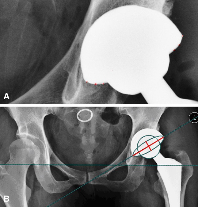
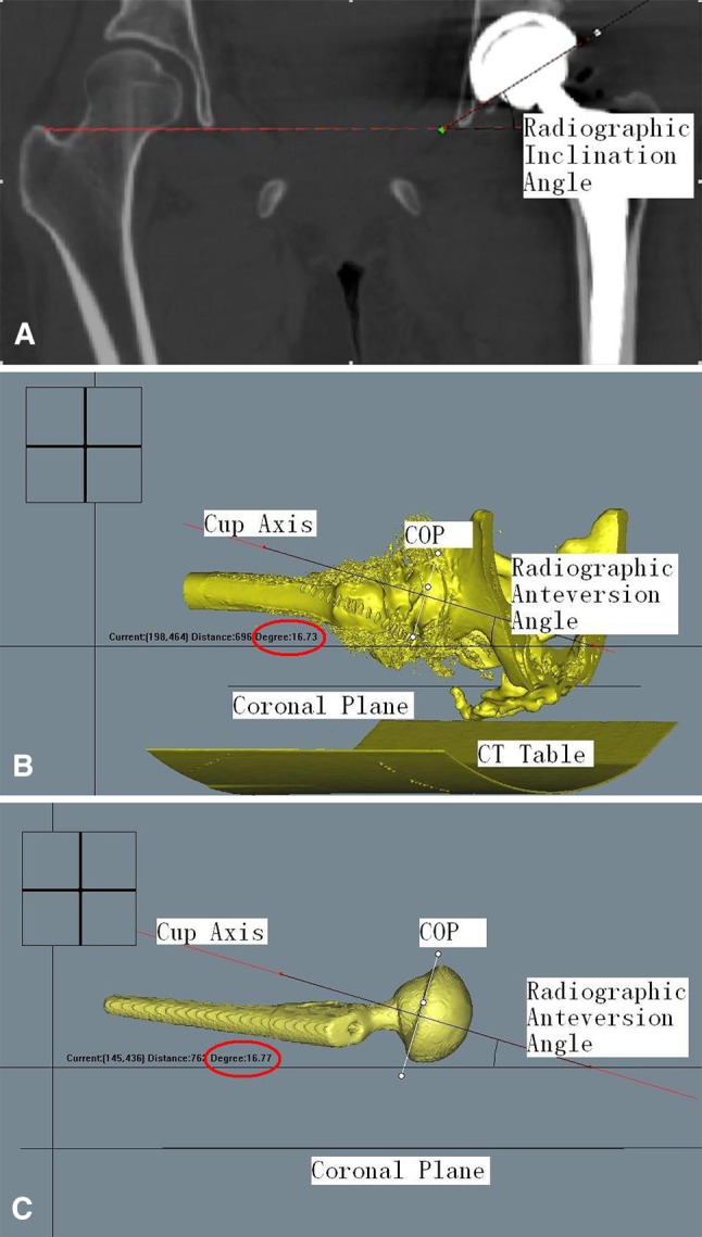
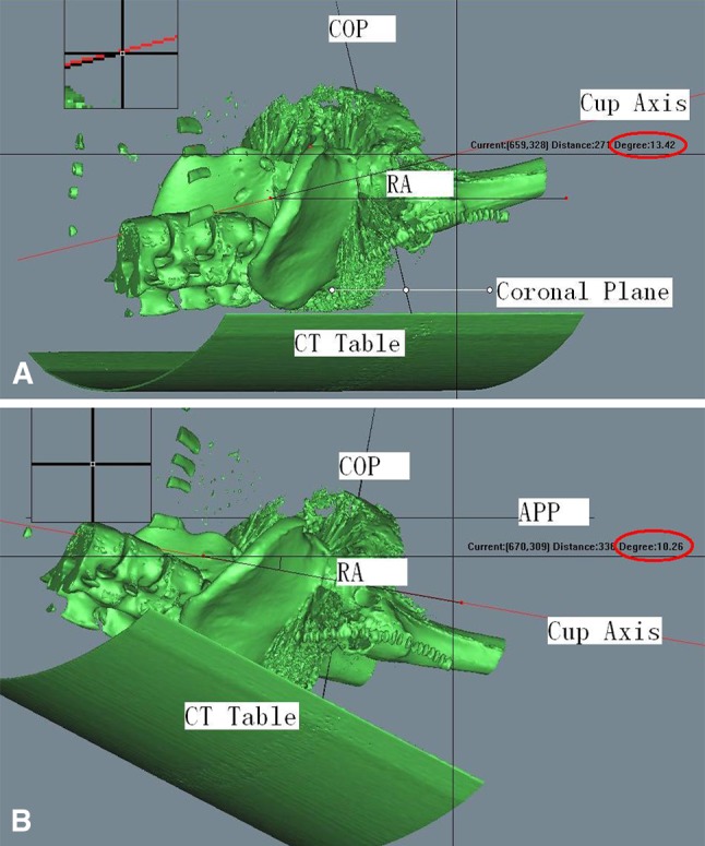
Methods: We obtained plain AP radiographs and CT scans for 60 patients who underwent 60 primary total hip arthroplasties (THAs). The method devised by Lewinnek et al. was used to measure the orientation of acetabular components on plain AP radiographs, and three-dimensional (3-D) CT scans were used to measure both the radiographic anteversion angle and the inclination angle. Reliability was evaluated by analysis of the agreement between inter- and intraobserver measurements using plain AP radiographs. Measurements on 3-D CT scans were regarded as the reference standard; validity was assessed by comparing radiographic measurements with the CT scans.

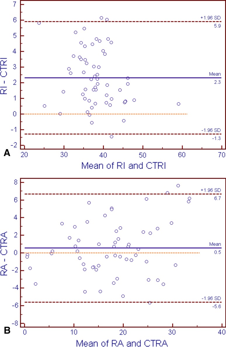
Results: Inter- and intraobserver reliability for measuring component orientation on plain AP radiographs was nearly perfect with intraclass correlation coefficients of 0.896 and 0.969 for anteversion and 0.984 and 0.993 for inclination. Measurement of cup inclination angles differed between plain radiographs and CT scans, but the difference was small, and the difference, although statistically significant, probably was not clinically important (2.3° ± 1.8°, p < 0.001). There was no significant difference between the anteversion as measured on CT scan versus that measured on plain radiographs (p = 0.19).

Conclusions: Measurement of the orientation of acetabular components on plain AP radiographs is reliable and accurate compared with measurement on CT.


Figures

Fig. 1A–B
Fig. 1A–B
(A) Sequence for drawing the elliptical appearance of the acetabular cup rim. First determine the two apexes of the cup, which is also the direction of the long axis. Then draw four random points on the cup opening rim, which is not obscured by the femoral head; the ellipse will be automatically completed. (B) Measuring the distance of the short and long axes of the ellipse (red lines) and inclination angle. Anteversion = arcsin (short axis/long axis); inclination is the angle between the line on which the long axis of the ellipse is located and the interteardrop line.
Fig. 2A–C
Fig. 2A–C
(A) Measurement of radiographic inclination on the CT coronal plane. (B) Measurement of radiographic anteversion on 3-D CT. The cup opening plane (COP) and the coronal plane (two lines coming together) are both perpendicular to the screen. Here, for the purposes of illustration, the coronal plane (the dotted line) is not quite parallel to the horizontal line; otherwise, it would not be visible. The screen here is the plane on which radiographic anteversion (the angle between the axis and the horizontal line) should be measured using screen-measuring software. (C) For the convenience of measurement, we have shown only the prosthesis and lines here by making the bony pelvis invisible.
Fig. 3A–B
Fig. 3A–B
Bland-Altman plots showed that the means of the measurements were spread evenly and randomly for inclination (RI) (A) and anteversion (B). CTRI = CT measurements of radiographic inclination; CTRA = CT measurements of radiographic anteversion.
Fig. 4A–B
Fig. 4A–B
(A) Hip with ankylosing spondylitis and severe pelvic posterior tilt. Radiographic orientation, measured using the functional coronal plane, was in 13.4° of anteversion. (B) Hip with ankylosing spondylitis and severe pelvic posterior tilt. Radiographic orientation, measured using the APP as the coronal plane, was in 10.2° of retroversion. RA = radiographic anteversion; COP = cup opening plane.

References

    1. Ackland MK, Bourne WB, Uhthoff HK. Anteversion of the acetabular cup: measurement of angle after total hip replacement. J Bone Joint Surg Br. 1986;68:409–413. - PubMed
    1. Babisch JW, Layher F, Amiot LP. The rationale for tilt-adjusted acetabular cup navigation. J Bone Joint Surg Am. 2008;90:357–365. doi: 10.2106/JBJS.F.00628. - DOI - PubMed
    1. Barrack RL, Lavernia C, Ries M, Thornberry R, Tozakoglou E. Virtual reality computer animation of the effect of component position and design on stability after total hip arthroplasty. Orthop Clin North Am. 2001;32:569–579. doi: 10.1016/S0030-5898(05)70227-3. - DOI - PubMed
    1. Del Schutte H, Jr, Lipman AJ, Bannar SM, Livermore JT, Ilstrup D, Morrey BF. Effects of acetabular abduction on cup wear rates in total hip arthroplasty. J Arthroplasty. 1998;13:621–626. doi: 10.1016/S0883-5403(98)80003-X. - DOI - PubMed
    1. DiGioia AM, 3rd, Jaramaz B, Plakseychuk AY, Moody JE, Jr, Nikou C, Labarca RS, Levison TJ, Picard F. Comparison of a mechanical acetabular alignment guide with computer placement of the socket. J Arthroplasty. 2002;17:359–364. doi: 10.1054/arth.2002.30411. - DOI - PubMed

Publication types