

Segmentation, feature extraction, and multiclass brain tumor classification
- PMID: 23645344
- PMCID: PMC3824920
- DOI: 10.1007/s10278-013-9600-0
Segmentation, feature extraction, and multiclass brain tumor classification
Abstract
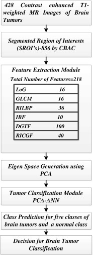
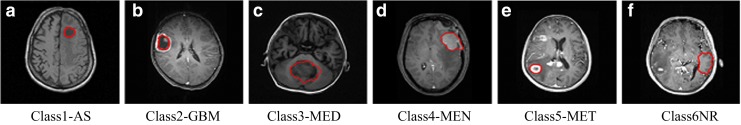
Multiclass brain tumor classification is performed by using a diversified dataset of 428 post-contrast T1-weighted MR images from 55 patients. These images are of primary brain tumors namely astrocytoma (AS), glioblastoma multiforme (GBM), childhood tumor-medulloblastoma (MED), meningioma (MEN), secondary tumor-metastatic (MET), and normal regions (NR). Eight hundred fifty-six regions of interest (SROIs) are extracted by a content-based active contour model. Two hundred eighteen intensity and texture features are extracted from these SROIs. In this study, principal component analysis (PCA) is used for reduction of dimensionality of the feature space. These six classes are then classified by artificial neural network (ANN). Hence, this approach is named as PCA-ANN approach. Three sets of experiments have been performed. In the first experiment, classification accuracy by ANN approach is performed. In the second experiment, PCA-ANN approach with random sub-sampling has been used in which the SROIs from the same patient may get repeated during testing. It is observed that the classification accuracy has increased from 77 to 91 %. PCA-ANN has delivered high accuracy for each class: AS-90.74 %, GBM-88.46 %, MED-85 %, MEN-90.70 %, MET-96.67 %, and NR-93.78 %. In the third experiment, to remove bias and to test the robustness of the proposed system, data is partitioned in a manner such that the SROIs from the same patient are not common for training and testing sets. In this case also, the proposed system has performed well by delivering an overall accuracy of 85.23 %. The individual class accuracy for each class is: AS-86.15 %, GBM-65.1 %, MED-63.36 %, MEN-91.5 %, MET-65.21 %, and NR-93.3 %. A computer-aided diagnostic system comprising of developed methods for segmentation, feature extraction, and classification of brain tumors can be beneficial to radiologists for precise localization, diagnosis, and interpretation of brain tumors on MR images.
Figures
References
-
- Caselles V. Geodesic active contours. Int J Comput Vision. 1997;22(3):61–79. doi: 10.1023/A:1007979827043. - DOI
-
- Dou W, Ruan S, Chen Y, Bloyet D, Constans J. A framework of fuzzy information fusion for the segmentation of brain tumor tissues on MR images. Image Vis Comput. 2007;25:164–171. doi: 10.1016/j.imavis.2006.01.025. - DOI
Publication types
MeSH terms
LinkOut - more resources
Full Text Sources
Other Literature Sources
Medical
Research Materials
Miscellaneous
