

Pulmonary cerium dioxide nanoparticle exposure differentially impairs coronary and mesenteric arteriolar reactivity
- PMID: 23645470
- PMCID: PMC3796163
- DOI: 10.1007/s12012-013-9213-3
Pulmonary cerium dioxide nanoparticle exposure differentially impairs coronary and mesenteric arteriolar reactivity
Abstract
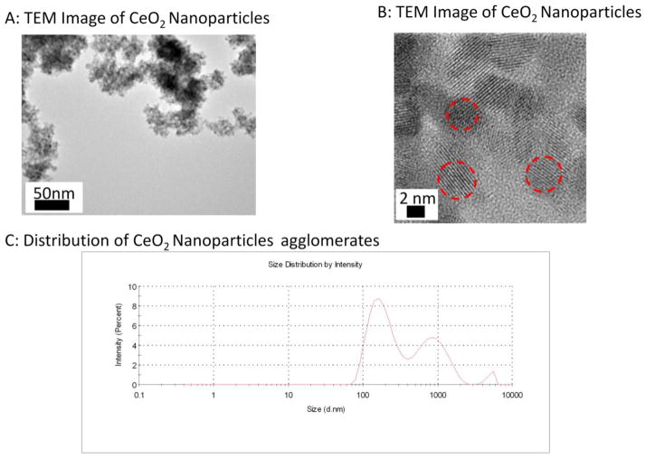
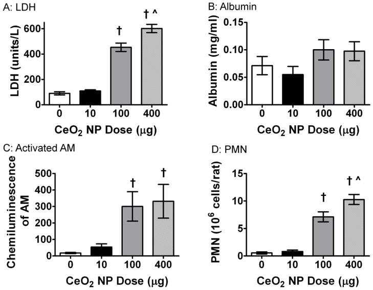
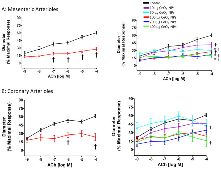
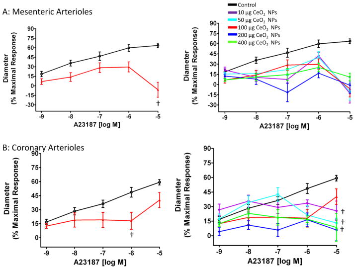
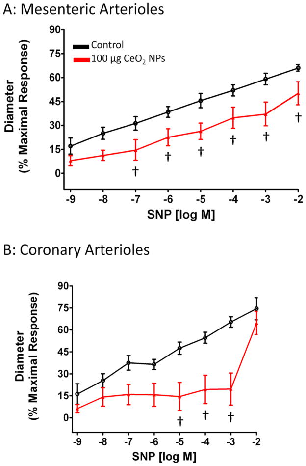
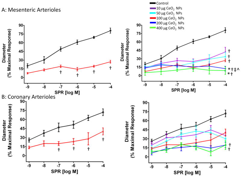
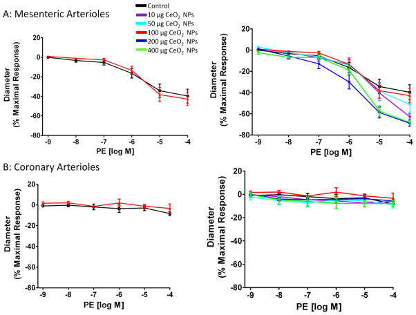
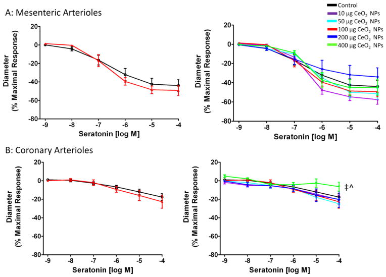
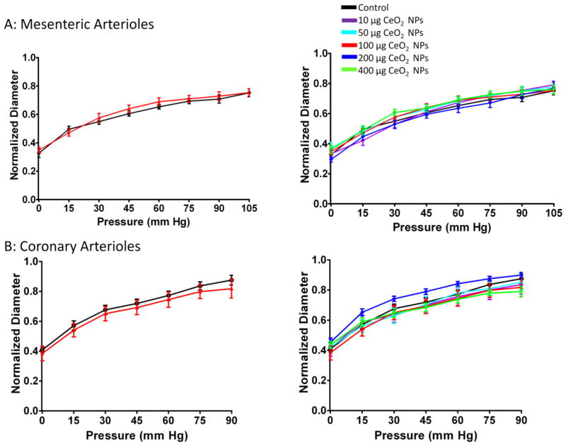
Cerium dioxide nanoparticles (CeO2 NPs) are an engineered nanomaterial (ENM) that possesses unique catalytic, oxidative, and reductive properties. Currently, CeO2 NPs are being used as a fuel catalyst but these properties are also utilized in the development of potential drug treatments for radiation and stroke protection. These uses of CeO2 NPs present a risk for human exposure; however, to date, no studies have investigated the effects of CeO2 NPs on the microcirculation following pulmonary exposure. Previous studies in our laboratory with other nanomaterials have shown impairments in normal microvascular function after pulmonary exposures. Therefore, we predicted that CeO2 NP exposure would cause microvascular dysfunction that is dependent on the tissue bed and dose. Twenty-four-hour post-exposure to CeO2 NPs (0-400 μg), mesenteric, and coronary arterioles was isolated and microvascular function was assessed. Our results provided evidence that pulmonary CeO2 NP exposure impairs endothelium-dependent and endothelium-independent arteriolar dilation in a dose-dependent manner. The CeO2 NP exposure dose which causes a 50 % impairment in arteriolar function (EC50) was calculated and ranged from 15 to 100 μg depending on the chemical agonist and microvascular bed. Microvascular assessments with acetylcholine revealed a 33-75 % reduction in function following exposure. Additionally, there was a greater sensitivity to CeO2 NP exposure in the mesenteric microvasculature due to the 40 % decrease in the calculated EC50 compared to the coronary microvasculature EC50. CeO2 NP exposure increased mean arterial pressure in some groups. Taken together, these observed microvascular changes may likely have detrimental effects on local blood flow regulation and contribute to cardiovascular dysfunction associated with particle exposure.
Figures
Similar articles
-
Cerium Dioxide Nanoparticle Exposure Improves Microvascular Dysfunction and Reduces Oxidative Stress in Spontaneously Hypertensive Rats.Front Physiol. 2015 Nov 17;6:339. doi: 10.3389/fphys.2015.00339. eCollection 2015. Front Physiol. 2015. PMID: 26635625 Free PMC article.
-
Intravenous and gastric cerium dioxide nanoparticle exposure disrupts microvascular smooth muscle signaling.Toxicol Sci. 2015 Mar;144(1):77-89. doi: 10.1093/toxsci/kfu256. Epub 2014 Dec 5. Toxicol Sci. 2015. PMID: 25481005 Free PMC article.
-
Group II innate lymphoid cells and microvascular dysfunction from pulmonary titanium dioxide nanoparticle exposure.Part Fibre Toxicol. 2018 Nov 9;15(1):43. doi: 10.1186/s12989-018-0280-2. Part Fibre Toxicol. 2018. PMID: 30413212 Free PMC article.
-
Recent insights into the impact, fate and transport of cerium oxide nanoparticles in the plant-soil continuum.Ecotoxicol Environ Saf. 2021 Sep 15;221:112403. doi: 10.1016/j.ecoenv.2021.112403. Epub 2021 Jun 17. Ecotoxicol Environ Saf. 2021. PMID: 34147863 Review.
-
Ceria nanoparticles: biomedical applications and toxicity.J Zhejiang Univ Sci B. 2024 May 15;25(5):361-388. doi: 10.1631/jzus.B2300854. J Zhejiang Univ Sci B. 2024. PMID: 38725338 Free PMC article. Review.
Cited by
-
Cerium Dioxide Nanoparticle Exposure Improves Microvascular Dysfunction and Reduces Oxidative Stress in Spontaneously Hypertensive Rats.Front Physiol. 2015 Nov 17;6:339. doi: 10.3389/fphys.2015.00339. eCollection 2015. Front Physiol. 2015. PMID: 26635625 Free PMC article.
-
Ultrafine Particulate Matter Increases Cardiac Ischemia/Reperfusion Injury via Mitochondrial Permeability Transition Pore.Cardiovasc Toxicol. 2017 Oct;17(4):441-450. doi: 10.1007/s12012-017-9402-6. Cardiovasc Toxicol. 2017. PMID: 28194639 Free PMC article.
-
Impact of pulmonary exposure to gold core silver nanoparticles of different size and capping agents on cardiovascular injury.Part Fibre Toxicol. 2016 Aug 24;13(1):48. doi: 10.1186/s12989-016-0159-z. Part Fibre Toxicol. 2016. PMID: 27558113 Free PMC article.
-
Diabetes Upregulates Oxidative Stress and Downregulates Cardiac Protection to Exacerbate Myocardial Ischemia/Reperfusion Injury in Rats.Antioxidants (Basel). 2020 Jul 29;9(8):679. doi: 10.3390/antiox9080679. Antioxidants (Basel). 2020. PMID: 32751309 Free PMC article.
-
Nanopolystyrene translocation and fetal deposition after acute lung exposure during late-stage pregnancy.Part Fibre Toxicol. 2020 Oct 24;17(1):55. doi: 10.1186/s12989-020-00385-9. Part Fibre Toxicol. 2020. PMID: 33099312 Free PMC article.
References
-
- Hanson N, Harris J, Joseph LA, Ramakrishnan K, Thompson T. EPA Needs to Manage Nanomaterial Risks More Effectively. 2011. Report No.: 12-P-0162.
-
- Borm PJ, Muller-Schulte D. Nanoparticles in drug delivery and environmental exposure: same size, same risks? Nanomedicine (Lond) 2006;1(2):235–49. - PubMed
-
- Aitken RJ, Chaudhry MQ, Boxall AB, Hull M. Manufacture and use of nanomaterials: current status in the UK and global trends. Occup Med (Lond) 2006;56(5):300–6. - PubMed
-
- Cassee FR, van Balen EC, Singh C, Green D, Muijser H, Weinstein J, Dreher K. Exposure, health and ecological effects review of engineered nanoscale cerium and cerium oxide associated with its use as a fuel additive. Crit Rev Toxicol. 2011;41(3):213–29. - PubMed
Publication types
MeSH terms
Substances
Grants and funding
LinkOut - more resources
Full Text Sources
Other Literature Sources
Miscellaneous
