

Intramural gas in stomach along with acute calculus cholecystitis: an unusual association
- PMID: 23645637
- PMCID: PMC3670034
- DOI: 10.1136/bcr-2012-007757
Intramural gas in stomach along with acute calculus cholecystitis: an unusual association
Abstract
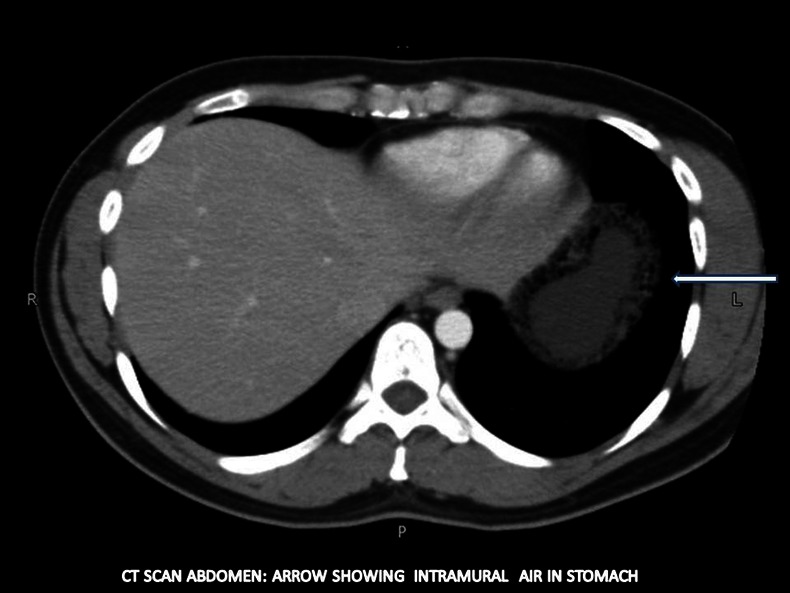
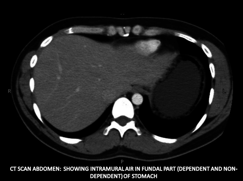
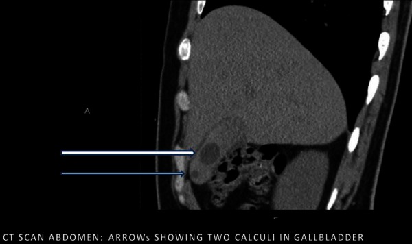
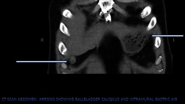
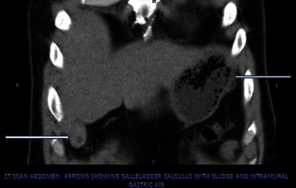
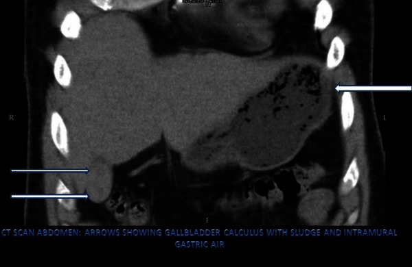
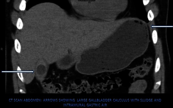
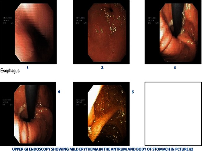
Intramural gas in stomach is a rare finding, but differential diagnosis of this condition into gastric emphysema and emphysematous gastritis is clinically important because of vastly different aetiologies and prognosis. Emphysematous gastritis is caused by gas producing micro-organisms inside the stomach wall and is a potentially fatal condition, while, on the other hand, gas enters stomach wall through mucosal breach in the case of gastric emphysema and prognosis is usually good with complete resolution. To date, no case has been reported in the literature showing gas in the stomach wall in a patient with acute calculus cholecystitis. We present a case of a young man with upper abdominal pain, and who, upon diagnostic work up was diagnosed with acute calculus cholecystitis with associated intramural gas in the stomach with no known aetiological factors to be positive. Conservative management with close observation resulted in complete symptomatic resolution.
Figures
Similar articles
-
A combination of intramural stomach and portal venous air: conservative treatment.J Community Hosp Intern Med Perspect. 2016 Feb 17;6(1):30519. doi: 10.3402/jchimp.v6.30519. eCollection 2016. J Community Hosp Intern Med Perspect. 2016. PMID: 26908389 Free PMC article.
-
Diagnostic dilemma of gastric intramural air.Ann R Coll Surg Engl. 2014 Oct;96(7):e11-3. doi: 10.1308/003588414X13946184901128. Ann R Coll Surg Engl. 2014. PMID: 25245715 Free PMC article.
-
The role of the Tokyo guidelines in the diagnosis of acute calculous cholecystitis.J Hepatobiliary Pancreat Sci. 2010 Nov;17(6):879-84. doi: 10.1007/s00534-010-0289-x. Epub 2010 Apr 24. J Hepatobiliary Pancreat Sci. 2010. PMID: 20419385
-
An Unusual Case of Acute Cholecystitis with Amyloidosis: A Case Report and Literature Review.Intern Med. 2019 Mar 15;58(6):803-807. doi: 10.2169/internalmedicine.1805-18. Epub 2018 Nov 19. Intern Med. 2019. PMID: 30449804 Free PMC article.
-
Emphysematous gastritis: case report and literature review.Int J Surg. 2008 Dec;6(6):e63-6. doi: 10.1016/j.ijsu.2007.02.007. Epub 2007 Mar 3. Int J Surg. 2008. PMID: 17446149 Review.
Cited by
-
Gastric Emphysema in a Critically Ill Patient Successfully Treated without Surgery.Case Rep Crit Care. 2019 Mar 18;2019:1824101. doi: 10.1155/2019/1824101. eCollection 2019. Case Rep Crit Care. 2019. PMID: 31011454 Free PMC article.
-
A combination of intramural stomach and portal venous air: conservative treatment.J Community Hosp Intern Med Perspect. 2016 Feb 17;6(1):30519. doi: 10.3402/jchimp.v6.30519. eCollection 2016. J Community Hosp Intern Med Perspect. 2016. PMID: 26908389 Free PMC article.
References
-
- Arezzo A, Famiglietti F, Garabello D, et al. Complete resolution of emphysematous gastritis after conservative management. Clin Gastroenterol Hepatol 2011;2013:e30. - PubMed
-
- Nasr D, Bernard P, Basile N. [Emphysematous gastritis]. J Chir (Paris) 2007;2013:439–40 - PubMed
-
- Hyun YS, Han DS, Lee HL, et al. Gastric emphysema after endoscopic submucosal dissection. Endoscopy 2011;2013(Suppl 2 UCTN):E83–4 - PubMed
-
- Zenooz NA, Robbin MR, Perez V. Gastric pneumatosis following nasogastric tube placement: a case report with literature review. Emerg Radiol 2007;2013:205–7 - PubMed
Publication types
MeSH terms
Substances
LinkOut - more resources
Full Text Sources
Other Literature Sources
Medical