. 2013 Jun 14;288(24):17202-13.
doi: 10.1074/jbc.M113.466920. Epub 2013 May 3.

A self-defeating anabolic program leads to β-cell apoptosis in endoplasmic reticulum stress-induced diabetes via regulation of amino acid flux

Affiliations

A self-defeating anabolic program leads to β-cell apoptosis in endoplasmic reticulum stress-induced diabetes via regulation of amino acid flux

Dawid Krokowski et al. J Biol Chem. .

Abstract

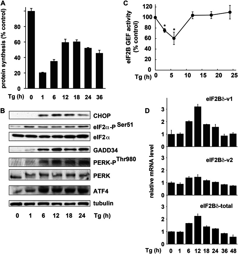
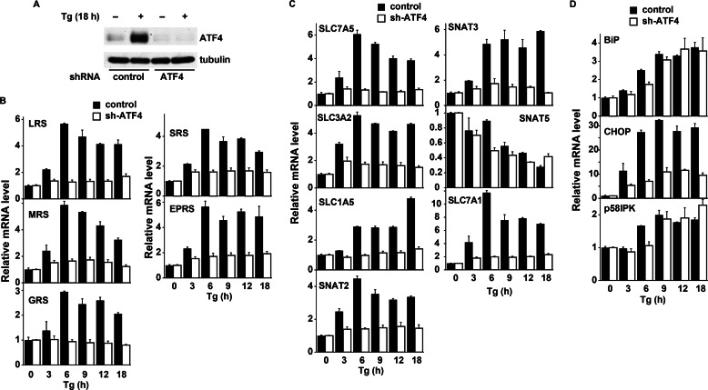
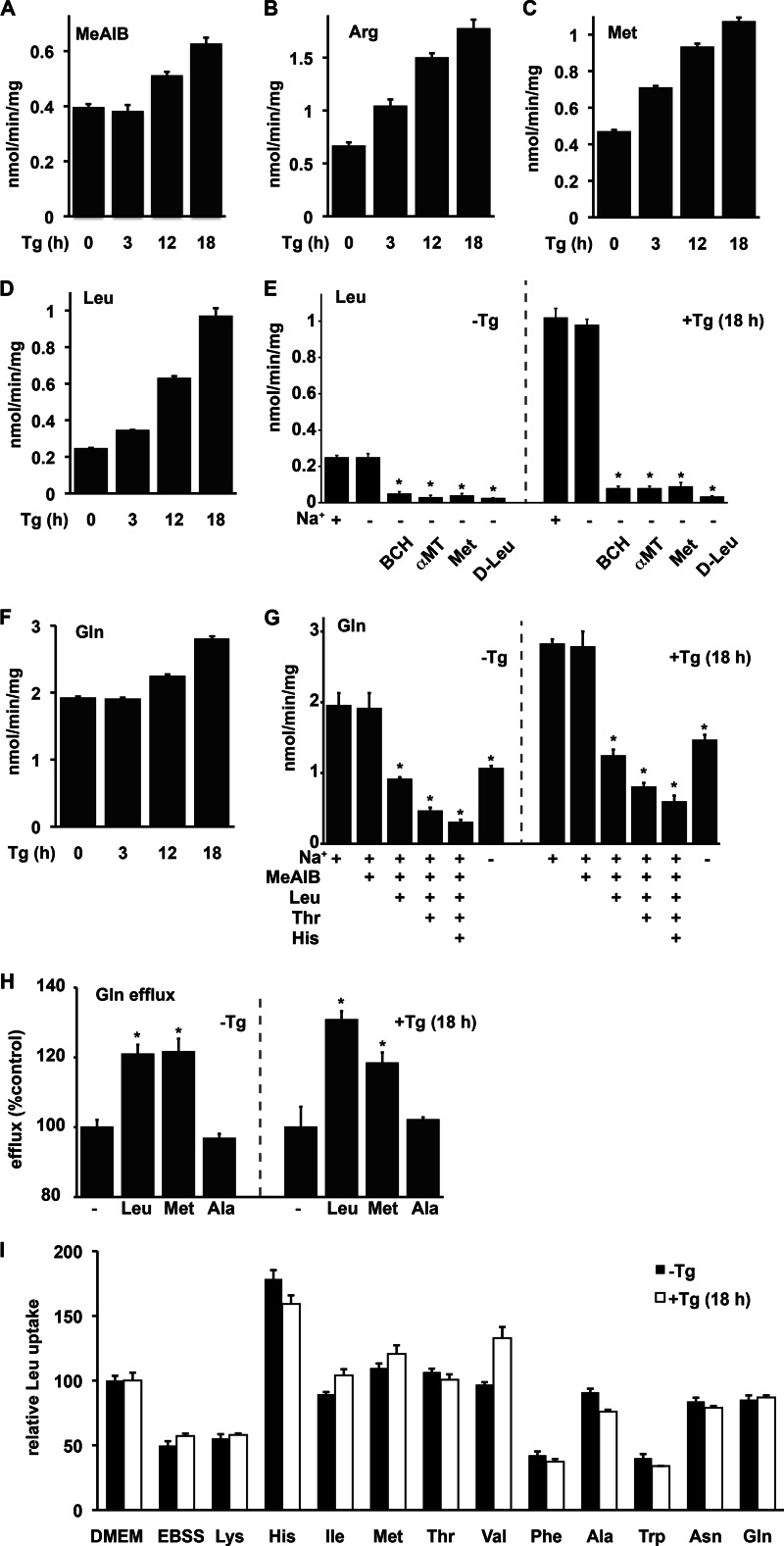
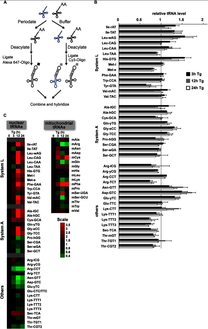
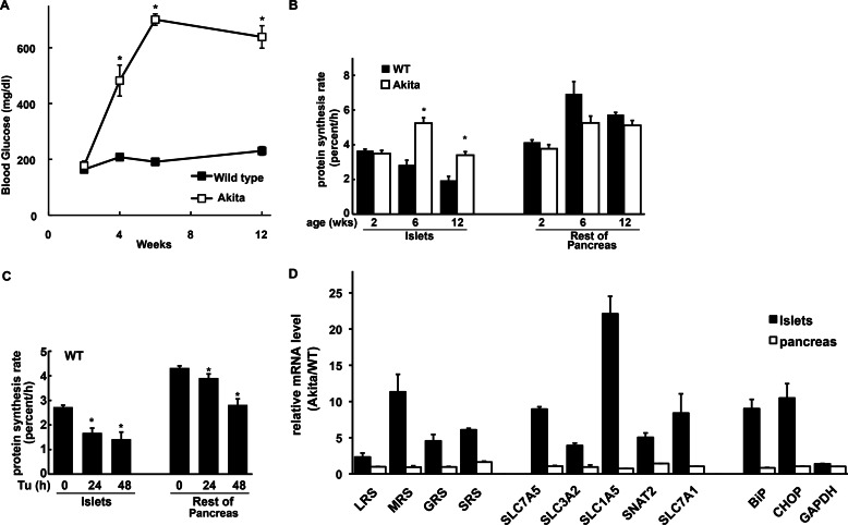
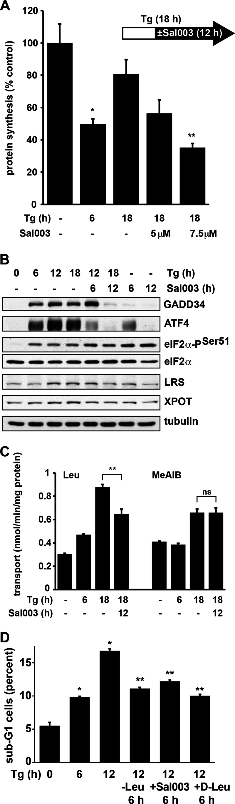
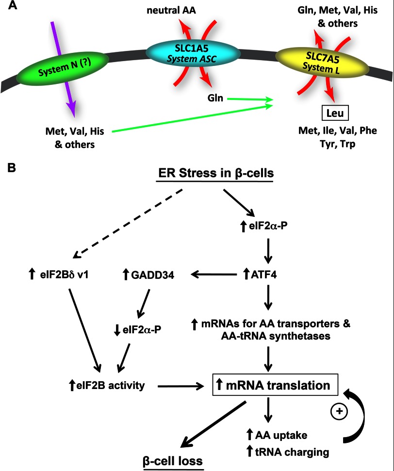
Endoplasmic reticulum (ER) stress-induced responses are associated with the loss of insulin-producing β-cells in type 2 diabetes mellitus. β-Cell survival during ER stress is believed to depend on decreased protein synthesis rates that are mediated via phosphorylation of the translation initiation factor eIF2α. It is reported here that chronic ER stress correlated with increased islet protein synthesis and apoptosis in β-cells in vivo. Paradoxically, chronic ER stress in β-cells induced an anabolic transcription program to overcome translational repression by eIF2α phosphorylation. This program included expression of amino acid transporter and aminoacyl-tRNA synthetase genes downstream of the stress-induced ATF4-mediated transcription program. The anabolic response was associated with increased amino acid flux and charging of tRNAs for branched chain and aromatic amino acids (e.g. leucine and tryptophan), the levels of which are early serum indicators of diabetes. We conclude that regulation of amino acid transport in β-cells during ER stress involves responses leading to increased protein synthesis, which can be protective during acute stress but can lead to apoptosis during chronic stress. These studies suggest that the increased expression of amino acid transporters in islets can serve as early diagnostic biomarkers for the development of diabetes.

Keywords: Amino Acid Transport; Aminoacyl tRNA Synthetase; Beta Cell; Diabetes; ER Stress; Glutamine.


Figures

FIGURE 1.
FIGURE 1.
Translational recovery in Min6 cells during prolonged ER stress. A, [35S]Met/Cys incorporation into proteins in Min6 cells treated with Tg for the indicated times. Results are the mean of triplicate determinations. All Tg-treated samples are significantly less than the untreated control p < 0.01. B, Western blot analysis of extracts from cells treated with Tg for the indicated times. Antibodies for the listed antigens were from the following vendors: eIF2a-PSer51, PERK-PThr980, and PERK from Cell Signaling; CHOP, GADD34, ATF4, and eIF2α from Santa Cruz Biotechnology; tubulin from Sigma. C, eIF2B-GEF activity measured in extracts from cells treated with Tg for the indicated times. Data are expressed as mean ± S.E. (error bars) of three independent experiments. *, significantly different from the untreated control (p < 0.01). D, qPCR analysis of mRNA isolated from cells treated with Tg for the indicated times. qPCR primers detect the indicated variants or total eIF2Bδ mRNA. Results of duplicate analyses were normalized to 18S rRNA and shown as -fold change over untreated cells. One-way ANOVA showed significant changes for each of the mRNAs over time (p < 0.01).
FIGURE 2.
FIGURE 2.
Induction of aminoacyl-tRNA synthetase and AA transporter genes during ER stress in Min6 cells involves the transcription factor ATF4. A, Western blot analysis of the ATF4 protein in Min6 cells infected with adenovirus expressing control and shRNA against ATF4. B–D, 5 days after infection, cells were treated with Tg for the indicated times and used for qPCR analysis. B, aminoacyl-tRNA synthetase genes. C, plasma membrane AA transporter genes. D, stress-induced marker genes. As expected, induction of the ER chaperone genes (BiP and p58IPK) was independent of ATF4, and induction of CHOP mRNA was severely inhibited in cells depleted of ATF4. Two-way ANOVA showed significant time-dependent changes for all samples and significant effects for the shRNA in all samples except BiP and p58IPK (p < 0.01).
FIGURE 3.
FIGURE 3.
Regulation of AA flux in Min6 cells during ER stress. A–D, uptake of MeAIB by system A (A), Arg by system y+ (B), and Met and Leu by system L (C and D) tested in unstressed cells and after Tg treatment for the indicated times. E, effect of Na+ ions and inhibitors of system L (BCH and α-methyltryptophan (α-MT)) and Met or d-Leu (5 mm) on Leu uptake in unstressed and Tg-treated cells. F, Na+-dependent uptake of Gln in cells treated with Tg for the indicated times. G, effect of Na+ ions and AA competitors (5 mm) on Gln uptake in untreated and Tg-treated cells. H, Gln efflux in the presence of 0.1 mm indicated AAs, in untreated or Tg-treated cells. I, Leu uptake in untreated and Tg-treated cells depleted of AAs (10-min incubation in EBSS), followed by 5 min re-feeding with individual AAs (5 mm). Data were normalized to Leu uptake by cells in DMEM. Results are expressed as mean ± S.E. (error bars) of three independent experiments. Tg caused significant increases in the uptake of MeAIB, Arg, Met, and Gln (A–D and F; one-way ANOVA (p < 0.01)). G and H, significant effects of added compounds (p < 0.05) are indicated (*). I, Leu uptake in all media was significantly different (p < 0.01) from EBSS, except media with Lys and Phe.
FIGURE 4.
FIGURE 4.
Genome-wide analysis of tRNA charging in Min6 cells during ER stress. A, tRNA was extracted from untreated and Tg-treated cells under acidic conditions to retain aminoacylated-tRNAs. One portion of each sample was treated with periodate, which oxidizes the 3′-acceptor stem of uncharged tRNAs. A second sample was kept in buffer solution. The tRNAs in both samples were deacylated and ligated to fluorescently tagged oligonucleotides with a stable stem-loop and a region complementary to the 3′-CCA sequence that is conserved among all tRNAs (Integrated DNA Technologies). This allowed the labeling of one sample with Cy3 and the other with Alexa Fluor 647. Samples were combined and hybridized to tRNA microarrays containing complementary probes for 40 nuclear encoded mouse tRNAs and 18 mouse mitochondria-encoded tRNAs. Microarrays were custom printed by Microarray Inc. (Nashville, TN). B, abundance of total tRNA in cells treated with Tg for the indicated times. The average of two independent experiments is shown. C, levels of charged tRNA shown as a TreeView image. All values are relative to unstressed cells. Assays were performed with Alexa Fluor 647-labeled charged tRNA and Cy3-labeled total tRNA or with Cy3-labeled charged tRNA and Alexa Fluor 647-labeled total tRNA. Data are the average from these two experiments. Green indicates a lower and red a higher level of charged tRNA during stress relative to control.
FIGURE 5.
FIGURE 5.
Regulation of protein synthesis and anabolic gene expression in pancreatic islets from Akita mice. A, blood glucose levels from Akita and WT (C57BL/6J) mice (n = 8). B, fractional protein synthesis rates measured as [2H]Ala enrichment in proteins from islets and rest of pancreas (remaining pancreatic tissue after removal of islets) in 2-, 6-, and 12-week-old male Akita (n = 6–8) and age/sex-matched WT littermates (n = 4–8). C, fractional protein synthesis rates in islets of 6-week-old male WT mice (n = 4) measured as [2H]Ala enrichment in proteins from islets and rest of pancreas after Tu injection (2 μg/g of body weight). A–C, *, significantly different from WT (p < 0.01). D, qPCR analysis of RNA from islets and whole pancreas from 6-week-old Akita male mice (n = 6) and age/sex-matched WT littermates (n = 4). The ratio of signals in Akita and WT mice is shown. For islets, all of the signals from Akita mice were significantly higher than WT (p < 0.05) for all mRNAs except GAPDH. No significant differences between Akita and WT were seen in the remaining pancreatic tissue.
FIGURE 6.
FIGURE 6.
Attenuation of translational recovery during ER stress in Min6 cells promotes survival. A, [35S]Met/Cys incorporation into proteins in cells treated with Tg and Sal003 for the indicated times. Sal003 was added 6 h after initiation of Tg treatment as indicated by the scheme. B, Western blot analysis of the indicated proteins from cells treated with Tg and Sal003 (7.5 μm for the last 6 or 12 h of Tg treatment). Antibodies against LRS and XPOT were from Abcam; others are listed in Fig. 1. C, Leu and MeAIB uptake in cells treated with Tg and Sal003 (7.5 μm) for the indicated times. D, cells were treated as indicated and apoptosis was assessed by measuring the percentage of sub-G1 cells by flow cytometry of propidium iodide-stained cells. Leu starvation was performed by replacing the media after 6 h of Tg treatment with Tg-containing Leu-free medium. d-Leu (40 mm) or Sal003 (7.5 μm) was added for the last 6 h of Tg treatment. Significant differences (p < 0.01) from untreated cells (*) or the appropriate Tg-treated cells (**) are indicated. Error bars, S.E.
FIGURE 7.
FIGURE 7.
Working model of the AA network and its contribution in regulation of protein synthesis in β-cells during ER stress. A, proposed mechanism for the increased uptake of Leu via system L during chronic ER stress. B, proposed model by which translational recovery leads to apoptosis during chronic ER stress.

References

    1. Samuel V. T., Shulman G. I. (2012) Mechanisms for insulin resistance: common threads and missing links. Cell 148, 852–871 - PMC - PubMed
    1. Back S. H., Kaufman R. J. (2012) Endoplasmic reticulum stress and type 2 diabetes. Annu. Rev. Biochem. 81, 767–793 - PMC - PubMed
    1. Back S. H., Scheuner D., Han J., Song B., Ribick M., Wang J., Gildersleeve R. D., Pennathur S., Kaufman R. J. (2009) Translation attenuation through eIF2α phosphorylation prevents oxidative stress and maintains the differentiated state in β- ‘cells. Cell Metab. 10, 13–26 - PMC - PubMed
    1. Papa F. R. (2012) Endoplasmic reticulum stress, pancreatic β-cell degeneration, and diabetes. Cold Spring Harb. Perspect. Med. 2, a007666. - PMC - PubMed
    1. Oslowski C. M., Urano F. (2011) The binary switch that controls the life and death decisions of ER-stressed β-cells. Curr. Opin. Cell Biol. 23, 207–215 - PMC - PubMed

Publication types

MeSH terms