

Renovascular hypertension in an 8-year-old girl
- PMID: 23645701
- PMCID: PMC3669994
- DOI: 10.1136/bcr-2013-009691
Renovascular hypertension in an 8-year-old girl
Abstract
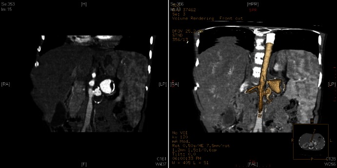
Secondary hypertension is the most common form of hypertension in childhood, particularly in the young age group: parenchymal disease and lesions of the renal artery account for the majority of such cases. Renal artery aneurysms (RAA) are rare and are usually diagnosed by Doppler ultrasonography or angiography performed in the investigation of specific clinical symptoms. We report herein a case of severe arterial hypertension in an 8-year-old girl arising from a large saccular RAA. Intravenous antihypertensive drugs were necessary to achieve blood pressure control and the final diagnosis was obtained from angio-CT scan and selective angiography that demonstrated a large saccular aneurysm of the left renal artery with parietal calcification. After confirmation of inexistent function of the entire left kidney by Tc99m-MAG3 renal isotope scan, nephrectomy was performed. The child's blood pressure further normalised and, 1 month after surgery, she had ceased any antihypertensive therapy.
Figures


Similar articles
-
Calcified renal artery aneurism in the right kidney causing hypertension.Saudi J Kidney Dis Transpl. 2020 Jan-Feb;31(1):266-270. doi: 10.4103/1319-2442.279951. Saudi J Kidney Dis Transpl. 2020. PMID: 32129223
-
Primary intimal fibroplasia with multiple aneurysms of renal artery in childhood.Child Nephrol Urol. 1990;10(1):51-5. Child Nephrol Urol. 1990. PMID: 2354468
-
[Calcified renal artery aneurism and high blood pressure. A case report and review of the literature].Cir Cir. 2004 May-Jun;72(3):217-20. Cir Cir. 2004. PMID: 15310449 Review. Spanish.
-
Aneurysmectomy with arterial reconstruction of renal artery aneurysms in the endovascular era: a safe, effective treatment for both aneurysm and associated hypertension.Ann Vasc Surg. 2010 May;24(4):503-10. doi: 10.1016/j.avsg.2009.07.030. Epub 2009 Dec 29. Ann Vasc Surg. 2010. PMID: 20036510
-
Renovascular hypertension.Annu Rev Med. 1984;35:665-92. doi: 10.1146/annurev.me.35.020184.003313. Annu Rev Med. 1984. PMID: 6232894 Review.
Cited by
-
High prevalence of elevated blood pressure among children with neurofibromatosis type 1.Pediatr Nephrol. 2016 Jan;31(1):131-6. doi: 10.1007/s00467-015-3191-6. Epub 2015 Aug 28. Pediatr Nephrol. 2016. PMID: 26314566
References
-
- Robitaille P, Lord H, Dubois J, et al. A large unilateral renal artery aneurysm in a young child. Pediatr Radiol 2004;2013:253–5 - PubMed
-
- Hobbs DJ, Barletta GM, Mowry JA, et al. Renovascular hypertension and intrarenal artery aneurysms in a preschool child. Pediatr Radiol 2009;2013:988–90 - PubMed
-
- Gumustas S, Ciftci E, Bircan Z. Renal artery aneurysm in a hypertensive child treated by percutaneous coil embolization. Pediatr Radiol 2010;2013:1285–7 - PubMed
-
- Oguzkurt L, Cekirge S, Balkanci F. Inferior suprarenal artery aneurysm in polyarteritis nodosa. Pediatr Radiol 1997;2013:234–5 - PubMed
-
- McCulloch M, Andronikou S, Goddard E, et al. Angiographic features of 26 children with Takayasu's arteritis. Pediatr Radiol 2003;2013:230–5 - PubMed
Publication types
MeSH terms
Substances
LinkOut - more resources
Full Text Sources
Other Literature Sources
Medical