Review
. 2013 Jun 1;3(6):a013433.
doi: 10.1101/cshperspect.a013433.

Cell-free hemoglobin and its scavenger proteins: new disease models leading the way to targeted therapies

Affiliations
Review

Cell-free hemoglobin and its scavenger proteins: new disease models leading the way to targeted therapies

Dominik J Schaer et al. Cold Spring Harb Perspect Med. .

Abstract

Hemoglobin (Hb) has multiple pathophysiologic effects when released into the intravascular space during hemolysis. The extracellular effects of Hb have resulted in novel models of toxicity, which help to explain endothelial dysfunction and cardiovascular complications that accompany genetic hemolytic anemias, malaria, blood transfusion, and atherosclerosis. The majority of models focus on nitric oxide (NO) depletion; however, in local tissue environments, Hb can also act as a pro-oxidant and inflammatory agent. This can alter cellular differentiation with the potential to deviate immune responses. The understanding of these mechanisms set in the context of natural scavenger and detoxification systems may accelerate the development of novel treatment strategies.


Figures
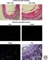
Figure 1.
Figure 1.
Vascular effects of old blood transfusion-associated hemolysis. The top panel shows regions of vascular necrosis appearing to initiate within the vascular lumen and progress to the tunica media. These observations were captured 24 h after massive transfusion to guinea pigs at 100× and 400× magnification. The injury in isolated regions of the aorta in transfused animals was consistent with coagulative necrosis and may be the result of hemoglobin and/or damaged red blood cells. The lower panels indicate macrophage accumulation to hemoglobin-rich regions within the adventitia consistent with HO-1, CD163, and nonheme iron accumulation.
Figure 2.
Figure 2.
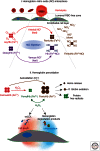
Schematic of hemoglobin-mediated toxicity initiated by intravascular hemolysis. (I) NO reactions with hemoglobin—Red blood cells (RBC) do not enter the vascular luminal RBC-free zone; however, free hemoglobin can enter the RBC-free zone and gain access to endothelial and perivascular spaces. When free oxyhemoglobin (red) enters the perivascular space it can react with NO rapidly via dioxygenation forming ferric hemoglobin (brown) and nitrate (NO3). Additionally, deoxyhemoglobin (purple) can react with NO to form iron nitrosyl hemoglobin. Additionally, ferric hemoglobin can react with NO, but at much slower rates. It remains unknown whether this reaction is of any biologic significance. (II) Hemoglobin peroxidative reactions—Oxyhemoglobin (red) undergoes peroxidation to ferryl hemoglobin (green) followed by a redox cycle that is driven by hydrogen peroxide. In the presence of large quantities of reducing agents ferryl stability in this reaction is reduced to an extent that the species cannot accumulate, and in most cases cannot be directly detected. The most prevalent oxidized Hb species in vivo is therefore ferric Hb, which can result from peroxidation but also from autoxidation and NO dioxygenation depicted above. In vitro, the result of redox cycling mediated by H2O2 can be release of heme, globin chain radicals, and oxidation of amino acids within the hemoglobin molecule. The protein may also degrade to precipitate formed by globin-heme adducts and cross-links that physically damage endothelial cells leading to inflammation. Additionally, free radical transfer from ferryl radical oxidizes lipid, leading to potential tissue oxidation as well as inflammation.
Figure 3.
Figure 3.
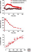
Plasma concentration blood pressure relationships. (Top) The blood pressure response to ferrous (red) and ferric (brown) hemoglobin following a 20% top load to conscious guinea pigs. Ferric hemoglobin shows no response, whereas ferrous hemoglobin shows a rapid and transient increase in mean arterial pressure. This is consistent with the hemoglobin plasma concentration time course shown (middle). These two time effects can be better visualized by plotting plasma concentration versus percent mean arterial blood pressure change from baseline (bottom).
Figure 4.
Figure 4.
Physiologic hemoglobin scavengers and detoxification systems. Hb can be released from red blood cells (RBC) into the plasma and extracellular environment during hemolysis or tissue injury. In the extracellular compartment, Hb reacts with peroxides (H2O2 and lipid hydroperoxides) and promotes oxidative tissue damage. In the plasma or extracellular space, Hb is sequestered in the Hb–Hp complex. Complex formation prevents Hb-induced hypertensive and oxidative reactions. The Hb–Hp complex is subsequently endocytosed by the macrophage Hb scavenger receptor CD163. Within the macrophage, heme is released from globin and degraded by HO-1 into bilirubin and carbon monoxide (CO). The released iron induces ferritin synthesis. Iron can either be exported for iron recycling or it can be stored in a ferritin complex. Backup systems such as hemopexin (Hpx) and the Hpx–heme complex receptor (LDL receptor-related protein, LRP) pathway can bind and detoxify free heme that is released from oxidized Hb.
Figure 5.
Figure 5.
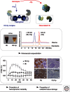
The haptoglobin paradigm of damage prevention during hemolysis. After hemolysis free Hb can dissociate into αβ dimers with a relatively small molecule size of ∼32 kD (eluting at 19.8 min in the shown plasma size exclusion chromatography [SEC] profile). Susceptible tissues are exposed to toxic Hb. The results are hemoglobinuria, hemodynamic instability with acute hypertension, and oxidative tissue damage. With haptoglobin (Hp) treatment 100% of the free Hb is captured within the large sized (>150 kD) Hb:Hp complex that elutes at 14.7 min in plasma SEC (note that there remains no free Hb in the Hp-treated plasma sample). As a result, free Hb remains compartmentalized within the circulation. Hemoglobinuria, hemodynamic instability, and oxidative tissue damage are prevented. The image in the left middle panel shows urine collections of Hb and Hb:Hp infused guinea pigs. The lower left panel shows blood pressure recordings of Hb, Hb:Hp, or starch (control) infused animals. The kidney sections in the lower right panel were stained for 4-HNE as a marker of oxidative tissue damage (brown color) in Hb-infused (but not in Hb transfused +Hp-treated) animals.

References

    1. Abraham NG, Kappas A 2008. Pharmacological and clinical aspects of heme oxygenase. Pharmacol Rev 60: 79–127 - PubMed
    1. Alayash AI 2004. Oxygen therapeutics: Can we tame haemoglobin? Nat Rev Drug Discov 3: 152–159 - PubMed
    1. Baek JH, D’Agnillo F, Vallelian F, Pereira CP, Williams MC, Jia Y, Schaer DJ, Buehler PW 2012. Hemoglobin-driven pathophysiology is an in vivo consequence of the red blood cell storage lesion that can be attenuated in guinea pigs by haptoglobin therapy. J Clin Invest 122: 1444–1458 - PMC - PubMed
    1. Baldwin AL, Wiley EB, Alayash AI 2002. Comparison of effects of two hemoglobin-based O(2) carriers on intestinal integrity and microvascular leakage. Am J Physiol Heart Circ Physiol 283: H1292–H1301 - PubMed
    1. Baldwin AL, Wiley EB, Summers AG, Alayash AI 2003. Sodium selenite reduces hemoglobin-induced venular leakage in the rat mesentery. Am J Physiol Heart Circ Physiol 284: H81–H91 - PubMed

Publication types

MeSH terms