Review
. 2013 Oct;34(5):691-724.
doi: 10.1210/er.2012-1072. Epub 2013 May 3.

Novel insights on thyroid-stimulating hormone receptor signal transduction

Affiliations
Review

Novel insights on thyroid-stimulating hormone receptor signal transduction

Gunnar Kleinau et al. Endocr Rev. 2013 Oct.

Abstract

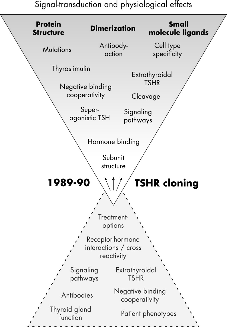
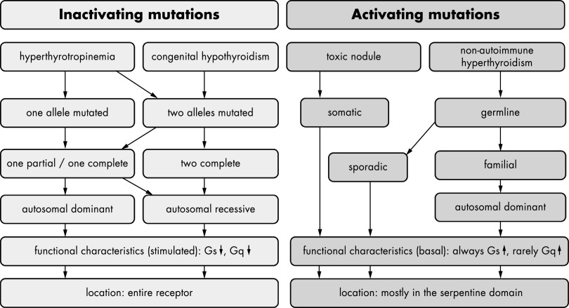
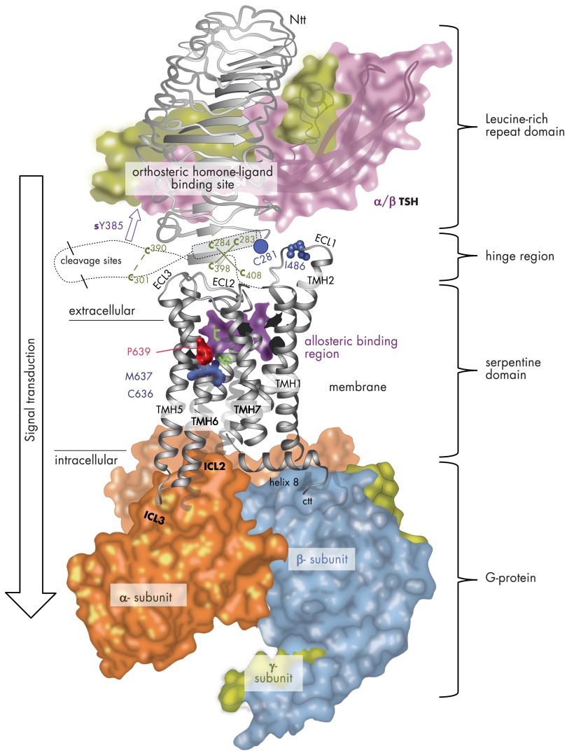
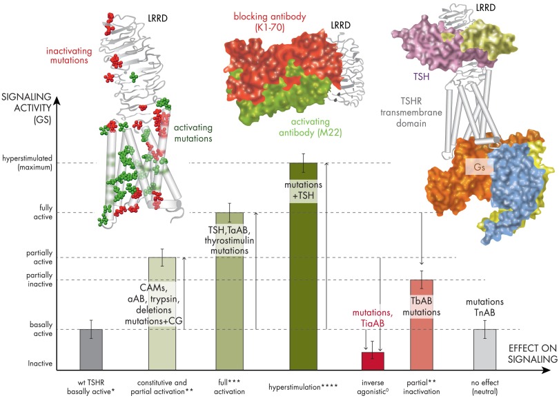
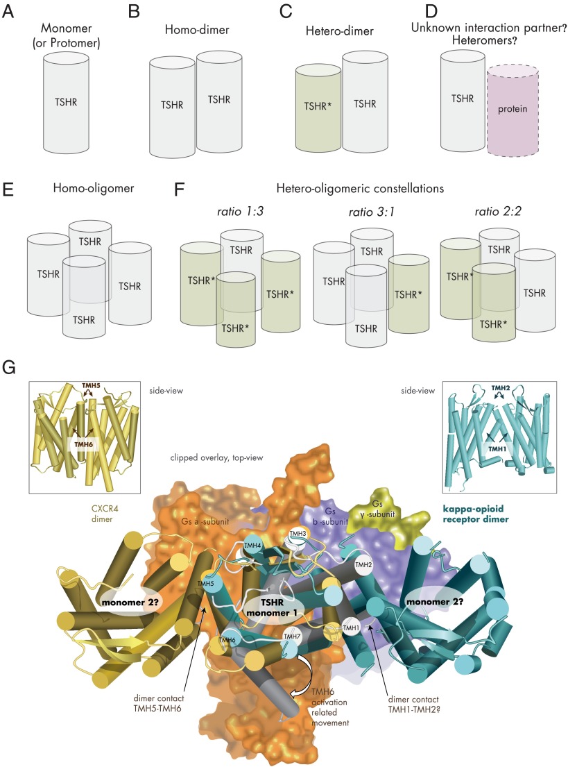
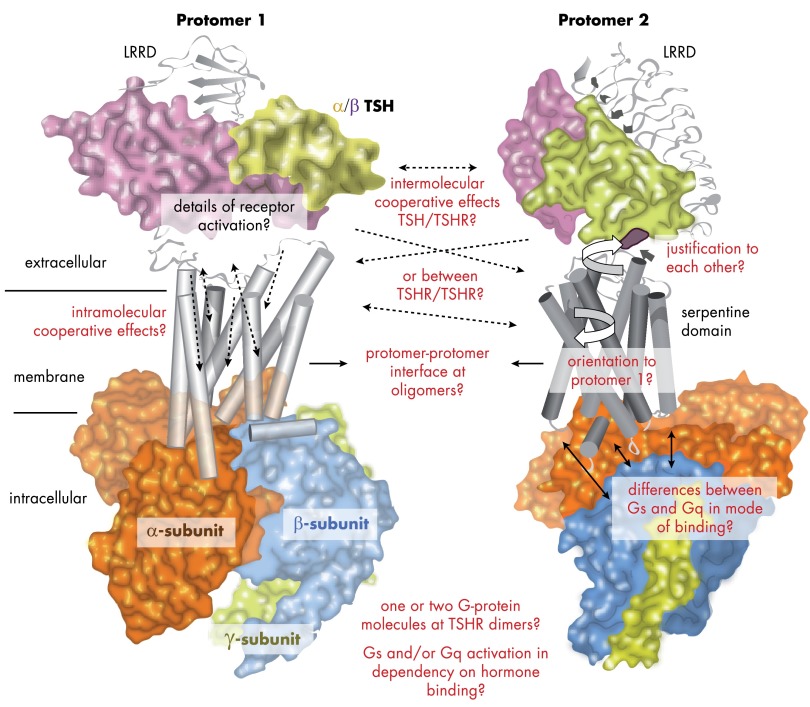
The TSH receptor (TSHR) is a member of the glycoprotein hormone receptors, a subfamily of family A G protein-coupled receptors. The TSHR is of great importance for the growth and function of the thyroid gland. The TSHR and its endogenous ligand TSH are pivotal proteins with respect to a variety of physiological functions and malfunctions. The molecular events of TSHR regulation can be summarized as a process of signal transduction, including signal reception, conversion, and amplification. The steps during signal transduction from the extra- to the intracellular sites of the cell are not yet comprehensively understood. However, essential new insights have been achieved in recent years on the interrelated mechanisms at the extracellular region, the transmembrane domain, and intracellular components. This review contains a critical summary of available knowledge of the molecular mechanisms of signal transduction at the TSHR, for example, the key amino acids involved in hormone binding or in the structural conformational changes that lead to G protein activation or signaling regulation. Aspects of TSHR oligomerization, signaling promiscuity, signaling selectivity, phenotypes of genetic variations, and potential extrathyroidal receptor activity are also considered, because these are relevant to an understanding of the overall function of the TSHR, including physiological, pathophysiological, and pharmacological perspectives. Directions for future research are discussed.


Figures

Figure 1.
Figure 1.
Principal topics of TSHR research. After cloning of the TSHR, scientific and medical investigation has developed exponentially, with the aim of characterizing the signal transduction process and the physiological impact of the TSHR. Many fields have been also rediscovered, but important basic questions still await answers, e.g., the relation between hormone binding and dimerization, activity regulation, or the physiological relevance of basal signaling activity. The genotype-phenotype relationship of TSHR dysfunctions cannot yet be clearly discriminated. The in vivo occurrence or physiological relevance of extracellular cleavage is still unknown, as are the tissue- and cell-specific functions and molecular characteristics of TSHR species, compared with experimental in vitro conditions. Finally, extrathyroidal TSHR expression is well supported, but the physiological impact in humans is still unknown.
Figure 2.
Figure 2.
Characteristics of inactivating and activating TSHR mutations. Phenotypic and functional implications of naturally occurring inactivating mutations are indicated in light gray. Such mutations were identified in patients suffering from hyperthyrotropinemia or congenital hypothyroidism. In the case of hyperthyrotropinemia, these mutations are either located on one (dominant inheritance) or on both alleles (recessive inheritance). In patients suffering from congenital hypothyroidism, mutations are located on both alleles. The mutations leading to congenital hypothyroidism (mostly investigated in heterologous overexpressing systems) are either complete loss-of-function mutations on both alleles or one complete loss-of-function mutation combined with a partial loss-of-function mutation. This combination of functional properties was also found in patients suffering from hyperthyrotropinemia; however, in that condition, the mutations are mostly partial loss-of-function mutations. The term “functional characteristics” refers to signaling properties after TSH simulation that resulted in a reduction of TSH-stimulated functionality. Inactivating mutations have been found in all receptor parts. Phenotypic and functional properties of activating mutations are indicated in dark gray. These mutations could occur either as somatic or as germline mutations. Germline mutations either are sporadic or were found as familial cases and were inherited in an autosomal dominant manner. Activating mutations are always characterized by an enhanced basal Gs signaling; a few mutations also show enhanced Gq signaling.
Figure 3.
Figure 3.
TSHR homology model with putative spatial orientations and interactions between receptor components, the hormone and G protein. This combined TSHR model highlights causal relationships between functional and structural data. This includes hormone and G protein binding at an activated TSHR monomer as well as mechanisms in the transmembrane region related to the activation process. GPHR crystal structures are available only for the LRRD and hinge region (Jul 2012: PDB entry codes 3G04, 2XWT, and 4AY0). The structure and binding of bTSH (backbone-ribbon and translucent, colored surface) at the LRRD (orthosteric binding site) is based on the assumption of similarity with the crystallized FSHR LRRD/FSH complexes. The hormone interacts with specific side chains at LRRs 2–9. The newly determined homologous FSHR extracellular region (LRRD and hinge region)/FSH crystal structures have shown 11 complete repeats at the extracellular region (140). It has been observed that the LRRD continues through specific amino acids of cysteine-box 2 (amino acids from Cys283 to Cys301) with a small helix conformation (cylinder) at repeat 11. A few important amino acids of cysteine-box 3 (amino acids Cys390 to Cys408) close to TMH1 are arranged as an additional β-strand, parallel to the LRRD. Six cysteines are disulfide bridged to each other between cysteine-box 2 (Cb-2) and Cb-3. In conclusion, the unit of 11 repeats and the C terminus of the extracellular region are located in close proximity as an interface to the transmembrane region. It is noteworthy that CAMs were identified at these receptor domains, e.g., at Ser281 (blue dot). Several amino acids with an impact on hormone binding and signaling have been detected between positions 289 and 410, including the sulfated tyrosine 385 (sTyr385, magenta arrow). Details of interaction between this residue and the hormone are now represented in the recent FSHR/FSH crystal structure (140) and also confirm that the hinge region is a central region for signal transduction from the extracellular site to further receptor components. However, the precise orientation of the LRRD/hormone, hinge region complex, and the SD to each other is not known yet (just exemplarily here). Newly identified naturally occurring side-chain substitutions, e.g., at wild-type position Pro639 (TMH6, red atom spheres, inactivating mutation) or Cys636 (TMH6, blue atom spheres) and Ile486 (ECL1, blue atom spheres), that lead to TSHR inactivation (red) or activation (blue) are mapped to the transmembrane-spanning SD. Mutations at Cys636, Met637, and Pro639 have provided details into TSHR because an activity switch in this conserved family A GPCR motif is localized at TMH6. The potential allosteric binding pocket for drug-like small-molecule ligands (417) between the helices and ECL2 (magenta) is highlighted by an inner pocket surface image (violet translucent surface). Finally, the activated SD is characterized by the capacity to activate G proteins. Orientation of the activated heterotrimeric Gs protein (backbones of α-, β-, and γ- subunits with translucent colored surface) at the active-state conformation of TSHR (white backbone ribbon) is in accordance with recently published data for G protein activation at the TSHR and the crystal structures of other GPCRs (101, 289, 324).
Figure 4.
Figure 4.
Scheme of TSHR activity states related to diverse effectors. This graphic visualizes the spectrum of putative signaling activity states (active, green; inactive, red) of the TSHR that are related to different structural features and effectors. The magnitudes of the bars are relative to each other and do not reflect exact data values. The TSHR wild type (wt) exhibits a permanent (basal) signaling activity (Gs activation) that is indicated by an asterisk (*) at the x-axis. CAMs, activating antibodies, treatment with trypsin, and specific mutations in combination with an increased CG affinity lead to an enhanced but partial signaling activity (**). TSH, thyrostimulin, or some thyroid-stimulating antibodies (TsABs) are able to activate the TSHR fully (***), but few cases are reported where an activation of more than 100% was observed (****). In contrast to receptor activation, specific mutations and thyroid-blocking antibodies (TbAB) reduce the maximum TSHR activation. Both blocking antibodies and mutations can have different mechanisms of inactivation such as suppression of the intrinsic signaling capacity (TSH independent), inhibition of hormone binding, or signaling activation (G protein). A specific case is inverse agonism (also called silencing) caused by mutations or antibodies (TiaAB). Here the activity is suppressed below the basal level of the wild type or lowered in case of a constitutively activated TSHR. To complete the potential scenarios, also neutral effects are known, where mutations or intermolecular effectors (neutral antibodies [TnAB]) show no primary effect on signaling. The picture on the left shows a structural TSHR model with mapped positions of naturally occurring activating (green atom spheres) and inactivating (red atom spheres) mutations. Activating mutations do support the active-state conformation of the TSHR. The picture in the middle shows crystal structures of activating (green surface) and inactivating (red surface) antibodies bound to the LRRD that were solved previously (134, 135). They are distinguished concerning their binding mode at the LRRD. The picture on the right shows a completed TSHR model with bound TSH (green surface) and bound Gs (subunit surfaces are differently colored). The comparison of structural insights of all 3 exemplary models (left, middle, and right) reveals why specific mutations or antibodies modulate particular steps of the signal transduction process at the TSHR in diverse modes (e.g., blocking of TSH binding, inhibition of G protein activation, and hampered signal transduction at the TSHR structure).
Figure 5.
Figure 5.
Potential TSHR constellations. A, TSHR as monomer (schematic cylinder indicates the entire receptor). B, Two monomers interacting as TSHR homodimer. C, Dimerization of wild-type and mutant TSHR (TSHR*), which can result in a dominant-negative effect for heterozygous inactivating mutations (57). D, Potentially, the TSHR might be able also to interact with as yet unknown membrane-spanning proteins. E, The TSHR may also form homo-oligomers with different interfaces between the monomers. F, Assuming tetrameric (or higher-order) TSHR oligomers (, 418), complexes with different monomer ratios could be formed from mutated variants and wild-type receptor: 1) wild type receptors only, 2) mutant receptors only, 3) 1 wild-type and 3 mutant receptors, 4) 2 wild-type and 2 mutant receptors or 3 wild-type and 1 mutant receptor. G, The crystallized complex between the ADRB2 and the heterotrimeric Gs protein pointed to significant structural differences between the inactive and active GPCR conformations (101). Especially, TMH6 is moved toward the membrane during G protein activation (active TSHR model [Figure 3], gray background, arrow at TMH6). Combining a monomeric active-state TSHR model coupled with Gs (subunits as surface), based on the crystallized ADRB2/Gs complex (101), with dimeric GPCR structures (CXCR4, dark-yellow) (388), and the κ-opioid receptor (light blue) (389) by structural overlay makes it obvious that in putative TSHR monomer-monomer complexes according to these crystal structure arrangements, coupling of 2 large activated bulky G protein molecules simultaneously at each receptor monomer would be sterically hindered, which would lead to asymmetric G protein binding.
Figure 6.
Figure 6.
Questions that shall be answered by additional studies on TSHR structure and function visualized at models of signaling complexes. As pointed out throughout this review, specific questions regarding the mechanisms of TSHR signaling are still awaiting answers. These topics are related to complex situations, i.e., binding of the G protein subtypes, the arrangement and justification of receptor components relative to each other, and the aspects of a dimeric or oligomeric state of the TSHR. So far, it is unknown how receptor components like the hinge region and the SD interrelate to each other or how they influence their function mutually. The same is true for the potential interaction interface between 2 receptor molecules. Strikingly, these missing data hamper a detailed determination of components that transfer known synergistic effects. The signal transduction process at the TSHR leads to intracellular activation of signaling pathways. Therefore, molecular events will be better understood if we will be able to answer the following questions. Why does the TSHR express a permanent basal signaling activity? Do 1 or 2 G protein molecules bind at a receptor dimer? Is there a relation between monomeric and dimeric constellation and G protein selectivity? Answers to these questions will allow a better explanation of pathogenic constellations on the molecular level of TSHR activation.

Similar articles

Cited by

References

    1. Vassart G, Pardo L, Costagliola S. A molecular dissection of the glycoprotein hormone receptors. Trends Biochem Sci. 2004;29:119–126 - PubMed
    1. Allgeier A, Offermanns S, Van Sande J, Spicher K, Schultz G, Dumont JE. The human thyrotropin receptor activates G-proteins Gs and Gq/11. J Biol Chem. 1994;269:13733–13735 - PubMed
    1. Laugwitz KL, Allgeier A, Offermanns S, Spicher K, Van Sande J, Dumont JE, Schultz G. The human thyrotropin receptor: a heptahelical receptor capable of stimulating members of all four G protein families. Proc Natl Acad Sci U S A. 1996;93:116–120 - PMC - PubMed
    1. Laurent E, Mockel J, Van Sande J, Graff I, Dumont JE. Dual activation by thyrotropin of the phospholipase C and cyclic AMP cascades in human thyroid. Mol Cell Endocrinol. 1987;52:273–278 - PubMed
    1. Van Sande J, Raspé E, Perret J, et al. Thyrotropin activates both the cyclic AMP and the PIP2 cascades in CHO cells expressing the human cDNA of TSH receptor. Mol Cell Endocrinol. 1990;74:R1–R6 - PubMed

Publication types