. 2013 Apr 30;8(4):e63205.
doi: 10.1371/journal.pone.0063205. Print 2013.

Ablation of C/EBP homologous protein does not protect T17M RHO mice from retinal degeneration

Affiliations

Ablation of C/EBP homologous protein does not protect T17M RHO mice from retinal degeneration

Sonali Nashine et al. PLoS One. .

Abstract

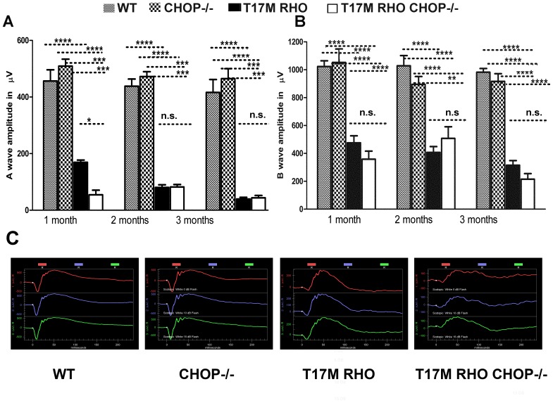
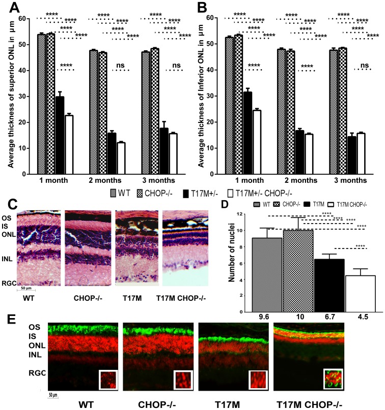
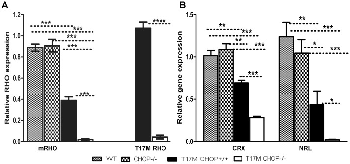
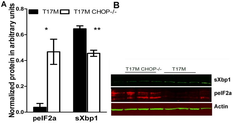
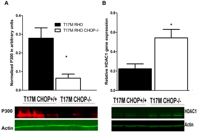
Despite the proposed link between ablation of the CHOP protein and delay of the onset of ER stress-mediated disorders including diabetes, Alzheimer Disease, and cardiac hypertrophy, the role of CHOP protein in photoreceptor cell death associated with Autosomal Dominant Retinitis Pigmentosa (ADRP) has not been investigated. T17M RHO transgenic mice carry a mutated human rhodopsin transgene, the expression of which in retina leads to protein misfolding, activation of UPR and progressive retinal degeneration. The purpose of this study is to investigate the role of CHOP protein in T17M RHO retina. Wild-type, CHOP-/-, T17M RHO and T17M RHO CHOP-/-mice were used in the study. Evaluation of the impact of CHOP ablation was performed using electroretinography (ERG), spectral-domain optical coherence tomography (SD-OCT), quantitative Real-Time PCR (qRT-PCR) and western blot analysis. Dark-adapted ERG analysis demonstrated that by 1 month, the T17M RHO CHOP-/- mice had a 70% reduction of the a-wave amplitude compared to the T17M RHO mice. The loss of function in T17M RHO CHOP-/- photoreceptors was associated with a 22-24% decline in the thickness of the outer nuclear layer. These mice had significant reduction in the expression of transcription factors, Crx and Nrl, and also in mouse Rho, and human RHO. The reduction was associated with an 8-fold elevation of the UPR marker, p-eIf2α protein and 30% down-regulation of sXbp1 protein. In addition, the histone deacetylase 1 (Hdac1) protein was 2-fold elevated in the T17M RHO CHOP-/- retina. The ablation of CHOP led to a reduction in the expression of photoreceptor-specific transcriptional factors, and both endogenous and exogenous RHO mRNA. Thus, despite its role in promoting apoptosis, CHOP protects rod photoreceptors carrying an ADRP mutation.


Conflict of interest statement

Competing Interests: The authors have declared that no competing interests exist.

Figures

Figure 1
Figure 1. Lack of CHOP protein does not protect T17M RHO retinas from degeneration, as measured by scotopic ERG responses at 10DB.
We analyzed 4 groups of animals (N = 6). A: The a-wave of the scotopic ERG amplitude was diminished in T17M RHO CHOP−/− mice at 1 month of age and the values of a-wave amplitudes were 456.3 µv ±39.7 in wild-type; 509.1 µv ±24.9 in CHOP−/−; 169.0 µv ±7.9 in T17M RHO; and 54.4 µv ±16.4 in T17M RHO CHOP−/−. This data reflected a 70% difference in the a-wave amplitudes between T17M RHO and T17M RHO CHOP−/− mice. The differences between all groups were statistically significant (P<0.0001). The difference between wild-type and CHOP−/− mice was not significant (n.s.). In the 2nd and the 3rd months, the a-wave amplitudes declined in T17M RHO mice but did not decline any further in T17M RHO CHOP−/− mice (90.7 µv ±13.5 in T17M RHO vs. 82.05 µv ±8.7 in T17M RHO CHOP−/− at 2 months and 39.7 µv ±5.7 in T17M RHO vs. 44.0 µv ±8.20 in T17M RHO CHOP−/− at 3 months). In wild-type mice, the a-wave amplitude was 438.2±25.4, vs. 472 µv ±17.3 in CHOP−/− mice at 2 months, while it was 416.2 µv ±45.5 in wild-type vs. 464.9 µv ±35.2 in CHOP−/− mice at 3 months. At 2 and 3 months, the differences between wild-type or CHOP−/− and T17M RHO or T17M RHO CHOP−/− groups were significant (P<0.001), but we did not register any differences between wild-type and CHOP−/− mice at 1, 2, or 3 months of age and difference between T17M RHO and T17M RHO CHOP−/− mice at 2 and 3 months of age. B: The b-wave of the scotopic ERG amplitude was decreased in T17M RHO CHOP−/− mice over the 3 examined months when compared to T17M RHO retinas. In 1-month-old animals, the b-wave amplitudes were 1023.0 µv ±41.6 in wild-type; 1052.0 µv ±96.5 in CHOP−/−; 475.6 µv ±50.7 in T17M RHO; and 358.2 µv ±58.2 in T17M RHO CHOP−/−. The differences between all groups were statistically significant (P<0.0001), except between T17M RHO and T17M RHO CHOP−/−, which was not significant. In the 2nd and 3rd months, the b-wave amplitudes in the T17M RHO mice declined. However, in the T17M RHO CHOP−/− mice, they were consistently low during the next 2 months. The b-wave amplitudes were 407.1 µv ±41.7 in T17M RHO vs. 428.7.5 µv ±67.0 in T17M RHO CHOP−/− at 2 months and 315.3 µv ±32.7 in T17M RHO vs. 214.9 µv ±40.0 in T17M RHO CHOP−/− at 3 months and were not significantly different. No difference was detected between the wild-type and CHOP−/− mice (1028.0 µv ±73.4 in wild-type vs. 894.7 µv ±56.9 in CHOP−/− at 2 months and 982.4 µv ±26.4 in wild-type vs. 916.2 µv ±55.5 in CHOP−/− at 3 months). However, at 2 months, the differences between the wild-type and T17M RHO or T17M RHO CHOP−/− mice were significant at the P<0.0001 level as were those between the CHOP−/− and T17M RHO mice, while the differences between the CHOP−/− and T17M RHO CHOP−/− mice were significant at the P<0.01 level. At 3 months, the differences between the wild-type and T17M RHO or T17M RHO CHOP−/− mice were significant at the P<0.00001 level, while those between CHOP−/− and T17M RHO or T17M RHO CHOP−/− were significant at the P<0.0001 level. The difference in the b-wave of the ERG amplitude between T17M RHO and T17M RHO CHOP−/− mice was not significant at 3 months. C: Images of the scotopic ERG amplitudes registered at 0 DB or 2.5 cd*s/m2 (in red), 10 DB or 25 cd*s/m2 (in blue) and 15 DB or 79.1 cd/m2 (in green) in four groups of animals.
Figure 2
Figure 2. Retinal structure measured by SD-OCT was altered in the T17M RHO CHOP−/− retina.
We analyzed 4 groups of animals (N = 6) by two-way ANOVA and found significant changes in the average thickness of the ONL in the inferior and superior hemispheres in 1,2 and 3-month-old mice. A: In the superior region, the average thickness of the ONL was 53.89 µm ±0.8 in wild-type, vs. 54.2 µm ±0.2 in CHOP−/− mice. A dramatic reduction of 22% in the ONL thickness was observed between the T17M RHO and T17M RHO CHOP−/− retinas (29.88 µm ±0.3 in T17M RHO vs. 23.7 µm ±0.3 in T17M RHO CHOP−/−), which was statistically significant (P<0.001). The differences between wild-type and T17M RHO or T17M RHO CHOP−/− and CHOP−/− and T17M RHO or T17M RHO CHOP−/− were also statistically significant (P<0.0001). No difference in the thickness of the superior ONL was observed when wild-type and CHOP−/− retinas were compared. At 2 months, the ONL thickness in the T17M RHO retina continued to decline and was 15.83 µm ±0.95 in T17M RHO vs. 12.19 µm ±0.43 in T17M RHO CHOP−/−), which was not statistically significant. In wild- type animals, the ONL thickness was 47.8 µm ±0.38 vs 46.92 µm ±0.37 in CHOP−/− mice. The differences between wild-type and T17M RHO or T17M RHO CHOP−/− and CHOP−/− and T17M RHO or T17M RHO CHOP−/− were also statistically significant (P<0.0001). No difference in the thickness of the superior ONL was observed when wild-type and CHOP−/− retinas were compared. At 3 months of age, the ONL thickness in the T17M RHO retina was 17.72 µm ±2.59 in T17M RHO vs. 15.68 µm ±0.43 in T17M RHO CHOP−/−), which was not statistically significant. In wild- type animals, the ONL thickness was 47.15 µm ±0.44 vs 48.47 µm ±0.37 in CHOP−/− mice. The differences between wild-type and T17M RHO or T17M RHO CHOP−/− and CHOP−/− and T17M RHO or T17M RHO CHOP−/− were also statistically significant (P<0.0001). No difference in the thickness of the superior ONL was observed when wild-type and CHOP−/− retinas were compared. B: The average thickness of the inferior ONL was also measured in the 4 groups of mice. We found that the average thickness was 52.5 µm ±0.51 in wild-type; 53.4 µm ±0.3 in CHOP−/− mice vs. 31.5 µm ±0.2 in T17M RHO; and 24.5 µm ±0.4 in T17M RHO CHOP−/−. The differences between wild-type and T17M RHO or T17M RHO CHOP−/− and CHOP−/− and T17M RHO or T17M RHO CHOP−/− were statistically significant (P<0.0001). The difference (24%) between T17M RHO and T17M RHO CHOP−/− was also statistically significant (P<0.001). No difference in the thickness of the inferior ONL was observed when wild-type and CHOP−/− retinas were compared. The average inferior ONL thickness in 2 month-old animals was different in all groups and was 48.0 µm ±0.46 in wild-type; 47.3 µm ±0.75 in CHOP−/− mice vs. 16.7 µm ±0.8 in T17M RHO; and 15.4 µm ±0.4 in T17M RHO CHOP−/−. The differences between wild-type and T17M RHO or T17M RHO CHOP−/− and CHOP−/− and T17M RHO or T17M RHO CHOP−/− were statistically significant (P<0.0001). The difference between T17M RHO and T17M RHO CHOP−/− was also statistically significant (P<0.001). No difference in the thickness of the inferior ONL was observed when wild-type and CHOP−/− retinas were compared. At 3 months of age the difference in the average inferior ONL thickness was not significant between T17M RHO and T17M RHO CHOP−/− mice (14.4 µm ±0.8 in T17M RHO vs 15.6 µm ±0.4 in T17M RHO CHOP−/−) while differences between wild-type (47.6 µm ±0.8) and T17M RHO or T17M RHO CHOP−/− and CHOP−/− (48.4 µm ±0.3) and T17M RHO or T17M RHO CHOP−/− were statistically significant (P<0.0001). No difference in the thickness of the inferior ONL was observed when wild-type and CHOP−/− retinas are compared. C: Histological analyses of wild-type, T17M RHO, T17M RHO CHOP−/− and CHOP−/− retinas: Images of wild-type, T17M RHO, T17M RHO CHOP−/− and CHOP−/− retinas stained with hematoxylin and eosin (H&E). Four animals in each group were used in this experiment. Histology of experimental mouse retinas at 1 month of age showed loss of photoreceptor cell nuclei, shortening of the outer segments, and general disorganization in the T17M RHO retina. Ablation of the CHOP protein in these retinas, however, led to more rapid retinal degeneration that resulted from the shortening of the outer and inner segments and more pronounced general retinal disorganization in the T17M RHO CHOP−/− mice. GCL, retinal ganglion cells; IPL, inner plexiform layer; INL, inner nuclear layer; ONL, outer nuclear layer; IS, inner segments; OS, outer segments. Scale bar indicates 50 µm. D: Photoreceptor cell nuclei in all 4 groups of animals. The number of nuclei was counted by a masked researcher. Two-way Anova with multiple comparison analysis demonstrated differences in all 4 groups of animals (****, P<0.0001) at 1 month of age with the exception of comparison between wild-type and CHOP−/− mice. For example, one-month-old T17M RHO mice had 6.7±0.15 rows whereas the T17M RHO CHOP−/− mice had more severe loss of photoreceptor cells with 4.5±0. 21 rows. These numbers were significantly different from those in the wild type and CHOP−/− animals (9.6±0.31 and 10.0±0.41 respectively). Histological analysis confirmed our OCT data suggesting first, that there is a decline in the number of photoreceptor cells in T17M RHO at 1month and second, the CHOP ablation expedites retinal degeneration in T17M RHO retina. Decline in the number of photoreceptors in these animals was 33% compared to T17M RHO mice. E: Immunohistological analyses of one-month-old wild-type, T17M RHO, T17M RHO CHOP−/− and CHOP−/− retinas. 12 micron cryostat sections of retinas were treated with anti-rhodopsin antibody (in green). The IHC analysis revealed normal localization of rhodopsin in the outer segments of photoreceptor cells in wild type and CHOP−/− retinas. The T17M RHO retinas demonstrated shortening of the OS of photoreceptors (propidium iodide -stained ONL nuclei in red). In the T17M RHO CHOP−/− retina we detected mislocalization of rhodopsin (in yellow) in addition to the shortening of the OS of photoreceptors. Rhodopsin was found in the cytoplasm of photoreceptors around the nuclei (in yellow ONL layer) and this, evidently indicates more severe retinal degeneration compared to T17M RHO mice. RGC, retinal ganglion cells; IPL, inner plexiform layer; INL, inner nuclear layer; ONL, outer nuclear layer; IS, inner segments; OS, outer segments. Scale bar indicates 50 µm.
Figure 3
Figure 3. CHOP ablation in P30 T17M RHO retinas modulates the expression of Rhodopsin (m-Rho and h-Rho) and photoreceptor-specific transcription factor (Crx and Nrl) genes.
We analyzed 4 groups of animals (N = 6) and found differences in the expression levels of Crx, Nrl and RHO (mRho and T17M RHO). A: mRho and T17M RHO expression was modulated in T17M RHO CHOP−/− retinas. The relative expression of endogenous mouse Rho was 0.9±0.03 in wild-type; 0.9±0.06 in CHOP−/−; 0.4±0.03 in T17M RHO; and 0.02±0.01 in T17M RHO CHOP−/− mice. The differences between wild-type and T17M RHO or T17M RHO CHOP−/− and CHOP−/− and T17M RHO or T17M RHO CHOP−/− were statistically significant (P<0.001). The 96% reduction of endogenous RHO gene expression, observed in T17M RHO CHOP−/− mice compared to T17M RHO mice was statistically significant (P<0.001). No difference in Crx gene expression was observed when the expression of mRho mRNA was compared in wild-type and CHOP retinas. The expression of the human T17M RHO transgene was also modulated in T17M RHO CHOP−/− mice, with a value of 1.1±0.06 in T17M RHO mice vs. 0.04±0.01 in T17M RHO CHOP−/− mice. The observed 96% reduction of transgene expression was statistically significant (P<0.0001). B: Modified Expressions of Crx and Nrl in the T17M RHO CHOP−/− retina. The relative Crx gene expression was 1.0±0.059 in wild-type; 1.1±0.1 in CHOP−/−; 0.7±0.03 in T17M RHO; and 0.3±0.02 in T17M RHO. The differences between the wild-type or CHOP−/− and T17M RHO groups were statistically significant (P<0.01), as were the differences between wild-type or CHOP−/− and T17M RHO CHOP−/− (P<0.001). The 60% reduction of Crx gene expression detected in T17M RHO CHOP−/− retinas compared to T17M RHO was statistically significant (P<0.001). No difference was observed when the wild-type and CHOP−/− retinas were compared. The level of Nrl gene expression was 1.2±0.2 in wild-type; 1.04±0.2 in CHOP−/−; 0.4±0.15 in T17M RHO; and 0.02±0.03 in T17M RHO CHOP−/−. The differences between wild-type and T17M RHO CHOP−/− and CHOP−/− and T17M RHO CHOP−/− were significant at the P<0.001 level, while those between wild-type and T17M RHO, T17M RHO and CHOP−/−, and T17M RHO and T17M RHO CHOP−/− were significant at the P<0.01 level. The 95% reduction of Nrl gene expression observed in the T17M RHO CHOP−/− compared to the T17M RHO retina was statistically significant (P<0.05). No difference in Nrl gene expression was observed when wild-type and CHOP−/− retinas were compared.
Figure 4
Figure 4. Ablation of CHOP protein in P30 T17M RHO retinas led to modulation of the PERK and IRE1 pathways of the UPR.
A: We analyzed T17M RHO and T17M RHO CHOP−/− retinas (N = 4) and found that the expression of phosphorylated eIF2α protein was increased by over 8 fold in T17M RHO CHOP−/− mice (0.5±0.1 a.u.vs. 0.04±0.1 a.u., P = 0.01). The level of spliced Xbp1 in the T17M RHO retina was decreased by 30%, presenting a value of 0.64±0.027 a.u. vs. 0.4±0.03 a.u.in T17M RHO CHOP−/− mice (P = 0.004). B: Representative images of western blots treated with antibodies against Xbp1, peIF2α and β-actin proteins.
Figure 5
Figure 5. Expressions of the histone deacetylase, Hdac1 and the transcriptional co-activator, P300 were modified in P30 T17M RHO CHOP−/− retinas (N = 3).
A: A 78% reduction in the P300 protein level was observed in the T17M RHO CHOP−/− retina compared to T17M RHO. The normalized level of P300 was 0.3±0.06 a.u. in T17M RHO vs. 0.1±0.02 a.u. in T17M RHO CHOP−/− (P = 0.02). B: A 245% increase in Hdac1 protein expression was observed in T17M RHO CHOP−/− retinas (0.22±0.05 arbitrary units in T17M RHO vs. 0.54±0.08 a. u. in T17M RHO CHOP−/−, P = 0.03). Bottom: Representative images of western blots treated with antibodies against P300, HDAC1 and β-actin proteins.

References

    1. Hartong DT, Berson EL, Dryja TP (2006) Retinitis pigmentosa. Lancet 368: 1795–1809. - PubMed
    1. Lee ES, Flannery JG (2007) Transport of truncated rhodopsin and its effects on rod function and degeneration. Invest Ophthalmol Vis Sci 48: 2868–2876. - PMC - PubMed
    1. Mendes HF, van der Spuy J, Chapple JP, Cheetham ME (2005) Mechanisms of cell death in rhodopsin retinitis pigmentosa: implications for therapy. Trends Mol Med 11: 177–185. - PubMed
    1. Kunte MM, Choudhury S, Manheim JF, Shinde VM, Miura M, et al... (2012) ER stress is Involved in T17M Rhodopsin-Induced Retinal Degeneration. Invest Ophthalmol Vis Sci. - PMC - PubMed
    1. Wang S, Kaufman RJ (2012) The impact of the unfolded protein response on human disease. J Cell Biol 197: 857–867. - PMC - PubMed

Publication types

MeSH terms