. 2013 Jul;169(6):1372-88.
doi: 10.1111/bph.12227.

Activation of adenosine A(2A) receptor reduces osteoclast formation via PKA- and ERK1/2-mediated suppression of NFκB nuclear translocation

Affiliations

Activation of adenosine A(2A) receptor reduces osteoclast formation via PKA- and ERK1/2-mediated suppression of NFκB nuclear translocation

Aránzazu Mediero et al. Br J Pharmacol. 2013 Jul.

Abstract

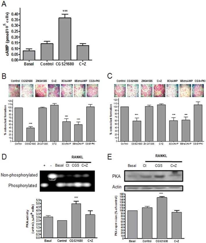
Background and purpose: We previously reported that adenosine, acting at adenosine A(2A) receptors (A(2A)R), inhibits osteoclast (OC) differentiation in vitro (A(2A)R activation OC formation reduces by half) and in vivo. For a better understanding how adenosine A(2A)R stimulation regulates OC differentiation, we dissected the signalling pathways involved in A(2A)R signalling.

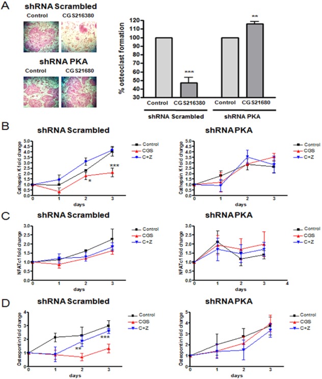
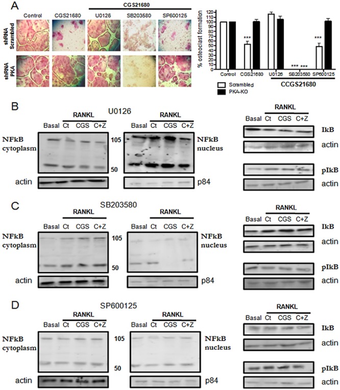
Experimental approach: OC differentiation was studied as TRAP+ multinucleated cells following M-CSF/RANKL stimulation of either primary murine bone marrow cells or the murine macrophage line, RAW264.7, in presence/absence of the A(2A)R agonist CGS21680, the A(2A)R antagonist ZM241385, PKA activators (8-Cl-cAMP 100 nM, 6-Bnz-cAMP) and the PKA inhibitor (PKI). cAMP was quantitated by EIA and PKA activity assays were carried out. Signalling events were studied in PKA knockdown (lentiviral shRNA for PKA) RAW264.7 cells (scrambled shRNA as control). OC marker expression was studied by RT-PCR.

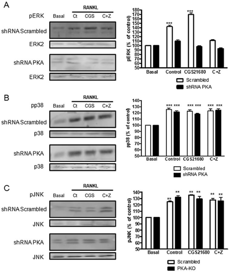
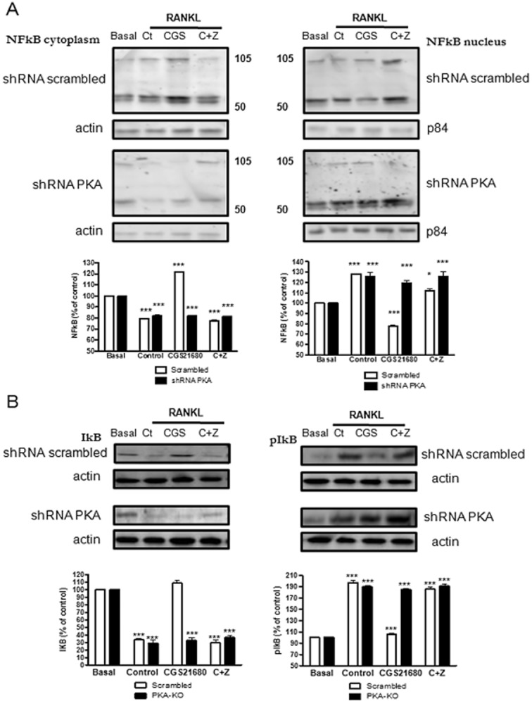
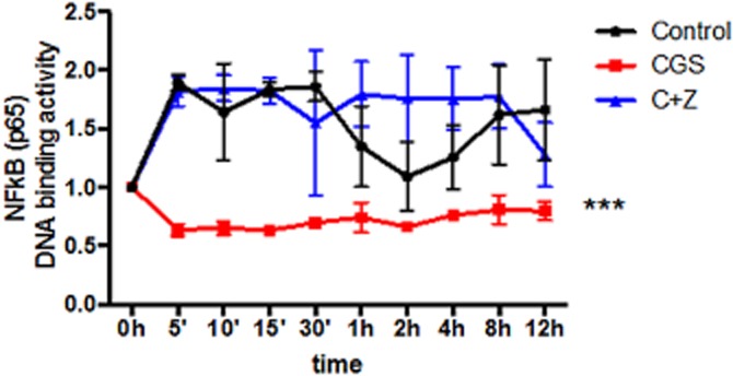
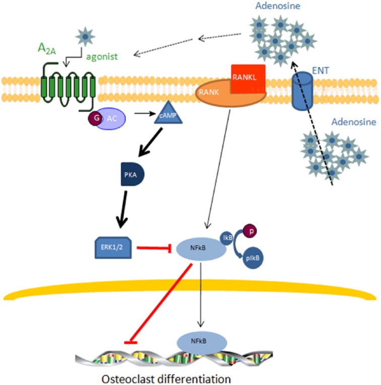
Key results: A(2A)R stimulation increased cAMP and PKA activity which and were reversed by addition of ZM241385. The direct PKA stimuli 8-Cl-cAMP and 6-Bnz-cAMP inhibited OC maturation whereas PKI increased OC differentiation. A(2A)R stimulation inhibited p50/p105 NFκB nuclear translocation in control but not in PKA KO cells. A(2A)R stimulation activated ERK1/2 by a PKA-dependent mechanism, an effect reversed by ZM241385, but not p38 and JNK activation. A(2A)R stimulation inhibited OC expression of differentiation markers by a PKA-mechanism.

Conclusions and implications: A(2A)R activation inhibits OC differentiation and regulates bone turnover via PKA-dependent inhibition of NFκB nuclear translocation, suggesting a mechanism by which adenosine could target bone destruction in inflammatory diseases like rheumatoid arthritis.


Figures

Figure 1
Figure 1
Adenosine A2AR activation increased cAMP production and increased PKA activation. BMCs and RAW264.7 cells were treated with 50 ng·mL−1 RANKL together with CGS21680 1 μM (CGS) alone or in the presence of ZM241385 1 μM (C + Z), 8-Cl-cAMP and 6-Bnz-cAMP 100 nM each and PKI 10 μg·mL−1. (A) Intracellular cAMP levels were measured after 10 min stimulation. cAMP values are expressed as the mean ± SEM pmol/105 cells (n = 6). (B) Primary bone marrow cell-derived osteoclasts were fixed and stained for TRAP. TRAP+ cells containing three or more nuclei were counted as osteoclasts. The results were expressed as the mean ± SEM (n = 6) (C) RAW264.7 derived osteoclast were fixed and stained for TRAP following the same conditions as primary cells. TRAP+ cells containing three or more nuclei were counted as osteoclasts. The results were expressed as the mean ± SEM (n = 6). PKI + CGS216580 TRAP+ cells are expressed as % of PKI-treated cells alone. (D) PKA activity was calculated 15 min after RAW264.7 cells stimulation. Gel image reflect separation of phosphorylated/non-phosphorylated peptide migration. Data were express as the mean ± SEM in units/2.5 × 106 cells considering the relation of phosphorylated Peptide A1 and PKA activity according to manufacturer's indication (n = 4). + indicate positive control and – indicate negative control. (E) PKA expression was analysed 8 h after RAW264.7 cell stimulation by Western blot. To normalize for protein loading, the membranes were reprobed with actin and results were normalized to the density of the actin bands. The results were expressed as the mean ± SEM of four independent experiments. ***P < 0.001 versus non-stimulated control.
Figure 2
Figure 2
Expression of osteoclast differentiation markers mRNA in PKA catalytic alpha subunit knockdown cells. (A) RAW264.7 cells were permanently transfected with scrambled or PKA catalytic alpha subunit shRNA, and treated with 50 ng·mL−1 RANKL together with CGS21680 (1 μM). TRAP-positive cells containing three or more nuclei were counted as osteoclasts. The results were expressed as the means of four different assays in duplicate. The y axis have been expanded to show the difference more clearly. (B) Changes in Cathepsin K mRNA in osteoclasts during the osteoclast differentiation process in the presence of CGS21680 1 μM (CGS) alone or with ZM241385 1 μM (C + Z) in PKA catalytic alpha subunit shRNA RAW264.7 cells compared to scrambled shRNA. (C) NFATc1 mRNA fold change in RANKL derived osteoclast during the three days osteoclast differentiation process in the presence of 1 μM (CGS) alone or with ZM241385 1 μM (C + Z) in PKA catalytic alpha subunit shRNA RAW264.7 cells compared to scrambled shRNA infected cells. (D) Changes in Osteopontin mRNA in RANKL derived osteoclast during the three days osteoclast differentiation process in the presence of CGS21680 1 μM (CGS) alone or with ZM241385 1 μM (C + Z) in PKA catalytic alpha subunit shRNA RAW264.7 cells compared to scrambled shRNA infected cells. ***P < 0.001, **P < 0.01, *P < 0.5 versus non-stimulated control.
Figure 3
Figure 3
MAPKs are activated during osteoclast differentiation in a PKA dependent mechanism. RAW264.7 cells were permanently transfected with scrambled or PKA catalytic alpha subunit shRNA, and treated with 50 ng·mL−1 RANKL together with CGS21680 1 μM (CGS) alone or in the presence of ZM241385 1 μM (C + Z). (A) ERK1/2 phosphorylation was analysed 10 min after stimulation by Western blot. (B) p38 phosphorylation was analysed 10 min after stimulation by Western blot. (C) JNK phosphorylation was analysed 2 h after stimulation by Western blot. To normalize for protein loading, the membranes were reprobed with ERK2, p38 or JNK respectively and results normalized appropriately. The results were expressed as the means of four independent experiments. ***P < 0.001, *P < 0.5 versus non-stimulated control. In all cases, the y axis has been expanded to show the difference more clearly.
Figure 4
Figure 4
A2A receptor activation inhibits p50/p105 NFκB nuclear translocation in a PKA-dependent mechanism. RAW264.7 cells were permanently transfected with scrambled or PKA catalytic alpha subunit shRNA, and treated with 50 ng·mL−1 RANKL together with CGS21680 1 μM (CGS) alone or in the presence of ZM241385 1 μM (C + Z). (A) p50/p105 NFκB was analysed by Western blot both in the cytoplasmic and nuclear cell fraction 15 min after stimulation. In data not shown, actin was not found in the nuclear fraction ruling out cytoplasmic contamination of the preparation. (B) IκB alpha and p-IκB alpha were analysed in the cytoplasmic cell fraction 5 min after stimulation by Western blot. To normalize for protein loading, the membranes were reprobed with actin in the cytoplasmic fraction and the specific nuclear membrane protein p84 in the nuclear fraction. The results were expressed as the means of four independent experiments. ***P < 0.001, **P < 0.01, *P < 0.5 versus non-stimulated control. In all cases, the y axis has been expanded to show the difference more clearly.
Figure 5
Figure 5
NFκB (p65) transcription factor DNA binding activity is decreased by activation of the A2AR. RAW264.7 cells were treated with 50 ng·mL−1 RANKL together with CGS21680 1 μM (CGS) alone or in the presence of ZM241385 1 μM (C + Z) and NFκB (p65) transcription factor DNA binding activity was calculated and normalized to non-stimulated control. Activity was measured for up to 12 h and NFκB (p65) transcription factor DNA binding activity is expressed as the mean ± SEM of five different assays. ***P < 0.001, versus non-stimulated control.
Figure 6
Figure 6
A2AR activation inhibits NFκB p50/p105 nuclear translocation by a PKA-ERK1/2 mechanism. (A) PKA catalytic alpha subunit and scrambled silenced RAW264.7-derived osteoclasts were fixed and stained for TRAP after being cultured in the presence of CGS21680 1 μM (CGS) alone or in the presence of U0126, SB203580 and SP600125 (10 μM each). TRAP-positive cells containing three or more nuclei were counted as osteoclasts. The results are expressed as the means of six different assays carried out in duplicate. (B) p50/p105 NFκB expression in the cytoplasmic and nuclear cell fractions and IκB alpha and p-IκB alpha expression after stimulation in the presence of U0126 10 μM by Western blot in scrambled cells. (C) p50/p105 NFκB expression in the cytoplasmic and nuclear cell fraction and IκB alpha and p-IκB alpha expression after stimulation in the presence of SB203580 (1 μM) by Western blot in scrambled cells. (D) p50/p105 NFκB expression in the cytoplasmic and nuclear cell fraction and IκB alpha and p-IκB alpha expression after stimulation in the presence of SP600125 (10 μM) by Western blot in scrambled cells. The results were expressed as the means of four independent experiments. ***P < 0.001, **P < 0.01, *P < 0.5 versus non-stimulated control.
Figure 7
Figure 7
Extracellular adenosine activates adenosine A2AR and inhibits osteoclast differentiation by inhibiting p50/p105 NFκB nuclear translocation. Adenosine release into the extracellular space by adenosine transporters (ENT) produces an increase in adenosine concentration that activates the adenosine A2AR resulting in an activation of adenylate cyclase and increased cAMP levels which further activate PKA producing ERK1/2 phosphorylation and the concomitant inhibition of p50/p105 NFκB nuclear translocation. These events result in inhibition of osteoclast differentiation.

References

    1. Blair HC, Robinson LJ, Zaidi M. Osteoclast signalling pathways. Biochem Biophys Res Commun. 2005;328:728–738. - PubMed
    1. Bohm C, Hayer S, Kilian A, Zaiss MM, Finger S, Hess A, et al. The alpha-isoform of p38 MAPK specifically regulates arthritic bone loss. J Immunol. 2009;183:5938–5947. - PubMed
    1. Boyce BF, Yao Z, Xing L. Osteoclasts have multiple roles in bone in addition to bone resorption. Crit Rev Eukaryot Gene Expr. 2009;19:171–180. - PMC - PubMed
    1. van Calker D, Muller M, Hamprecht B. Adenosine regulates via two different types of receptors, the accumulation of cyclic AMP in cultured brain cells. J Neurochem. 1979;33:999–1005. - PubMed
    1. Chan ES, Cronstein BN. Methotrexate – how does it really work? Nat Rev Rheumatol. 2010;6:175–178. - PubMed

Publication types

MeSH terms