. 2013 Jun;88(6):1083-92.
doi: 10.1111/mmi.12241. Epub 2013 May 7.

Identification of ClpP substrates in Caulobacter crescentus reveals a role for regulated proteolysis in bacterial development

Affiliations

Identification of ClpP substrates in Caulobacter crescentus reveals a role for regulated proteolysis in bacterial development

Nowsheen H Bhat et al. Mol Microbiol. 2013 Jun.

Abstract

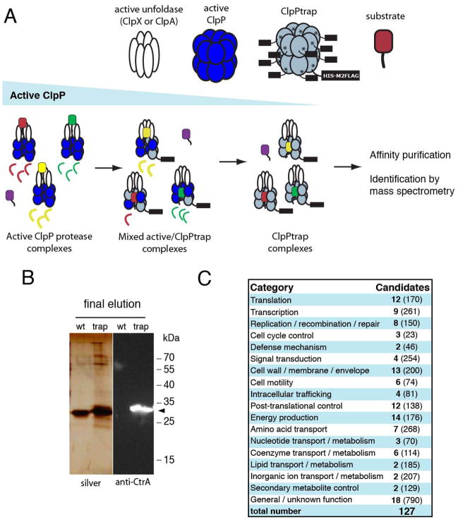
Energy-dependent proteases ensure the timely removal of unwanted proteins in a highly selective fashion. In Caulobacter crescentus, protein degradation by the ClpXP protease is critical for cell cycle progression; however, only a handful of substrates are currently known. Here, we use a trapping approach to identify putative substrates of the ClpP associated proteases in C. crescentus. Biochemical validation of several of these targets reveals specific protease recognition motifs and suggests a need for ClpXP-specific degradation beyond degradation of known cell cycle regulators. We focus on a particular instance of regulated proteolysis in Caulobacter by exploring the role of ClpXP in degrading the stalk synthesis transcription factor TacA. We show that TacA degradation is controlled during the cell cycle dependent on the ClpXP regulator CpdR and that stabilization of TacA increases degradation of another ClpXP substrate, CtrA, while restoring deficiencies associated with prolific CpdR activity. Together, our work reveals a number of new validated ClpXP substrates, clarifies rules of protease substrate selection, and demonstrates how regulated protein degradation is critical for Caulobacter development and cell cycle progression.


Figures

Figure 1
Figure 1
ClpP trapping strategy and substrate characterization. A. Inactive oligomers of ClpP can capture substrates delivered to them by active unfoldases. Depletion of endogenous, active ClpP enriches for inactive ClpPtrap oligomers. Affinity purification followed by mass spectrometry identifies candidate substrates. B. Substrates are co-purified with ClpPtrap, but are absent when an affinity tagged active ClpP is used. Western blotting with antibodies recognizing the known ClpXP substrate CtrA (black markers) confirms trapping procedure. Confirmation of additional substrates by Western blotting is shown in Figure S1. C. Candidate substrates are widely distributed across many functional categories as annotated by COG groups (NCBI), total numbers of proteins in each category shown in parentheses.
Figure 2
Figure 2
Trapped substrates reveal conserved motif requirements for ClpXP proteolysis. A. Select substrates were cloned, expressed recombinantly and purified, then assayed for in vitro degradation by ClpXP. Table lists CC annotation / gene name, sequence of the C-terminal six residues, and whether the candidate substrate was degraded by ClpXP in standard conditions (see Methods and Figure S2). B. IbpA and FlaF are both degraded by ClpXP and mutation of their C-terminal Ala-Ala motif eliminates degradation. DnaK is not degraded by ClpXP in vitro.
Figure 3
Figure 3
The N-terminal domain of ClpX is a critical modulator of protease specificity. A. CC0360 degradation relies on the N-terminal domain of ClpX even though ssrA tagged substrates (GFP-ssrA) are degraded readily by both constructs. B. CtrA is recognized by both full length and ΔNClpX. C. The N-terminal domain of ClpX is essential for viability. Cells expressing a xylose inducible copy of clpX as the sole chromosomal copy and plasmids constitutively expressing either full length (WT) or ΔNClpX variants plated on inducing (xylose) or noninducing (no xylose) media. D. Western blot analysis using antibodies specific to C. crescentus ClpX confirm the constitutive expression of ΔNClpX from the plasmid in both inducing (xylose) and noninducing (glucose) conditions. Blots are registered so that upper bands (full length ClpX) are aligned.
Figure 4
Figure 4
TacA is degraded by ClpXP. A. TacA is recognized by ClpXP in vitro and mutating C-terminal residues to Asp-Asp inhibits proteolysis. In these gels, overlapping bands corresponding to ClpX and creatine kinase are marked along with the purified TacA proteins. B. M2-FLAG epitope tagged TacA (M2-TacA) expressed from an inducible plasmid is degraded in vivo in a CpdR-dependent fashion following shift to a noninducing media. Representative western blot is shown here; quantification of replicates can be found in Figure S4. C. Western blots against the M2FLAG epitope and CtrA in synchronized population of wildtype cells shows that M2-TacA is degraded in a cell-cycle dependent fashion, while M2-TacADD is not degraded. Additional replicate illustrating M2-TacA degradation is shown in Figure S4. D. Loss of CtrA after antibiotic mediated shutoff of synthesis in cells expressing M2-TacADD compared to cells expressing M2-TacA. Upper panel shows a representative western blot, lower graph represents averages of biological replicates (n=4). Error bars are standard errors of the mean.
Figure 5
Figure 5
Stabilization of TacA partially rescues stalk formation in ΔpleC cells. A. Cartoon of PleC dependent TacA degradation. PleC dephosphorylates DivK. Dephosphorylated DivK inhibits CpdR dephosphorylation indirectly through CckA/ChpT (not illustrated here). Dephosphorylated CpdR promotes ClpXP degradation of TacA. Thus, loss of PleC would result in more dephosphorylated CpdR and faster TacA degradation. B. Representative images of wildtype cells (upper) with stalks marked with white arrows (predivisional cell) or black arrows (stalked cell) and stalkless ΔpleC predivisional cells (lower). C. Stalk formation is partially recovered in ΔpleC cells expressing stabilized TacA. Quantification of predivisional wildtype cells, ΔpleC strains expressing TacA as the sole variant, or ΔpleC strains expressing TacA-DD as the sole variant. Error bars are standard errors, p-value is calculated from a two-tailed Welch’s t-test. Stalk elongation during phosphate limitation is also more pronounced in ΔpleC cells expressing stabilized TacA (Figure S5).

Similar articles

Cited by

References

    1. Abel S, Chien P, Wassmann P, Schirmer T, Kaever V, Laub MT, Baker TA, Jenal U. Regulatory cohesion of cell cycle and cell differentiation through interlinked phosphorylation and second messenger networks. Molecular cell. 2011;43:550–560. - PMC - PubMed
    1. Biondi EG, Reisinger SJ, Skerker JM, Arif M, Perchuk BS, Ryan KR, Laub MT. Regulation of the bacterial cell cycle by an integrated genetic circuit. Nature. 2006a;444:899–904. - PubMed
    1. Biondi EG, Skerker JM, Arif M, Prasol MS, Perchuk BS, Laub MT. A phosphorelay system controls stalk biogenesis during cell cycle progression in Caulobacter crescentus. Molecular microbiology. 2006b;59:386–401. - PubMed
    1. Bissonnette SA, Rivera-Rivera I, Sauer RT, Baker TA. The IbpA and IbpB small heat-shock proteins are substrates of the AAA+ Lon protease. Molecular microbiology. 2010;75:1539–1549. - PMC - PubMed
    1. Britos L, Abeliuk E, Taverner T, Lipton M, McAdams H, Shapiro L. Regulatory response to carbon starvation in Caulobacter crescentus. PloS one. 2011;6:e18179. - PMC - PubMed

Publication types

MeSH terms

LinkOut - more resources