. 2013 Aug;126(3):338-48.
doi: 10.1111/jnc.12290. Epub 2013 May 20.

Distinct intrinsic and synaptic properties of pre-sympathetic and pre-parasympathetic output neurons in Barrington's nucleus

Affiliations

Distinct intrinsic and synaptic properties of pre-sympathetic and pre-parasympathetic output neurons in Barrington's nucleus

Yue-Xian Guo et al. J Neurochem. 2013 Aug.

Abstract

Barrington's nucleus (BN), commonly known as the pontine micturition center, controls micturition and other visceral functions through projections to the spinal cord. In this study, we developed a rat brain slice preparation to determine the intrinsic and synaptic mechanisms regulating pre-sympathetic output (PSO) and pre-parasympathetic output (PPO) neurons in the BN using patch-clamp recordings. The PSO and PPO neurons were retrogradely labeled by injecting fluorescent tracers into the intermediolateral region of the spinal cord at T13-L1 and S1-S2 levels, respectively. There were significantly more PPO than PSO neurons within the BN. The basal activity and membrane potential were significantly lower in PPO than in PSO neurons, and A-type K(+) currents were significantly larger in PPO than in PSO neurons. Blocking A-type K(+) channels increased the excitability more in PPO than in PSO neurons. Stimulting μ-opioid receptors inhibited firing in both PPO and PSO neurons. The glutamatergic EPSC frequency was much lower, whereas the glycinergic IPSC frequency was much higher, in PPO than in PSO neurons. Although blocking GABAA receptors increased the excitability of both PSO and PPO neurons, blocking glycine receptors increased the firing activity of PPO neurons only. Furthermore, blocking ionotropic glutamate receptors decreased the excitability of PSO neurons but paradoxically increased the firing activity of PPO neurons by reducing glycinergic input. Our findings indicate that the membrane and synaptic properties of PSO and PPO neurons in the BN are distinctly different. This information improves our understanding of the neural circuitry and central mechanisms regulating the bladder and other visceral organs.

Keywords: autonomic nervous system; micturition reflex; pontine micturition center; synaptic transmission; voltage-activated K+ channels.


Conflict of interest statement

The authors declare that they have no conflict of interest.

Figures
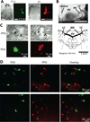
Figure 1
Figure 1. Identification of retrogradely labeled PPO and PSO neurons in the BN
A: representative light and fluorescence images show the microinjection site of fluorescent microspheres in the intermediolateral region of the spinal cord at the T13 (green) and S1 level (red). B: original brain slice image and schematic diagram depict the location of the BN in the brainstem. 4V, fourth ventricle; LC, locus coeruleus. C: fluorescent microsphere-labeled PPO (red) and PSO (green) neurons in the BN in fresh brain slices viewed under DIC and fluorescence illumination. * and ϖ show the labeled neuron and attached recording electrode, respectively. D: confocal images show retrogradely labeled PSO (green) and PPO (red) neurons in the BN in two separate brain sections. Digitally merged images are shown on the right.
Figure 2
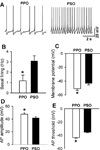
Figure 2. Comparison of the firing property of PPO and PSO neurons in the BN
A: representative recording traces show the spontaneous firing activity of one PPO and one PSO neurons. B-E: summary data show the basal firing rate (B), resting membrane potential (C), amplitude of action potentials (D), and threshold of action potentials (E) in 20 PPO and 16 PSO neurons. Data are presented as mean ± SEM. * P < 0.05 compared with PSO neurons.
Figure 3
Figure 3. Responses of PPO and PSO neurons to depolarizing currents and blocking A-type K+ channels
A: representative recordings show responses of one PPO neuron and one PSO neuron to injection of depolarizing currents. The onset of the first action potential is indicated by an arrow at the beginning of the trace, and the return of the membrane potential to the baseline after the repolarization pulse is indicated by an arrow at the end of the trace. B: original traces show the effect of 4-AP (5 mM) on the firing activity of one PPO neuron and one PSO neuron. C and D: summary data show that 4-AP caused a greater increase in the firing activity in PPO (n = 8) than in PSO (n = 9) neurons. * P < 0.05, compared with the baseline control. # P < 0.05, compared with PSO neurons.
Figure 4
Figure 4. Comparison of A-type K+ currents in PPO and PSO neurons in the BN
A: representative traces show the A-type K+ currents in one PPO neuron and one PSO neuron. B and C: I-V relationship of the total K+ currents (B) and A-type K+ currents (C) in 7 PPO and 8 PSO neurons tested. * P < 0.05, compared with corresponding values in PSO neurons.
Figure 5
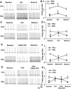
Figure 5. Comparison of the influence of inhibitory and excitatory synaptic inputs on the firing activity of PPO and PSO neurons
A and B: original recordings (A) and group data (B) show the effect of bicuculline (20 µM) on the firing activity of PPO (n = 9) and PSO (n = 8) neurons. C and D: representative traces (C) and summary data (D) show that 2 µM strychnine increased the firing activity of PPO (n = 8), but not PSO (n = 7), neurons. E and F: original traces (E) and summary data (F) show that CNQX (20 µM) and AP-5 (50 µM) decreased the firing activity of PSO (n = 7), but increased the firing of PPO (n = 7), neurons. G and H: original traces (G) and group data (H) show that CNQX and AP-5 failed to alter the firing activity of PPO (n = 8) neurons in the presence of strychnine. Note that CNQX and AP-5 still decreased the firing rate of 7 PSO neurons in the presence of strychnine. * P < 0.05, compared with the baseline control. # P < 0.05, compared with PSO neurons. BIC, bicuculline; STR, strychnine.
Figure 6
Figure 6. Comparison of glutamatergic input in PPO and PSO neurons
A: representative traces show that glutamatergic sEPSCs recorded from one PPO neuron and one PSO neuron. B and C: summary data show the frequency (D) and amplitude (E) of sEPSCs of PPO (n = 12) and PSO (n = 15) neurons. * P < 0.05, compared with PSO neurons.
Figure 7
Figure 7. Comparison of GABAergic and glycinergic inputs in PPO and PSO neurons and the effect of glutamatergic input on GABAergic and glycinergic sIPSCs
A-C: representative traces (A) and group data (B,C) show that CNQX and AP-5 decreased the frequency, but not the amplitude, of GABAergic sIPSCs in both PPO (n = 9) and PSO (n = 8) neurons. D-F: original traces (D) and summary data (E, F) show the differences in the baseline glycinergic sIPSC frequency and the differential effect of CNQX and AP-5 on the frequency of glycinergic sIPSCs of PPO (n = 8) and PSO (n = 7) neurons. * P < 0.05, compared with the baseline control. # P < 0.05, compared with PSO neurons.
Figure 8
Figure 8. Effect of the µ-opioid receptor agonist DAMGO on the firing activity of PPO and PSO neurons
A and B: representative traces and histogram (A) and group data (B) show that 1 µM DAMGO decreased the firing activity of PPO neurons (n = 8). C and D: original recordings and histogram (C) and summary data (D) show that DAMGO application inhibited the firing activity of PSO neurons (n = 9). * P < 0.05, compared with the baseline control.

Similar articles

Cited by

References

    1. Blok BF, Holstege G. Ultrastructural evidence for a direct pathway from the pontine micturition center to the parasympathetic preganglionic motoneurons of the bladder of the cat. Neurosci Lett. 1997;222:195–198. - PubMed
    1. Cano G, Card JP, Rinaman L, Sved AF. Connections of Barrington's nucleus to the sympathetic nervous system in rats. J Auton Nerv Syst. 2000;79:117–128. - PubMed
    1. de Groat WC, Saum WR. Sympathetic inhibition of the urinary bladder and of pelvic ganglionic transmission in the cat. J Physiol. 1972;220:297–314. - PMC - PubMed
    1. de Groat WC, Saum WR. Synaptic transmission in parasympathetic ganglia in the urinary bladder of the cat. J Physiol. 1976;256:137–158. - PMC - PubMed
    1. de Groat WC, Booth AM, Yoshimura N. The Autonomic Nervous System. Nervous Control of the Urogenital System. In: Maggi CA, editor. Neurophysiology of micturition and its modification in animal models of human disease. Vol. 3. London: Hartwood Academic; 1993. pp. 227–289.

Publication types