. 2013 Oct 9;168(4):3291-9.
doi: 10.1016/j.ijcard.2013.04.045. Epub 2013 May 3.

Protein-L-isoaspartate (D-aspartate) O-methyltransferase protects cardiomyocytes against hypoxia induced apoptosis through inhibiting proapoptotic kinase Mst1

Affiliations

Protein-L-isoaspartate (D-aspartate) O-methyltransferase protects cardiomyocytes against hypoxia induced apoptosis through inhibiting proapoptotic kinase Mst1

Guijun Yan et al. Int J Cardiol. .

Abstract

Background: Mammalian sterile 20-like kinase 1 (Mst1) is a mammalian homolog of Hippo kinase from Drosophila and it is a critical component of the Hippo signaling pathway, which regulates a variety of biological processes ranging from cell contact inhibition, organ size control, apoptosis and tumor suppression in mammals. Mst1 plays essential roles in heart disease since its activation causes cardiomyocyte apoptosis and dilated cardiomyopathy. However, the mechanism underlying Mst1 activation in the heart is not known.

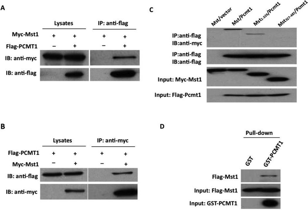
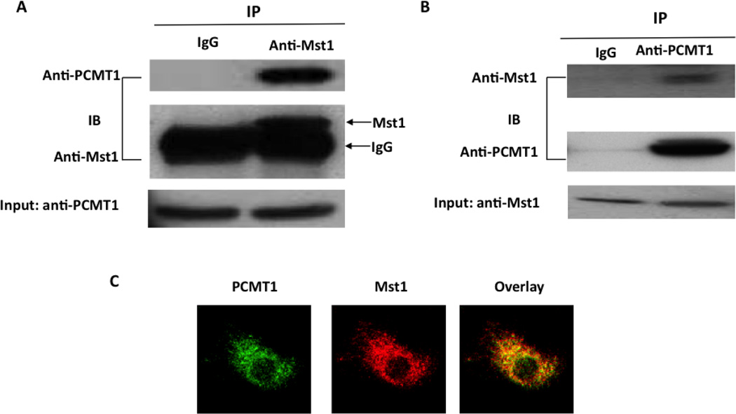
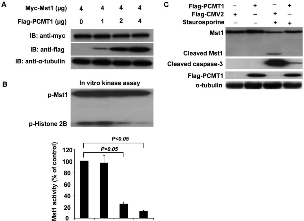
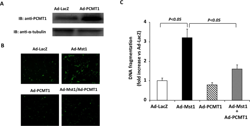
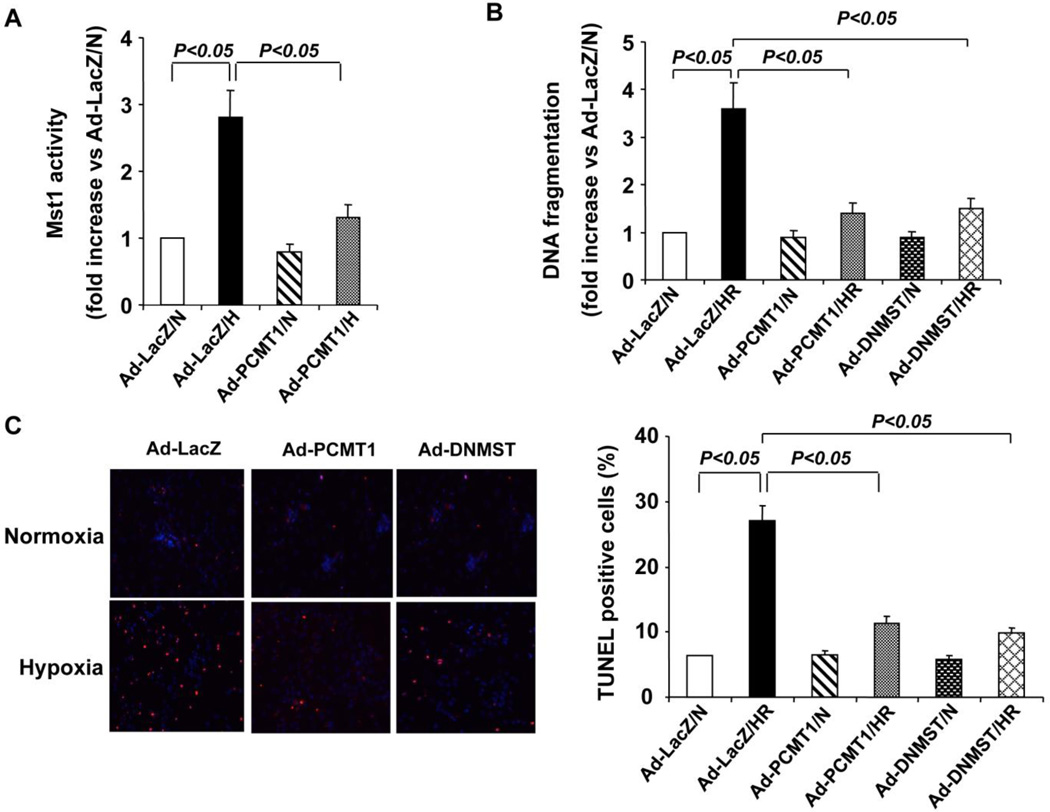
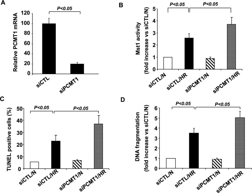
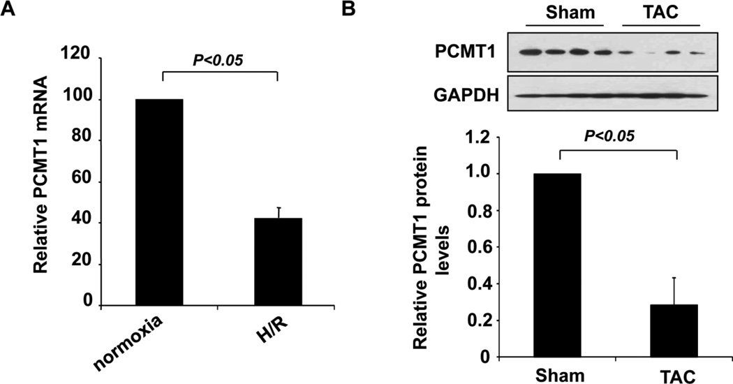
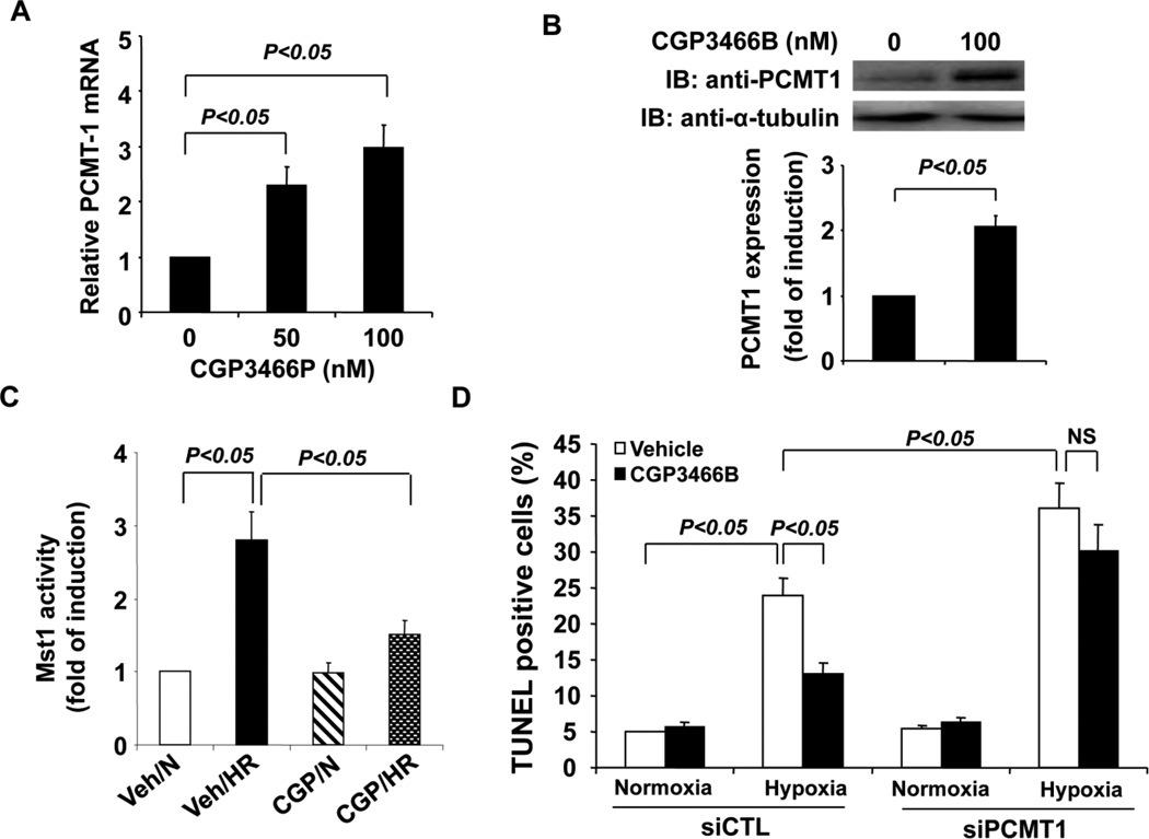
Methods and results: To identify novel cardiac proteins that may regulate Mst1 activity in the heart under pathophysiological conditions, a yeast two-hybrid screening of a human heart cDNA library with a dominant-negative Mst1 (K59R) mutant used as bait was performed. As a result, protein-L-isoaspartate (D-aspartate) O-methyltransferase (PCMT1) was identified as an Mst1-interacting protein. The interaction of PCMT1 with Mst1 was confirmed by co-immunoprecipitation in both co-transfected HEK293 cells and native cardiomyocytes, in which PCMT1 interacted with the kinase domain of Mst1, but not with its C-terminal regulatory domain. Overexpression of PCMT1 did not affect the Mst1 expression, but significantly attenuated the Mst1 activation and its apoptotic effects in response to the hypoxia/reoxygenation induced injury in cardiomyocytes. Indeed, upregulation of PCMT1 by CGP3466B, a compound related to the anti-Parkinson's drug R-(-)-deprenyl with potent antiapoptotic effects, inhibited the hypoxia/reoxygenation induced Mst1 activation and cardiomyocyte apoptosis.

Conclusions: These findings implicate PCMT1 as a novel inhibitor of Mst1 activation in cardiomyocytes and suggest that targeting PCMT1 may prevent myocardial apoptosis through inhibition of Mst1.

Keywords: Apoptosis; Cardiac myocytes; Hypoxia/reoxygenation; Mst1 kinase; PCMT1.


Conflict of interest statement

Potential conflicts of interest: none

Figures

Figure 1
Figure 1
Physical interaction between Mst1 and PCMT1 by immunoprecipitation analysis. A, Myc-Mst1 expression vector in combination of either empty vector or pFlag-PCMT1 were co-transfected into HEK293 cells. Extracted proteins were precipitated by anti-FLAG antibody and then separated by 12% SDS-PAGE. The transferred membrane was immunoblotted with either HRP conjugated anti-Myc or HRP conjugated anti-FLAG antibody. B, Flag-PCMT1 expression vector in combination of either empty vector, pMT2-Myc-Mst1 were co-transfected into HEK293 cells. Extracted proteins were precipitated by anti-Myc antibody and then separated by 12% SDS-PAGE. The transferred membrane was immunoblotted with either HRP conjugated anti-Myc or HRP conjugated anti-FLAG antibody. C. FLAG-PCMT1 expression vector in combination with either empty vector or expression vectors of Myc-Mst1 mutants were co-transfected into HEK293 cells. Extracted proteins were precipitated by anti-Flag antibody and then separated by 15% SDS-PAGE. The transferred membrane was immunoblotted with either HRP conjugated anti-FLAG or HRP conjugated anti-Myc antibody. Lysates were also immunoblotted with anti-FLAG antibody to show the expression levels of FLAG-PCMT1 in HEK293 cells. D, Recombinant Flag-Mst1 incubated with either GST or GST-PCMT1 and proteins were precipitated with glutathione-Sepharose 4B beads. The precipitated proteins were separated through SDS-PAGE, and Western blot analysis was done with anti-Mst1 antibody.
Figure 2
Figure 2
Association of Mst1 with PCMT1 in cardiomyocytes. A. Cell lysates obtained from neonatal rat cardiomyoctes were immunoprecipitated with anti-Mst1 antibody and then separated by 12 % SDS-PAGE. Transferred membrane was immunoblotted with either anti-PCMT1 or Mst1 antibody. B, Cell lysates obtained from neonatal rat cardiomyoctes were immunoprecipitated with anti-PCMT1 antibody and then separated by 12 % SDS-PAGE. Transferred membrane was immunoblotted with either anti-PCMT1 or Mst1 antibody. C, fixed cardiomyocytes were stained with anti-PCMT1 monoclonal antibody and rabbit polyclonal anti-Mst1 antibody and processed for confocal imaging. The merged image shows clear colocalization of these two proteins in cytoplasm of cardiomyocytes.
Figure 3
Figure 3
Inhibition of Mst1 activity by PCMT1. A, Myc-Mst1 expression vector together with increasing concentration of Flag-PCMT1 vector were co-transfected into HEK293 cells. Forty-eight hours after transfection, cell lysates were subjected to Western blot analysis of Mst1 and PCMT1 expression. The transferred membrane was immunoblotted with either HRP conjugated anti-Myc or HRP conjugated anti-FLAG antibody. B, HEK293 cells were transfected with Myc-Mst1 expression vector in conjuction with increasing concentration of Flag-PCMT1 expression vector. Forty-eight hours after transfection, equal amount of cell lysates were subjected to immunoprecipitate with anti-Myc antibody, and immunoprecipitates were then subjected to in gel kinase assay using Histone 2B as a substrate. C, HEK293 cells were transfected with either Flag-PCMT1 expression vector or empty vector. Forty-eight hours after transfection, cells were treated with 0.5 µM staurosporine (STS) for 6 hours. Full-length MST1 and a 36-kDa N-terminal fragment (Cleaved MST1), cleaved caspase-3, Flag-PCMT1, and α-tubulin were detected by western blot analysis. The data are representatives of 4 independent experiments.
Figure 4
Figure 4
PCMT1 overexpression Inhibits Mst1 induced cardiomyocyte apoptosis. A, NRCMs were transduced with either Ad-LacZ or Ad-PCMT1 (MOI=50). Forty-eight hours after transduction, PCMT1 expression was analyzed by Western blot analysis. B, NRCMs were transduced with Ad-LacZ, Ad-Mst1, or Ad-PCMT1 with different combinations (total MOI=100). Forty-eight hours after transduction, cell apoptosis was quantitated by Annexin-V (AV)–fluorescein isothiocyanate (FITC) stain detection kit (BioVision). C, Histone-associated DNA fragments were quantitated by the Cell Death Detection ELISA. The data are representatives of 4 independent experiments.
Figure 5
Figure 5
PCMT1 overexpression inhibits hypoxia/reoxygenation induced Mst1 activation and cell apoptosis in cardiomyoctes. A, NRCMs were transduced with either Ad-LacZ or Ad-PCMT1 (MOI=50). 48 hr after transduction, cells were subjected to hypoxia/reoxygenation injury. Mst1 was immunoprecipitated and subjected to an in vitro kinase assay using Histone 2B as a substrate. B, NRCMs were transduced with either Ad-LacZ or Ad-PCMT1 or Ad-DNMST1 (MOI=50). Forty-eight hours after transduction, cells were subjected to hypoxia/reoxygenation. DNA fragments were quantitated by the Cell Death Detection ELISA. C, NRCMs were transduced with either Ad-LacZ or Ad-PCMT1 or Ad-DNMST (MOI=50). Forty-eight hours after transduction, cells were subjected to hypoxia/reoxygenation injury. Cell apoptosis was quantitated by In Situ Cell Death Detection kit (Roche). N indicates normoxia; HR, hypoxia/reoxygenation. N indicates normoxia; HR, hypoxia/reoxygenation. The data are representatives of 4 independent experiments.
Figure 6
Figure 6
Knockdown of PCMT1 augments Mst1 activation and cell apoptosis under hypoxic conditions. A, Total RNA extracted from control siRNA (siCTL) and PCMT1 siRNA transfected cells was analyzed for the expression of PCMT1 in NRCMs by qRT-PCR. B, NRCMs were transfected with either control siRNA or PCMT1 siRNA. Forty-eight hours after transfection, cells were subjected to hypoxia/reoxygenation. Cell lysates were immunoprecipitated with anti-Mst1 antibody and Mst1 activity was then determined by an in vitro kinase assay using histone H2B as a substrate. N indicates normoxia; HR, hypoxia/reoxygenation. C, NRCMs were transfected with either control siRNA or PCMT1 siRNA. Forty-eight hours after transfection, cells were subjected to hypoxia/reoxygenation injury. Cell apoptosis was quantitated by In Situ Cell Death Detection kit (Roche). N indicates normoxia; HR, hypoxia/reoxygenation. D, NRCMs were transfected with either control siRNA or PCMT1 siRNA. Forty-eight hours after transfection, cells were subjected to hypoxia/reoxygenation injury. DNA fragments were quantitated by the Cell Death Detection ELISA. The data are representatives of 4 independent experiments. N indicates normoxia; HR, hypoxia/reoxygenation. The data are representatives of 4 independent experiments.
Figure 7
Figure 7
Down-regulation of PCMT1 in apoptotic cardiac cells. A, NRCMs were subjected to hypoxia/reoxygenation injury and the expression of PCMT1 in hypoxic cardiomyocytes was determined by qRT-PCR. B, mice were subjected to transverse aortic constriction (TAC) for 4 weeks and the expression of PCMT1 in the hearts of mice was determined by western blot and quantitated by Densitometric analysis.
Figure 8
Figure 8
Upregulation of PCMT1 attenuates Mst1 activation and cell apoptosis under hypoxic conditions. A, NRCMs were treated with either vehicle or CGP3466P as indicated for twelve hours. Total RNA was extracted and then subjected to the analysis for the expression of PCMT1 by qRT-PCR. B, Cardiomyoctes were treated with either vehicle or CGP3466P for twenty-four hours. Extracted cell lysates were subjected to the analysis of the expression of PCMT1 by Western blot. C, NRCMs were pretreated with either vehicle or CGP3466P for twenty-four hours and then subjected to hypoxia/reoxygenation. Mst1 was then immunoprecipitated and its activity was determined by an in vitro kinase assay using histone H2B as a substrate. N indicates normoxia; HR, hypoxia/reoxygenation. D, Forty-eight hours after transfection with either control siRNA or PCMT1 siRNA, NRCMs were treated with either vehicle or CGP3466B for twenty-four hours and then subjected to hypoxia/reoxygenation injury as indicated. Cell apoptosis was quantitated by In Situ Cell Death Detection kit (Roche). The data are representatives of 4 independent experiments.

Similar articles

Cited by

References

    1. Chan SW, Lim CJ, Chen L, Chong YF, Huang C, Song H, et al. The hippo pathway in biological control and cancer development. J Cell Physiol. 2011;226:928–939. - PubMed
    1. Avruch J, Zhou D, Fitamant J, Bardeesy N. Mst1/2 signalling to Yap: gatekeeper for liver size and tumour development. Br J Cancer. 2011;104:24–32. - PMC - PubMed
    1. Creasy CL, Ambrose DM, Chernoff J. The Ste20-like protein kinase, Mst1, dimerizes and contains an inhibitory domain. J Biol Chem. 1996;271:21049–21053. - PubMed
    1. Ura S, Masuyama N, Graves JD, Gotoh Y. Caspase cleavage of MST1 promotes nuclear translocation and chromatin condensation. Proc Natl Acad Sci U S A. 2001;98:10148–10153. - PMC - PubMed
    1. Lin Y, Khokhlatchev A, Figeys D, Avruch J. Death-associated protein 4 binds MST1 and augments MST1-induced apoptosis. J Biol Chem. 2002;277:47991–48001. - PubMed

Publication types

MeSH terms

Substances

LinkOut - more resources