

The cytotoxicity evaluation of magnetic iron oxide nanoparticles on human aortic endothelial cells
- PMID: 23647620
- PMCID: PMC3651330
- DOI: 10.1186/1556-276X-8-215
The cytotoxicity evaluation of magnetic iron oxide nanoparticles on human aortic endothelial cells
Abstract
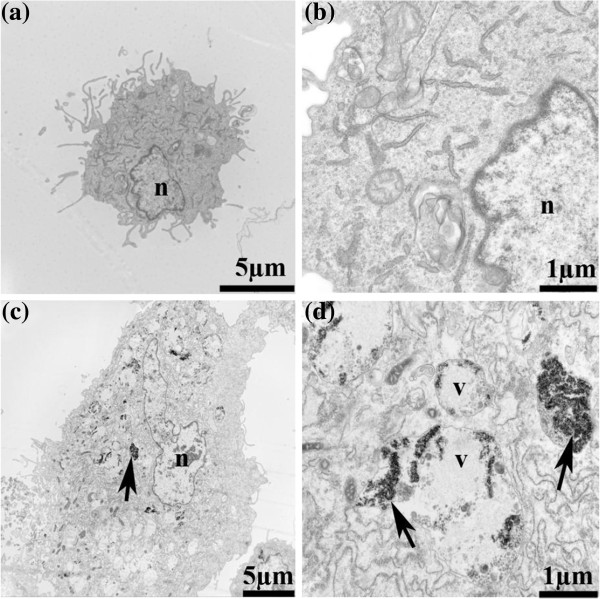
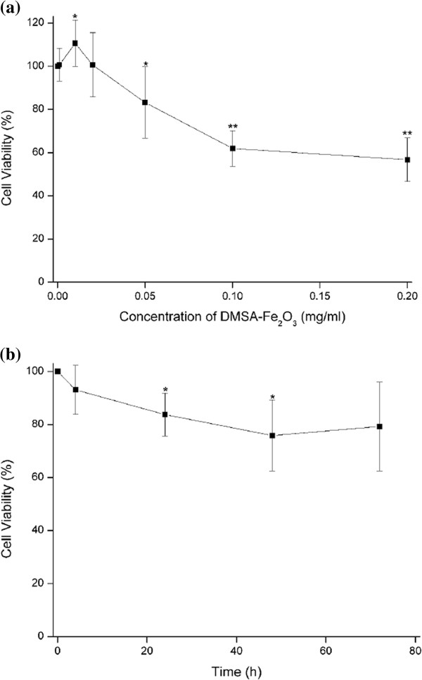
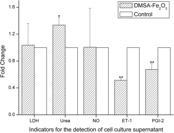
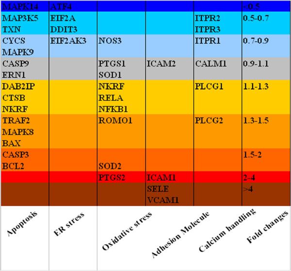
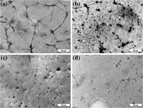
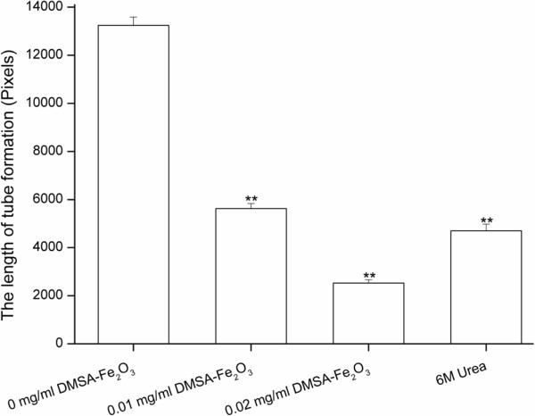
One major obstacle for successful application of nanoparticles in medicine is its potential nanotoxicity on the environment and human health. In this study, we evaluated the cytotoxicity effect of dimercaptosuccinic acid-coated iron oxide (DMSA-Fe2O3) using cultured human aortic endothelial cells (HAECs). Our results showed that DMSA-Fe2O3 in the culture medium could be absorbed into HAECs, and dispersed in the cytoplasm. The cytotoxicity effect of DMSA-Fe2O3 on HAECs was dose-dependent, and the concentrations no more than 0.02 mg/ml had little toxic effect which were revealed by tetrazolium dye assay. Meanwhile, the cell injury biomarker, lactate dehydrogenase, was not significantly higher than that from control cells (without DMSA-Fe2O3). However, the endocrine function for endothelin-1 and prostacyclin I-2, as well as the urea transporter function, was altered even without obvious evidence of cell injury in this context. We also showed by real-time PCR analysis that DMSA-Fe2O3 exposure resulted in differential effects on the expressions of pro- and anti-apoptosis genes of HAECs. Meanwhile, it was noted that DMSA-Fe2O3 exposure could activate the expression of genes related to oxidative stress and adhesion molecules, which suggested that inflammatory response might be evoked. Moreover, we demonstrated by in vitro endothelial tube formation that even a small amount of DMSA-Fe2O3 (0.01 and 0.02 mg/ml) could inhibit angiogenesis by the HAECs. Altogether, these results indicate that DMSA-Fe2O3 have some cytotoxicity that may cause side effects on normal endothelial cells.
Figures
Similar articles
-
In Vitro Effects of Hollow Gold Nanoshells on Human Aortic Endothelial Cells.Nanoscale Res Lett. 2016 Dec;11(1):397. doi: 10.1186/s11671-016-1620-5. Epub 2016 Sep 13. Nanoscale Res Lett. 2016. PMID: 27624340 Free PMC article.
-
Synergic fabrication of succimer coated titanium dioxide nanomaterials delivery for in vitro proliferation and in vivo examination on human aortic endothelial cells.Drug Deliv. 2021 Dec;28(1):1785-1794. doi: 10.1080/10717544.2021.1960925. Drug Deliv. 2021. PMID: 34470555 Free PMC article.
-
Iron oxide nanoparticles cause surface coating- and core chemistry-dependent endothelial cell ferroptosis.Nanotoxicology. 2022 Nov-Dec;16(9-10):829-843. doi: 10.1080/17435390.2022.2154176. Epub 2023 Jan 20. Nanotoxicology. 2022. PMID: 36660964
-
Labeling mesenchymal cells with DMSA-coated gold and iron oxide nanoparticles: assessment of biocompatibility and potential applications.J Nanobiotechnology. 2016 Jul 18;14(1):59. doi: 10.1186/s12951-016-0213-x. J Nanobiotechnology. 2016. PMID: 27431051 Free PMC article.
-
Dimercaptosuccinic acid (succimer; DMSA) in inorganic lead poisoning.Clin Toxicol (Phila). 2009 Aug;47(7):617-31. doi: 10.1080/15563650903174828. Clin Toxicol (Phila). 2009. PMID: 19663612 Review.
Cited by
-
Characterization of interaction of magnetic nanoparticles with breast cancer cells.J Nanobiotechnology. 2015 Feb 26;13:16. doi: 10.1186/s12951-015-0073-9. J Nanobiotechnology. 2015. PMID: 25880445 Free PMC article.
-
Potential Use of DMSA-Containing Iron Oxide Nanoparticles as Magnetic Vehicles against the COVID-19 Disease.ChemistrySelect. 2021 Aug 20;6(31):7931-7935. doi: 10.1002/slct.202101900. Epub 2021 Aug 17. ChemistrySelect. 2021. PMID: 34541297 Free PMC article.
-
Disruption of Cell Adhesion and Cytoskeletal Networks by Thiol-Functionalized Silica-Coated Iron Oxide Nanoparticles.Int J Mol Sci. 2020 Dec 8;21(24):9350. doi: 10.3390/ijms21249350. Int J Mol Sci. 2020. PMID: 33302486 Free PMC article.
-
Cobalt Zinc Ferrite Nanoparticles as a Potential Magnetic Resonance Imaging Agent: An In vitro Study.Avicenna J Med Biotechnol. 2015 Apr-Jun;7(2):64-8. Avicenna J Med Biotechnol. 2015. PMID: 26140183 Free PMC article.
-
Evaluation the toxic effects of Cobalt-Zinc Ferrite nanoparticles in experimental mice.Sci Rep. 2025 Feb 26;15(1):6903. doi: 10.1038/s41598-025-90043-x. Sci Rep. 2025. PMID: 40011491 Free PMC article.
References
-
- Alexiou C, Arnold W, Klein RJ, Parak FG, Hulin P, Bergemann C, Erhardt W, Wagenpfeil S, Lubbe AS. Locoregional cancer treatment with magnetic drug targeting. Cancer Res. 2000;8:6641–6648. - PubMed
LinkOut - more resources
Full Text Sources
Other Literature Sources
