

Characterization of clinical photosensitivity in cutaneous lupus erythematosus
- PMID: 23648190
- PMCID: PMC3928014
- DOI: 10.1016/j.jaad.2013.03.015
Characterization of clinical photosensitivity in cutaneous lupus erythematosus
Abstract
Background: Photosensitivity (PS) in lupus erythematosus (LE) is frequently determined by patient report.
Objective: We sought to characterize self-reported PS in cutaneous LE (CLE).
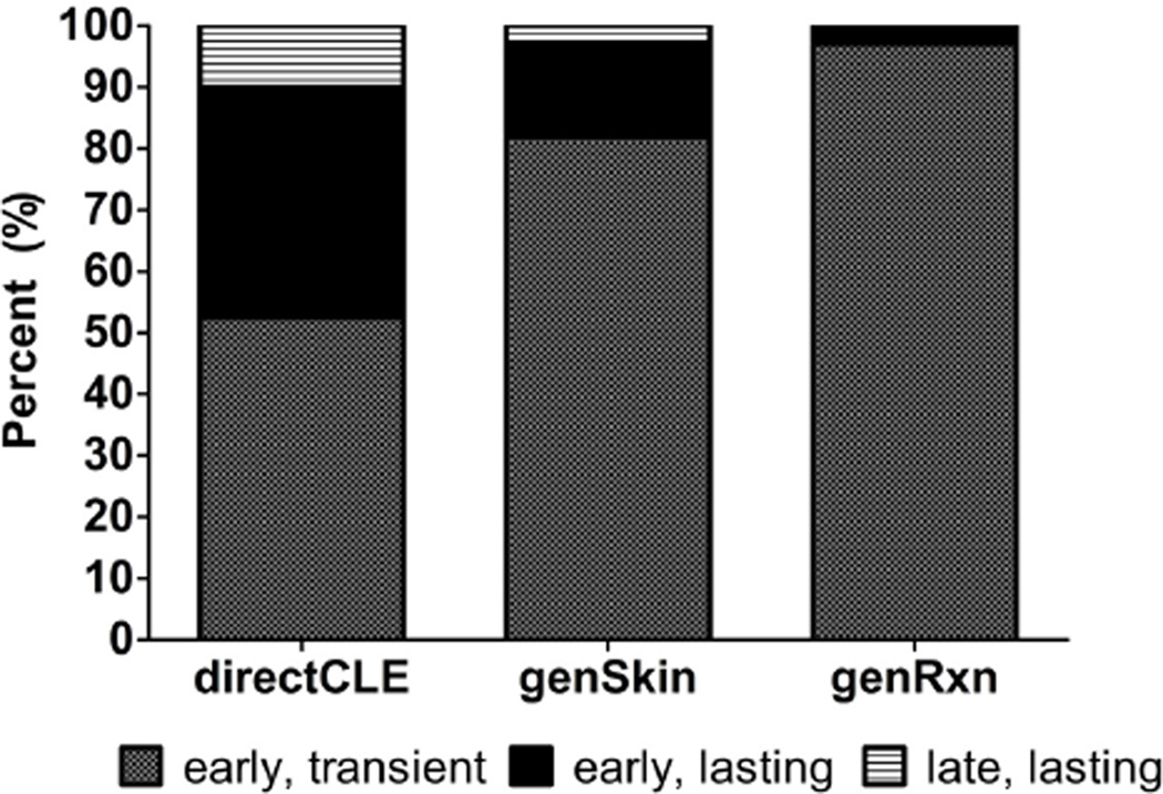
Methods: The PS survey was used to classify subject responses into 5 phenotypes: direct sun-induced CLE flare (directCLE); general exacerbation of CLE (genCLE); polymorphic light eruption-like reactions (genSkin); general pruritus/paresthesias (genRxn); and sun-induced systemic symptoms (genSys). In all, 91 subjects with CLE alone or with CLE and systemic LE were interviewed.
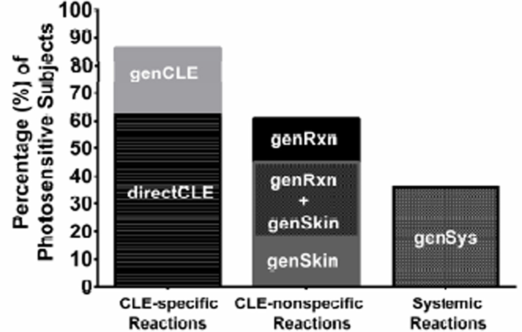
Results: In all, 81% ascribed to 1 or more PS phenotypes. CLE-specific reactions (direct sun-induced CLE flare or general exacerbation of CLE) were reported by 86% of photosensitive subjects. Higher CLE disease activity (measured by CLE Disease Area and Severity Index activity scores) was suggestive of direct sun-induced CLE flare reactions (P = .09). In all, 60% of photosensitive subjects described CLE-nonspecific reactions: polymorphic light eruption-like rash and general pruritus/paresthesias. These phenotypes often co-occurred with CLE-specific reactions and were predicted by more systemic disease activity as measured by Physicians Global Assessment (PGA) scores in regression analyses (genSkin, P = .02) and (genRxn, P = .05). In all, 36% of subjects reported systemic reactions and higher PGA scores were predictive of the sun-induced systemic symptoms phenotype (P = .02); a diagnosis of systemic LE was not (P = .14).
Limitations: PS was inferred from patient report and not directly observed.
Conclusions: Characterization of self-reported PS in LE reveals that patients experience combinations of CLE-specific, CLE-nonspecific, and systemic reactions to sunlight. Sun-induced CLE flares are associated with more active CLE disease. Polymorphic light eruption-like, generalized pruritus/paresthesias, and systemic reactions are associated with more active systemic disease. Recognition of PS phenotypes will permit improved definitions of clinical PS and allow for more precise investigation into its pathophysiology.
Keywords: CLASI; CLE; Cutaneous Lupus Erythematosus Disease Area and Severity Index; LE; PGA; PMLE; PS; Physicians Global Assessment; SELENA; SLE; SLEDAI; Safety of Estrogens in Lupus Erythematosus National Assessment; Systemic Lupus Erythematosus Disease Activity Index; cutaneous lupus erythematosus; dendritic cells; direct sun-induced cutaneous lupus erythematosus flare; directCLE; genCLE; genRxn; genSkin; genSys; general exacerbation of cutaneous lupus erythematosus; general pruritus/paresthesias; immunohistochemistry; lupus erythematosus; mDC; myeloid dendritic cells; photosensitivity; polymorphic light eruption; polymorphic light eruption-like; sun-induced systemic symptoms; systemic lupus erythematosus.
Copyright © 2013 American Academy of Dermatology, Inc. Published by Mosby, Inc. All rights reserved.
Conflict of interest statement
The authors declare no conflict of interest.
Figures
References
-
- Scheinfeld N, Deleo VA. Photosensitivity in lupus erythematosus. Photodermatol Photoimmunol Photomed. 2004 Oct;20(5):272–279. - PubMed
-
- Tan EM, Cohen AS, Fries JF, Masi AT, McShane DJ, Rothfield NF, Schaller JG, Talal N, Winchester RJ. The 1982 revised criteria for the classification of systemic lupus erythematosus. Arthritis Rheum. 1982 Nov;25(11):1271–1277. - PubMed
-
- Kind P, Lehmann P, Plewig G. Phototesting in lupus erythematosus. J Invest Dermatol. 1993 Jan;100(1):53S–57S. - PubMed
-
- Kuhn A, Sonntag M, Richter-Hintz D, Oslislo C, Megahed M, Ruzicka T, Lehmann P. Phototesting in lupus erythematosus: A 15-year experience. J Am Acad Dermatol. 2001;45(1):86–95. - PubMed
-
- Schornagel IJ, Knol EF, van Weelden H, Guikers CL, Bruijnzeel-Koomen CA, Sigurdsson V. Diagnostic phototesting in polymorphous light eruption: The optimal number of irradiations. Br J Dermatol. 2005 Dec;153(6):1234–1236. - PubMed
Publication types
MeSH terms
Grants and funding
LinkOut - more resources
Full Text Sources
Other Literature Sources
