

Muscular senescence in cetaceans: adaptation towards a slow muscle fibre phenotype
- PMID: 23648412
- PMCID: PMC3646281
- DOI: 10.1038/srep01795
Muscular senescence in cetaceans: adaptation towards a slow muscle fibre phenotype
Abstract
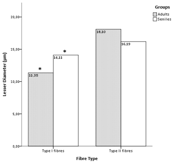
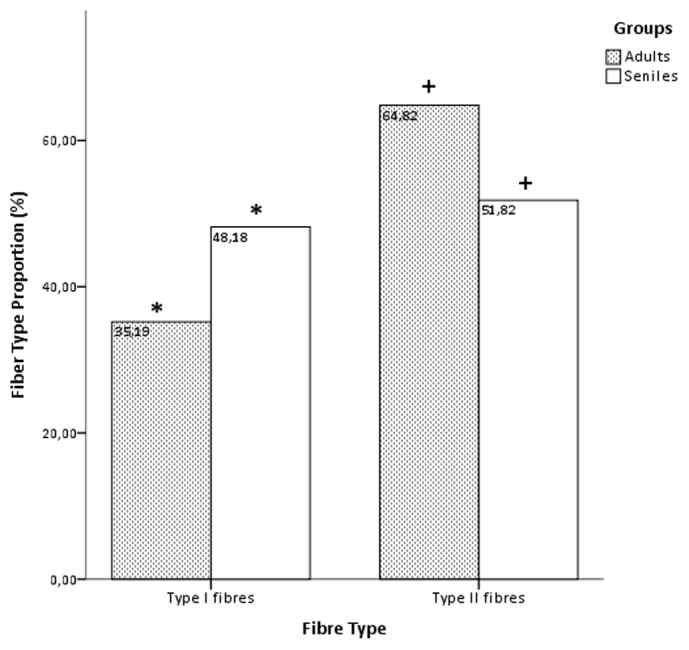
Sarcopenia, or senile muscle atrophy, is the slow and progressive loss of muscle mass with advancing age that constitutes the most prevalent form of muscle atrophy. The effects of ageing on skeletal muscle have been extensively studied in humans and laboratory animals (mice), while the few reports on wild animals are based on short-lived mammals. The present study describes the age-related changes in cetacean muscles regarding the three factors that determine muscle mass: fibre size, fibre number, and fibre type. We show that the skeletal muscle fibres in cetaceans change with advancing age, evolving towards a slower muscle phenotype. We suggest that this physiological evolution constitutes an adaptation that allows these marine mammals to perform prolonged, deep dives.
Figures
Similar articles
-
Drastic increase of myosin light chain MLC-2 in senescent skeletal muscle indicates fast-to-slow fibre transition in sarcopenia of old age.Eur J Cell Biol. 2009 Nov;88(11):685-700. doi: 10.1016/j.ejcb.2009.06.004. Epub 2009 Jul 19. Eur J Cell Biol. 2009. PMID: 19616867
-
Dihydrotestosterone treatment rescues the decline in protein synthesis as a result of sarcopenia in isolated mouse skeletal muscle fibres.J Cachexia Sarcopenia Muscle. 2017 Feb;8(1):48-56. doi: 10.1002/jcsm.12122. Epub 2016 Apr 25. J Cachexia Sarcopenia Muscle. 2017. PMID: 27239418 Free PMC article.
-
Differential activation of the calpain system involved in individualized adaptation of different fast-twitch muscles in hibernating Daurian ground squirrels.J Appl Physiol (1985). 2019 Aug 1;127(2):328-341. doi: 10.1152/japplphysiol.00124.2019. Epub 2019 Jun 20. J Appl Physiol (1985). 2019. PMID: 31219776
-
The age-related loss of skeletal muscle mass and function: Measurement and physiology of muscle fibre atrophy and muscle fibre loss in humans.Ageing Res Rev. 2018 Nov;47:123-132. doi: 10.1016/j.arr.2018.07.005. Epub 2018 Jul 23. Ageing Res Rev. 2018. PMID: 30048806 Free PMC article. Review.
-
Fibre types in skeletal muscles.Adv Anat Embryol Cell Biol. 2002;162:III-XV, 1-109. doi: 10.1007/978-3-642-59399-4. Adv Anat Embryol Cell Biol. 2002. PMID: 11892240 Review.
Cited by
-
High-precision isotopic analysis sheds new light on mercury metabolism in long-finned pilot whales (Globicephala melas).Sci Rep. 2019 May 13;9(1):7262. doi: 10.1038/s41598-019-43825-z. Sci Rep. 2019. PMID: 31086275 Free PMC article.
-
A procession of metabolic alterations accompanying muscle senescence in Manduca sexta.Sci Rep. 2018 Jan 17;8(1):1006. doi: 10.1038/s41598-018-19630-5. Sci Rep. 2018. PMID: 29343811 Free PMC article.
-
Single skeletal muscle fiber mechanical properties: a muscle quality biomarker of human aging.Eur J Appl Physiol. 2022 Jun;122(6):1383-1395. doi: 10.1007/s00421-022-04924-4. Epub 2022 Mar 6. Eur J Appl Physiol. 2022. PMID: 35249139 Review.
-
Histopathological muscle findings may be essential for a definitive diagnosis of suspected sharp trauma associated with ship strikes in stranded cetaceans.PLoS One. 2014 Feb 13;9(2):e88780. doi: 10.1371/journal.pone.0088780. eCollection 2014. PLoS One. 2014. PMID: 24551162 Free PMC article.
References
-
- WHO. What are the public health implications of global ageing? Online Q&A 29 September 2011 Retrieved from http://www.who.int/features/qa/42/en/index.html (2011).
-
- Walston J. et al. Research agenda for frailty in older adults: toward a better understanding of physiology and etiology: summary from the American Geriatrics Society/National Institute on Aging Research Conference on Frailty in Older Adults. J Am Geriatr Soc 54, 991–1001 (2006). - PubMed
-
- Rogers M. A. & Evans W. J. Changes in skeletal muscle with aging: effects of exercise training. Exerc Sport Sci Rev 21, 65–102 (1993). - PubMed
-
- Lexell J., Taylor C. C. & Sjostrom M. What is the cause of the ageing atrophy? Total number, size and proportion of different fiber types studied in whole vastus lateralis muscle from 15- to 83-year-old men. J Neurol Sci 84, 275–294 (1988). - PubMed
-
- Hagen J. L. et al. Skeletal muscle aging in F344BN F1-hybrid rats: I. Mitochondrial dysfunction contributes to the age-associated reduction in VO2max. J Gerontol A Biol Sci Med Sci 59, 1099–1110 (2004). - PubMed
Publication types
MeSH terms
LinkOut - more resources
Full Text Sources
Other Literature Sources
Medical
