. 2013 Jul;33(13):2603-13.
doi: 10.1128/MCB.01672-12. Epub 2013 May 6.

The protein level of PGC-1α, a key metabolic regulator, is controlled by NADH-NQO1

Affiliations

The protein level of PGC-1α, a key metabolic regulator, is controlled by NADH-NQO1

Yaarit Adamovich et al. Mol Cell Biol. 2013 Jul.

Abstract

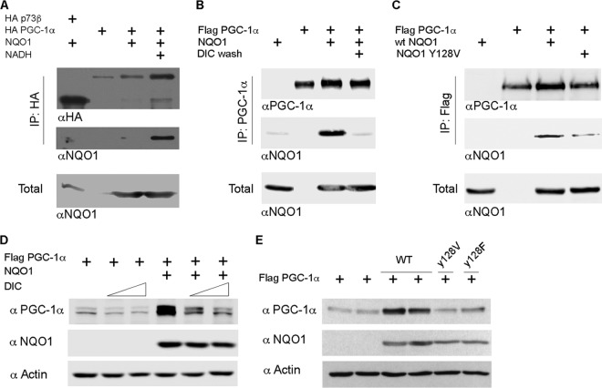
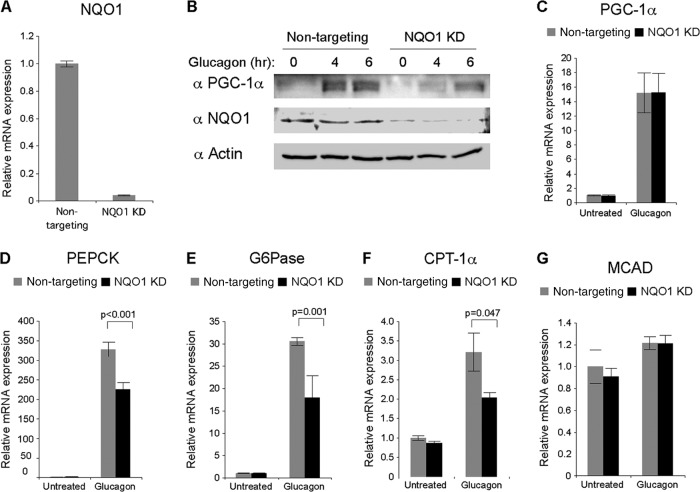
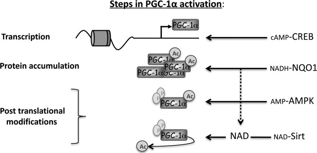
PGC-1α is a key transcription coactivator regulating energy metabolism in a tissue-specific manner. PGC-1α expression is tightly regulated, it is a highly labile protein, and it interacts with various proteins--the known attributes of intrinsically disordered proteins (IDPs). In this study, we characterize PGC-1α as an IDP and demonstrate that it is susceptible to 20S proteasomal degradation by default. We further demonstrate that PGC-1α degradation is inhibited by NQO1, a 20S gatekeeper protein. NQO1 binds and protects PGC-1α from degradation in an NADH-dependent manner. Using different cellular physiological settings, we also demonstrate that NQO1-mediated PGC-1α protection plays an important role in controlling both basal and physiologically induced PGC-1α protein level and activity. Our findings link NQO1, a cellular redox sensor, to the metabolite-sensing network that tunes PGC-1α expression and activity in regulating energy metabolism.


Figures

Fig 1
Fig 1
PGC-1α is an intrinsically disordered protein by prediction, susceptible to in vitro degradation by the 20S PC and protected by NQO1. (A) Analysis of PGC-1α and PCNA amino acid sequences by the FoldIndex prediction program. (B) In vitro-transcribed and -translated [35S]methionine-labeled PGC-1α and PCNA proteins were incubated with or without purified 20S PC for the indicated time points. Immediately after incubation, the mixture was subjected to analysis by SDS-PAGE. (C) In vitro-transcribed and -translated [35S]methionine-labeled PGC-1α was incubated with purified 20S PC with or without recombinant NQO1 protein for the indicated time periods. Immediately after incubation, the extracts were analyzed by SDS-PAGE.
Fig 2
Fig 2
NQO1 inhibits PGC-1α proteasomal degradation and increases its protein half-life. (A) HEK-293T cells and HEK-293T cells stably expressing the Flag-tagged β4 (PSMB2) ring subunit were transiently transfected with untagged PGC-1α- or NQO1-expressing vectors as indicated. Treatment with 25 μM MG132 was done 48 h postinfection, and cells were incubated for 2 h. Then, cells were harvested for immunoprecipitation (IP) using Flag beads. Analysis of the α3 (PSMA4) ring subunit indicates that the intact proteasome chamber was immunoprecipitated. (B) Flag-tagged PGC-1α was overexpressed in HEK-293 cells alone or together with an NQO1 expression vector. Cells were treated 24 h later with 25 μM MG132 for 3 or 6 h as indicated, after which cells from all points were harvested for protein analysis. (C) HEK-293T cells were transfected as indicated. GFP was cotransfected as a transfection and loading control. Cycloheximide (CHX) was added to the cells 48 h posttransfection, and cells were treated for the indicated time periods. MG132 (25 μM) was added together with CHX at the last time point where PGC-1α was expressed alone. S.E, short exposure; L.E, long exposure. The dashed line indicates an area in the final image in which an irrelevant lane was omitted. (D) HEK-293T cells were transfected as indicated. Cell extracts were immunoprecipitated using Flag beads and immunoblotted with anti-HA to visualize polyubiquitination. (E) Quantification of the ratio between polyubiquitinated PGC-1α ladder and PGC-1α from 3 independent biological triplicates.
Fig 3
Fig 3
NQO1 protects PGC-1α by NADH-dependent interaction. (A) HEK-293 cells were transfected as indicated and harvested for analysis 24 h posttransfection. HA beads were used to immunoprecipitate (IP) HA-tagged PGC-1α, whereas HA-p73β served as a negative control for the binding to NQO1. Where indicated, 1 mM NADH was added to the extraction buffer. (B) HEK-293 cells were transfected as indicated and harvested for analysis 24 h posttransfection. A/G beads conjugated to PGC-1α antibody (H-300) were used to immunoprecipitate Flag–PGC-1α. Dicoumarol (DIC; 150 μM) was added to the last two wash steps. (C) HEK-293T cells were transfected as indicated and harvested 24 h later. Flag beads were used to immunoprecipitate Flag-tagged PGC-1α. wt, wild type. (D) Flag-tagged PGC-1α was expressed in HEK-293 cells alone or together with NQO1. Twenty-four hours posttransfection, the cells were treated with dicoumarol (100 and 150 μM) for an additional 2 h, after which cells were harvested for protein analysis. (E) Flag-tagged PGC-1α was expressed in HEK-293 cells alone (2 individual transfections) or together with either wild-type NQO1 (WT; 2 individual transfections) or 2 different NQO1 mutants (Y128V and Y128F). Protein expression was analyzed 24 h posttransfection.
Fig 4
Fig 4
NQO1 and PGC-1α steady-state levels in myoblasts and myotubes. (A) Analysis of PGC-1α and NQO1 protein and mRNA levels in C2C12 myoblast (MB) cells and at sequential days after the cells were put in differentiation medium. (B) Same as for panel A, except that the analysis was done in primary mouse muscle cells. (C) Bortezomib (1 μM) was added 12 h before harvesting to primary mouse muscle cells either grown in growth medium as myoblasts (MB) or grown in differentiation medium for 1, 2, or 3 days. Due to the effect of proteasome inhibition on PGC-1α solubility (see above), in order to obtain the total amount of PGC-1α we analyzed the soluble and insoluble fractions together as the total. (Left) Quantification of total PGC-1α from bortezomib-treated cells relative to total PGC-1α from untreated cells. (Right) Western blot analysis. (D) Protein extracts obtained from extensor digitorum longus (EDL), soleus (SOL), and gastrocnemius (GAS) of an 8-week-old male C57 black mouse and subjected to Western blot analysis with the indicated antibodies. Immunoprecipitation (IP) was used to detect PGC-1α protein. IB, immunoblotting.
Fig 5
Fig 5
NQO1 knockdown in myoblasts reduces steady-state PGC-1α protein levels and activity. (A) NQO1 was knocked down in C2C12 cells using a lentivirus-based approach (3 individual experiments); all were run on the same gel, and cells were analyzed for NQO1 and PGC-1α protein expression. The right panel represents quantification of PGC-1α protein. (B) PGC-1α mRNA expression level was analyzed from C2C12 cells as described for panel A. (C) NADH and NAD+ content measured from basic and acidic extracts obtained from freshly prepared control (Non-targeting) and NQO1 knockdown (KD) C2C12 cells. (D) mRNA expression analysis of different genes from freshly prepared control (Non-targeting) and NQO1 knockdown (KD) C2C12 cells.
Fig 6
Fig 6
NQO1 regulates PGC-1α accumulation in response to induction by starvation-mimicking conditions in mouse primary hepatocytes. (A) Primary hepatocytes were infected with either GFP- or NQO1-expressing adenoviruses. Expression levels of NQO1 mRNA were analyzed 48 h postinfection to verify the efficiency of the knockdown. (B) Same as in panel A. Twenty-four hours after infection, cells were treated with 25 nM glucagon for either 4 or 6 h. Protein expression was analyzed by Western blotting. (C) Same as in panel B. Expression levels of PGC-1α mRNA were analyzed 4 h after addition of glucagon. (D to G) Same as in panel B. Expression levels of PEPCK (D), G6Pase (E), CPT-1α (F), and MCAD (G) mRNA were analyzed 4 h after addition of glucagon.
Fig 7
Fig 7
A role for NQO1 activity in regulating PGC-1α in fasting liver. (A) Mice were either fed ad libitum or fasted during the indicated time points (starting from the beginning of the night phase) and then sacrificed for hepatic analysis of NADH and NAD+ content. (B) Mice were i.p. injected with either dicoumarol (DIC) or vehicle and were divided into groups of feeding and fasting for 7 h (during the night). Immunoprecipitation was used to detect PGC-1α protein. (C) Mice were i.p. injected with either dicoumarol or vehicle and were divided into groups of feeding and fasting for 14 h (during the night). Blood glucose level was measured (n = 5). (D) Insulin measurements from the experiment described for panel C (n = 5).
Fig 8
Fig 8
Schematic model illustrating the convergent actions of CREB, NQO1, AMPK, and Sirt1 on PGC-1α. The scheme summarizes PGC-1α transcription and posttranslational regulation by different known metabolite-sensing proteins, including NQO1.

References

    1. Baar K, Wende AR, Jones TE, Marison M, Nolte LA, Chen M, Kelly DP, Holloszy JO. 2002. Adaptations of skeletal muscle to exercise: rapid increase in the transcriptional coactivator PGC-1. FASEB J. 16:1879–1886 - PubMed
    1. Michael LF, Wu Z, Cheatham RB, Puigserver P, Adelmant G, Lehman JJ, Kelly DP, Spiegelman BM. 2001. Restoration of insulin-sensitive glucose transporter (GLUT4) gene expression in muscle cells by the transcriptional coactivator PGC-1. Proc. Natl. Acad. Sci. U. S. A. 98:3820–3825 - PMC - PubMed
    1. Wu Z, Puigserver P, Andersson U, Zhang C, Adelmant G, Mootha V, Troy A, Cinti S, Lowell B, Scarpulla RC, Spiegelman BM. 1999. Mechanisms controlling mitochondrial biogenesis and respiration through the thermogenic coactivator PGC-1. Cell 98:115–124 - PubMed
    1. Herzig S, Long F, Jhala US, Hedrick S, Quinn R, Bauer A, Rudolph D, Schutz G, Yoon C, Puigserver P, Spiegelman B, Montminy M. 2001. CREB regulates hepatic gluconeogenesis through the coactivator PGC-1. Nature 413:179–183 - PubMed
    1. Yoon JC, Puigserver P, Chen G, Donovan J, Wu Z, Rhee J, Adelmant G, Stafford J, Kahn CR, Granner DK, Newgard CB, Spiegelman BM. 2001. Control of hepatic gluconeogenesis through the transcriptional coactivator PGC-1. Nature 413:131–138 - PubMed

Publication types

MeSH terms

LinkOut - more resources