. 2013 Jul:112:106-17.
doi: 10.1016/j.exer.2013.04.021. Epub 2013 May 3.

JUN regulates early transcriptional responses to axonal injury in retinal ganglion cells

Affiliations

JUN regulates early transcriptional responses to axonal injury in retinal ganglion cells

Kimberly A Fernandes et al. Exp Eye Res. 2013 Jul.

Abstract

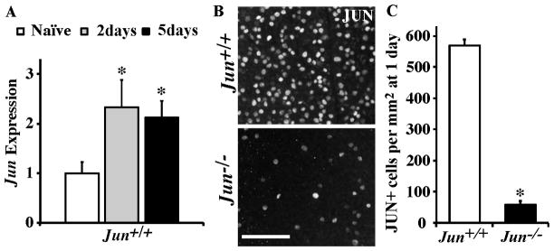
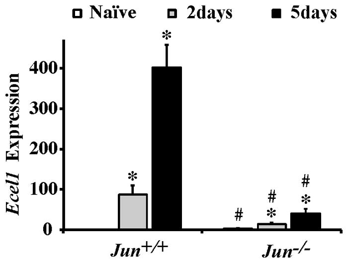
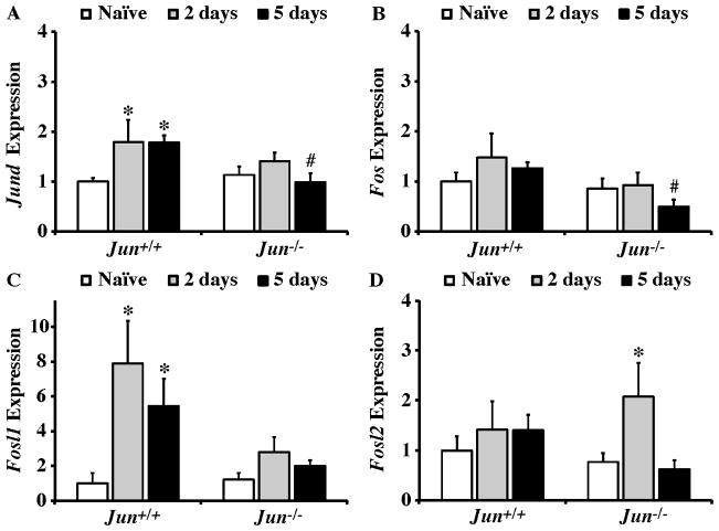
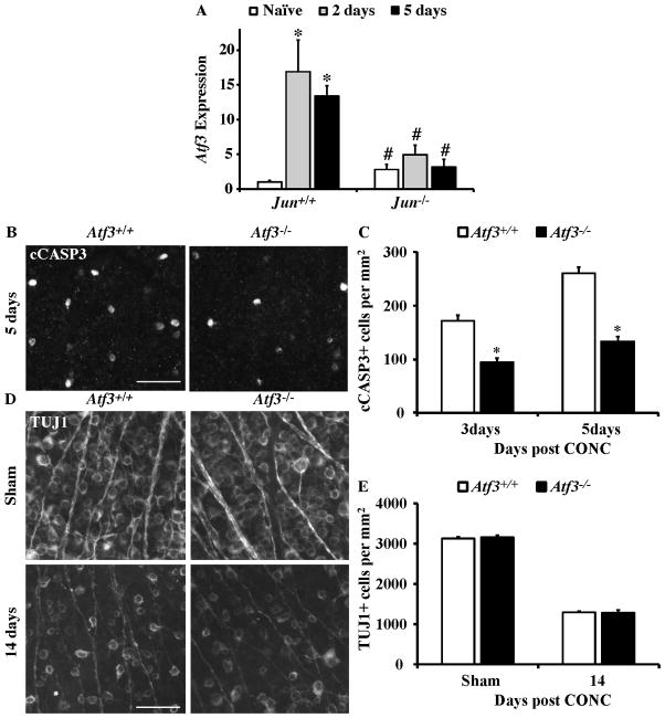
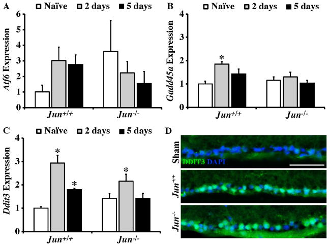
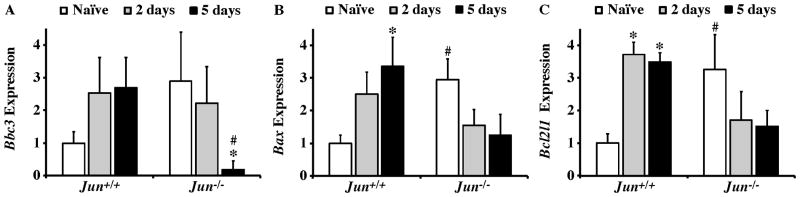
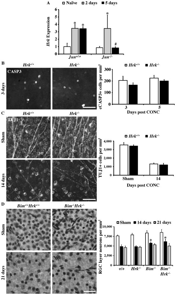
The AP1 family transcription factor JUN is an important molecule in the neuronal response to injury. In retinal ganglion cells (RGCs), JUN is upregulated soon after axonal injury and disrupting JUN activity delays RGC death. JUN is known to participate in the control of many different injury response pathways in neurons, including pathways controlling cell death and axonal regeneration. The role of JUN in regulating genes involved in cell death, ER stress, and regeneration was tested to determine the overall importance of JUN in regulating RGC response to axonal injury. Genes from each of these pathways were transcriptionally controlled following axonal injury and Jun deficiency altered the expression of many of these genes. The differentially expressed genes included, Atf3, Ddit3, Ecel1, Gadd45α, Gal, Hrk, Pten, Socs3, and Sprr1a. Two of these genes, Hrk and Atf3, were tested for importance in RGC death using null alleles of each gene. Disruption of the prodeath Bcl2 family member Hrk did not affect the rate or amount of RGC death after axonal trauma. Deficiency in the ATF/CREB family transcription factor Atf3 did lessen the amount of RGC death after injury, though it did not provide long term protection to RGCs. Since JUN's dimerization partner determines its transcriptional targets, the expression of several candidate AP1 family members were examined. Multiple AP1 family members were induced by axonal injury and had a different expression profile in Jun deficient retinas compared to wildtype retinas (Fosl1, Fosl2 and Jund). Overall, JUN appears to play a multifaceted role in regulating RGC response to axonal injury.


Figures

Figure 1
Figure 1. Axonal injury induces Jun upregulation
(A) The expression of Jun is significantly increased at both 2 and 5 days following CONC in Jun+/+ mice (represented as normalized fold expression). (B,C) To determine the recombination efficiency of the Jun floxed (Junfl/fl ) allele using Six3-cre, the number of JUN positive cells were counted in retinal flat mounts (RGC layer up) after CONC at a time when there is robust expression of JUN and before RGC cell death, 1 day after CONC. In Junfl/fl Six3cre+ (Jun−/−) retinas, the number of JUN+ cells was significantly reduced. *, P<0.05. Scale bar, 20 μm.
Figure 2
Figure 2. Jun deficiency alters the expression of an axon injury response gene, Ecel1, after CONC
Realtime PCR analysis for Ecel1 from retinas of Jun+/+ and Jun−/− mice at the indicated time-points following CONC shown as normalized fold change. Expression of the neuronal injury responsive gene Ecel1 increased progressively in both Jun+/+ and Jun−/− animals following CONC (*, intra-genotype comparison, P < 0.001). However, comparing the change in gene expression between Jun+/+ and Jun−/− mice showed that in Jun deficient mice the CONC-induced expression of Ecel1 was attenuated at both time points (#, inter-genotype comparison, P < 0.05). Also, Ecel1 expression was significantly increased in Jun−/− retinas compared to Jun+/+ retinas prior to injury.
Figure 3
Figure 3. JUN and FOS transcription factor expression after axonal-injury
Realtime PCR analysis of JUN and FOS family members in Jun+/+ and Jun−/− mice expressed as normalized fold change. (A) Jund expression significantly increased at both 2 and 5 days after CONC in Jun+/+ mice (*, intra-genotype comparison, P < 0.05) and did not change in Jun−/− mice. Comparing expression level changes between genotypes showed that Jund expression was significantly attenuated in Jun−/− retinas compared to wildtype retinas at 5 days after CONC (#, inter-genotype comparison, P < 0.05) (B) Fos expression remained unchanged in Jun+/+ and Jun−/− mice at both time points examined, however, Fos expression was attenuated in Jun−/− retinas compared to Jun+/+ retinas 5 days after CONC. (C) A significant increase in Fosl1 expression was observed in Jun+/+ mice at 2 and 5 days after CONC. Fosl1 expression was not significantly changed at either time point examined in Jun−/− retinas. (D) Fosl2 expression did not change after CONC in wildtype mice at 2 and 5 days compared to naïve retinas. However, in Jun deficient retinas Fosl2 expression was significantly increased 2 days after CONC. *, P < 0.05 comparing 2 or 5 day time points to Naïve retinas of same genotype (Intra-genotype); #, P < 0.05, comparing same time points across genotypes (Inter-genotype).
Figure 4
Figure 4. The Jun target, Atf3, suppresses RGC death following CONC
(A) The expression of Atf3 significantly increased after CONC in Jun+/+ retinas at 2 and 5 days (shown as normalized fold change; *, intra-genotype comparison, P < 0.05). Atf3 expression was not significantly increased after CONC in Jun−/− retinas at 2 and 5 days. In fact, comparing the change in gene expression of Atf3 between Jun+/+ and Jun−/− mice showed that Atf3 expression was significantly attenuated in Jun−/− at both time points after CONC (#, inter-genotype comparison, P < 0.05). Also, Atf3 expression was significantly increased in Jun−/− retinas compared to Jun+/+ retinas prior to injury. (B) Representative images of cleaved caspase-3 (cCASP3) labeled, dying RGCs in Atf3+/+ and Atf3−/− retinal whole mounts. (C) The number of cCASP3 labeled cells was significantly reduced in Atf3−/− retinas at both 3 days and 5 days after CONC (*, P < 0.05 for both time points; N≥5 for both genotypes and time points). (D) Representative images of anti-βIII tubulin (TUJ1) positive RGCs in retinal whole mounts from sham injured and CONC injured eyes 14 days following the insult. (E) Despite the small decrease in cell death in Atf3 deficient mice, Atf3 deficiency did not increase the number of surviving RGCs 14 days after CONC (P = 0.82, N= 6 for genotypes). Scalebar, 25 μm.
Figure 5
Figure 5. JUN contributes to transcriptional control of RGC regeneration potential
Realtime PCR analysis of genes involved in axon regeneration in Jun+/+ and Jun−/− mice shown as normalized fold change. (A,B) The expression of both Gal and Sprr1a positively correlate with regeneration and both significantly increased in Jun+/+ and Jun−/− mice after CONC (*, intra-genotype comparisons, P < 0.05). However, the increase in expression of both of these genes was significantly attenuated at least one time point in the Jun deficient mice (#, inter-genotype comparison, P < 0.05). (C–E) Genes that are known to suppress regeneration in injured RGCs were also examined. (C) The expression of Klf4 was not altered following CONC in either genotype. (D) Socs3 expression was not altered transcriptionally at 2 days or 5 days following CONC in Jun+/+ retinas but was significantly increased in Jun−/− retinas 2 days post CONC. (E) Pten was significantly upregulated at 5 days following CONC in Jun+/+ retinas. Pten expression did not change in Jun+/+ retinas, however, Pten expression was significantly attenuated at 5 days in Jun−/− deficient retinas compared to wildtype. Also, Pten expression was significantly increased in Jun−/− retinas compared to Jun+/+ retinas prior to injury. *, P < 0.05 comparing 2 or 5 day time points to Naïve retinas of same genotype (Intra-genotype); #, P < 0.05, comparing same time points across genotypes (Inter-genotype).
Figure 6
Figure 6. After axonal injury ER stress occurs in the absence of Jun
(A–C) Realtime PCR analysis of genes involved in ER stress response in Jun+/+ and Jun−/− mice shown as normalized fold change. (A) The expression of ER stress sensor Atf6 was not altered following CONC in either Jun+/+ or Jun−/− retinas. (B) The expression of the ER stress marker Gadd45a significantly increased at 2 days following CONC in Jun+/+ retinas (*, intra-genotype comparisons, P < 0.05). No other changes in Gadd45a expression were detected. (C) The expression of Ddit3, an ER stress target gene, significantly increased in Jun+/+ at both 2 and 5 days after CONC. In Jun−/− retinas Ddit3 expression was significantly increased only at 2 days following CONC. (D) Representative immunofluorescence staining for DDIT3 3 days following CONC confirms induction of DDIT3 in both Jun+/+ and Jun−/− retinas (DDIT3, green; DAPI, blue, was used to stain nuclei; the experiment was performed on 3 different mice for each genotype and condition). *, P < 0.05 comparing 2 or 5 day time points to naïve retinas of same genotype (Intra-genotype).
Figure 7
Figure 7. Bcl2 family expression after axonal-injury
Results of realtime PCR analysis showing the normalized fold expression of a subset of Bcl2 family members that have been implicated in axonal injury induced RGC death in Jun+/+ and Jun−/− mice. (A) Bbc3 expression was not significantly altered after CONC in either genotype, though the expression was significantly attenuated in Jun−/− retinas compared to Jun+/+ retinas 5 days after CONC (#, inter-genotype comparison, P < 0.05). (B) Bax was significantly increased 5 days after CONC in Jun+/+ (*, intra-genotype comparisons, P < 0.05). There were no differences in Bax expression in Jun−/− animals after CONC. Also, Bax levels did not differ between Jun+/+ and Jun−/− retinas after CONC, but Bax levels were significantly higher in naïve Jun−/− retinas compared to Jun+/+ naïve retinas. (C) Interestingly, Bcl2l1 had a similar expression pattern to Bax after CONC even though they have opposite effects on the probability of a cell undergoing apoptosis. The expression of Bcl2l1 was significantly increased after CONC in Jun+/+ but not Jun−/− retinas. Though as with Bax, Bcl2l1 expression is significantly higher to begin with in Jun−/− retinas compared to controls. *, P < 0.05 comparing 2 or 5 day time points to Naïve retinas of same genotype (intra-genotype); #, P < 0.05, comparing same time points across genotypes (inter-genotype).
Figure 8
Figure 8. The proapoptotic gene Hrk is not required for RGC death after axonal injury
(A) Expression of Hrk (a proapoptotic gene previously identified as a target of Jun) was significantly increased at both 2 days and 5 days following CONC (*, intra-genotype comparisons, P < 0.05). However, in Jun−/− retinas, Hrk expression was only significantly increased at 2 days following CONC and appeared to return to baseline levels of expression by 5 days after CONC. In fact, Hrk expression was significantly attenuated in Jun−/− retinas 5 days after CONC compared to Jun−/− retinas (#, inter-genotype comparison, P < 0.05). (B) Representative images of cleaved caspase 3 positive cells in retinal whole mounts from Hrk+/+ and Hrk−/− animals. At both 3 days and 5 days following CONC, the number of cleaved caspase-3 positive cells was unchanged by Hrk deficiency (N=4 for each genotype and time point; P ≥ 0.3). (C) Counts of anti-βIII tubulin (TUJ1) labeled RGCs in retinal whole mounts from sham injured and CONC injured eyes. Hrk deficiency does not alter the number of RGCs surviving 14 days following CONC (N=4 for each genotype; P = 0.37). (D) Counts of Nissl stained neurons in retinal whole mounts confirm the previous reported short term protection observed in Bim deficient mice compared to wildtype mice after CONC (*, P<0.01; Harder et al., 2012b). Combined deficiency of Bim and Hrk did not enhance RGC survival following CONC in comparison to single deficiency of Bim alone (P > 0.28 for both time points). Note, approximately 50% of RGC layer neurons are amacrine cells in mice (Jeon et al., 1998; Li et al., 1999; Li et al., 2007; Quigley et al., 2011) and do not die after CONC injury (Kielczewski et al., 2005), therefore a loss of 50% of RGC layer neurons reflects complete RGC loss. At least 4 retinas were examined at each time point for each genotype for the Nissl counts. Scale bar, 25 μm.

Similar articles

Cited by

References

    1. Aguilera C, Nakagawa K, Sancho R, Chakraborty A, Hendrich B, Behrens A. c-Jun N-terminal phosphorylation antagonises recruitment of the Mbd3/NuRD repressor complex. Nature. 2011;469:231–235. - PubMed
    1. Anderson DR, Hendrickson A. Effect of intraocular pressure on rapid axoplasmic transport in monkey optic nerve. Invest Ophthalmol. 1974;13:771–783. - PubMed
    1. Bahr M. Live or let die - retinal ganglion cell death and survival during development and in the lesioned adult CNS. Trends Neurosci. 2000;23:483–490. - PubMed
    1. Bakiri L, Matsuo K, Wisniewska M, Wagner EF, Yaniv M. Promoter specificity and biological activity of tethered AP-1 dimers. Mol Cell Biol. 2002;22:4952–4964. - PMC - PubMed
    1. Behrens A, Sibilia M, David JP, Mohle-Steinlein U, Tronche F, Schutz G, Wagner EF. Impaired postnatal hepatocyte proliferation and liver regeneration in mice lacking c-jun in the liver. The EMBO journal. 2002;21:1782–1790. - PMC - PubMed

Publication types

MeSH terms