Review
. 2013 May 2;13(2):197-211.
doi: 10.1102/1470-7330.2013.0022.

Molecular targeted therapies in non-GIST soft tissue sarcomas: what the radiologist needs to know

Affiliations
Review

Molecular targeted therapies in non-GIST soft tissue sarcomas: what the radiologist needs to know

Sree Harsha Tirumani et al. Cancer Imaging. .

Abstract

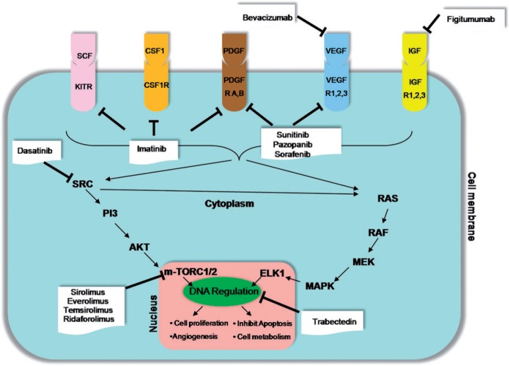
Non-gastrointestinal stromal soft tissue sarcomas are uncommon neoplasms that have a dismal prognosis due to a high incidence of metastases and a poor response to conventional chemotherapy. The identification of characteristic genetic alterations in several of these tumors has opened the window for molecular targeted therapies in patients who have failed conventional chemotherapy. Imaging plays a critical role in assessing the response to these novel therapeutic agents. Just like the response of gastrointestinal stromal tumors to imatinib, the response of non-gastrointestinal stromal soft tissue sarcomas to molecular targeted drugs is better evaluated on imaging by alternate tumor response criteria such as the Choi criteria. In addition, these drugs are associated with distinct class-specific drug toxicities that can come to attention for the first time on imaging. The purpose of this article is to provide a primer for the radiologist on the various molecular targeted therapies in advanced/metastatic non-gastrointestinal stromal soft tissue sarcomas with emphasis on the role of imaging in assessing treatment response and complications.


Figures

Figure 1
Figure 1
Cancer pathways in non-GIST STS and potential targets for therapies. VEGF/VEGFR, vascular endothelial growth factor/receptor; PDGF/PDGFR, platelet-derived growth factor/receptor; CSF1/CSF1R, colony-stimulating factor 1/receptor, IGF/IGFRm insulin-like growth factor/receptor.
Figure 2
Figure 2
A 33-year-old woman with familial adenomatous polyposis (FAP)-associated desmoid fibromatosis. (a) Axial fat-suppressed post-gadolinium T1-weighted MR image of the pelvis demonstrates 2 large heterogeneously enhancing pelvic desmoid masses (arrows and asterisk). (b) Follow-up axial fat-suppressed post-gadolinium T1-weighted MR image 6 months after treatment with imatinib shows a mild increase in the size of the anteriorly located mass (arrows) with marked central necrosis and a mild decrease in the size and enhancement of the posterior mass (asterisk) consistent with treatment response.
Figure 3
Figure 3
A 56-year-old woman with pigmented villonodular synovitis (PVNS) metastatic to the inguinal nodes and muscles of the thigh after resection of the primary tumor in the knee joint 1 year before. (a) Coronal [18F]FDG-PET/CT image of the right lower limb demonstrates an intensely FDG-avid (maximum standardized uptake value (SUVmax) of 22) mass in the anterior compartment of the right thigh (arrows). (b) Coronal [18F]FDG-PET/CT image obtained 1 month after treatment with imatinib shows decreased FDG uptake of the mass with central photopenia (arrows), suggesting interval necrosis and treatment response.
Figure 4
Figure 4
A 67-year-old woman with metastatic leiomyosarcoma. (a) Coronal contrast-enhanced CT image before the start of treatment demonstrates multiple heterogeneous solid liver lesions almost replacing the hepatic parenchyma (arrows) and a large mesenteric mass (asterisk) representing widespread metastatic disease. (b) Coronal contrast-enhanced CT image after 1 month of treatment with pazopanib reveals significantly decreased density and size of the liver metastases (arrows), many of which have become cystic, representing treatment response according to the Choi criteria. Similar changes are noted in the mesenteric mass (asterisk).
Figure 5
Figure 5
A 78-year-old man with metastatic leiomyosarcoma. (a) Axial contrast-enhanced CT image demonstrates a lobulated heterogeneous mass along the left pelvic sidewall (arrow) representing metastatic disease. (b) Axial contrast-enhanced CT image after 6 months of treatment with pazopanib reveals increased size of the mass with a concurrent significant decrease in the density (arrow). (c) Axial contrast-enhanced CT image of the abdomen during an episode of acute abdominal pain during the course of treatment reveals a bulky and edematous pancreas with peripancreatic stranding consistent with acute pancreatitis (arrowhead), a class-specific drug toxicity of tyrosine kinase inhibitors (TKIs).
Figure 6
Figure 6
A 55-year-old woman with a metastatic solitary fibrous tumor. (a) Axial contrast-enhanced CT image demonstrates large, heterogeneous and hypervascular metastases in the liver (white arrows). (b) Axial contrast-enhanced CT image obtained 12 months after initiating treatment with pazopanib shows progressive decrease in the enhancement of the lesions, which have become almost cystic in appearance (white arrows). (c) Follow-up axial contrast-enhanced CT image 18 months after the start of treatment shows new nodular enhancement within the cystic-appearing lesions (black arrows) termed nodule within a cyst development representative of tumor recurrence according to the Choi criteria.
Figure 7
Figure 7
A 24-year-old man with metastatic synovial sarcoma. (a) Baseline contrast-enhanced CT image of the chest demonstrates a large, heterogeneous left-sided pleural-based metastasis (arrow). (b) Follow-up CT after 6 months of treatment with dasatanib shows treatment response, with a decrease in the size and soft tissue component of the mass (arrow). Note the interval development of small bilateral pleural effusions (arrowheads), a common side effect of dasatinib therapy.
Figure 8
Figure 8
A 68-year-old man with metastatic malignant renal PEComa 3 years after nephrectomy. (a,b) Axial unenhanced CT images of the pelvis before (a) and 1 month after the start of treatment with everolimus (b) reveals a decrease in the size of a metastatic deposit in the rectovesical pouch (arrows). (c) Axial CT image of the lung bases at baseline demonstrates no abnormality. (d) Axial CT image of the lung bases 1 month after the start of everolimus treatment shows rapid interval development of interlobar septal thickening and ground glass opacities with basilar and peripheral distribution consistent with everolimus-induced interstitial pneumonitis, a class-specific drug toxicity of mTOR inhibitors. The patient was symptomatic, with shortness of breath. (e) Follow-up CT of the chest 3 months after discontinuing everolimus and initiating steroid therapy demonstrates improvement in the interstitial lung toxicity.
Figure 9
Figure 9
A 51-year-old woman with metastatic myxoid liposarcoma. (a) Axial contrast-enhanced CT image of the abdomen demonstrates a large heterogeneous metastatic mass in the liver (white arrow) and the pancreas (black arrow). Note the surgical clip related to previous cholecystectomy (arrowhead). Note the incidental renal cortical cysts. (b) Axial contrast-enhanced CT image 6 months after treatment with trabectedin reveals a mild increase in the size of both the lesions with homogeneous decreased internal density suggestive of treatment effect (white and black arrows). The increase in size represents pseudo-progression rather than true progression, wherein a lesion may increase in size but is in fact responding to therapy as shown by the density changes.
Figure 10
Figure 10
A 54-year-old woman with well-differentiated liposarcoma containing dedifferentiated components. (a) Axial contrast-enhanced CT image before the start of treatment demonstrates well-differentiated fat anteriorly (white arrows). The dedifferentiated component is seen as ill-defined soft tissue attenuation areas posteriorly (white arrowheads) with enhancing solid nodules (black arrow). (b) Axial contrast-enhanced CT image 12 months after treatment with trabectedin reveals replacement of the dedifferentiated component (white arrowheads) as well as the soft tissue nodule (black arrow) with fatty tissue suggestive of treatment effect or further differentiation in previously dedifferentiated components. The well-differentiated component (white arrow) is unchanged. (c) Axial CT image of the chest during the course of treatment when the patient developed an episode of fever and cough demonstrates interlobar interstitial thickening in the right lung base representing unilateral pulmonary edema, seen with capillary leak syndrome, a known complication of trabectedin therapy.

Similar articles

Cited by

References

    1. Teicher BA. Searching for molecular targets in sarcoma. Biochem Pharmacol. 2012;84:1–10. doi: 10.1016/j.bcp.2012.02.009. . PMid:22387046. - DOI - PubMed
    1. Demetri GD, Antonia S, Benjamin RS, et al. Soft tissue sarcoma. J Natl Compr Cancer Network. 2010;8:630–674. - PubMed
    1. Italiano A, Mathoulin-Pelissier S, Cesne AL, et al. Trends in survival for patients with metastatic soft-tissue sarcoma. Cancer. 2011;117:1049–1054. doi: 10.1002/cncr.25538. . PMid:20945333. - DOI - PubMed
    1. Casali PG, Blay JY. Soft tissue sarcomas: ESMO clinical practice guidelines for diagnosis, treatment and follow-up. Ann Oncol. 2010;21(Suppl 5):198–203. doi: 10.1093/annonc/mdq209. - DOI - PubMed
    1. Verma S, Younus J, Stys-Norman D, Haynes AE, Blackstein M. Meta-analysis of ifosfamide-based combination chemotherapy in advanced soft tissue sarcoma. Cancer Treat Rev. 2008;34:339–347. doi: 10.1016/j.ctrv.2008.01.005. . PMid:18313854. - DOI - PubMed

LinkOut - more resources