. 2013 Jun;121(6):676-82.
doi: 10.1289/ehp.1205693. Epub 2013 May 6.

Interlaboratory evaluation of rodent pulmonary responses to engineered nanomaterials: the NIEHS Nano GO Consortium

Affiliations

Interlaboratory evaluation of rodent pulmonary responses to engineered nanomaterials: the NIEHS Nano GO Consortium

James C Bonner et al. Environ Health Perspect. 2013 Jun.

Abstract

Background: Engineered nanomaterials (ENMs) have potential benefits, but they also present safety concerns for human health. Interlaboratory studies in rodents using standardized protocols are needed to assess ENM toxicity.

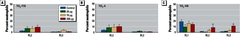
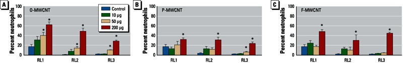
Methods: Four laboratories evaluated lung responses in C57BL/6 mice to ENMs delivered by oropharyngeal aspiration (OPA), and three labs evaluated Sprague-Dawley (SD) or Fisher 344 (F344) rats following intratracheal instillation (IT). ENMs tested included three forms of titanium dioxide (TiO2) [anatase/rutile spheres (TiO2-P25), anatase spheres (TiO2-A), and anatase nanobelts (TiO2-NBs)] and three forms of multiwalled carbon nanotubes (MWCNTs) [original (O), purified (P), and carboxylic acid "functionalized" (F)]. One day after treatment, bronchoalveolar lavage fluid was collected to determine differential cell counts, lactate dehydrogenase (LDH), and protein. Lungs were fixed for histopathology. Responses were also examined at 7 days (TiO2 forms) and 21 days (MWCNTs) after treatment.

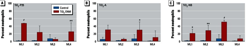
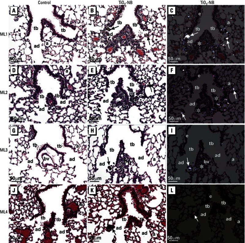
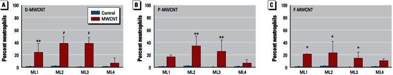
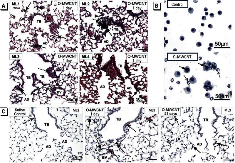
Results: TiO2-A, TiO2-P25, and TiO2-NB caused significant neutrophilia in mice at 1 day in three of four labs. TiO2-NB caused neutrophilia in rats at 1 day in two of three labs, and TiO2-P25 and TiO2-A had no significant effect in any of the labs. Inflammation induced by TiO2 in mice and rats resolved by day 7. All MWCNT types caused neutrophilia at 1 day in three of four mouse labs and in all rat labs. Three of four labs observed similar histopathology to O-MWCNTs and TiO2-NBs in mice.

Conclusions: ENMs produced similar patterns of neutrophilia and pathology in rats and mice. Although interlaboratory variability was found in the degree of neutrophilia caused by the three types of TiO2 nanoparticles, similar findings of relative potency for the three types of MWCNTs were found across all laboratories, thus providing greater confidence in these interlaboratory comparisons.


Conflict of interest statement

The authors declare they have no actual or potential competing financial interests.

Figures

Figure 1
Figure 1
Stimulation of neutrophilic inflammation in the lungs of C57BL/6 mice by TiO2‑P25 (A), TiO2-A (B), or TiO2‑NB (C) evaluated in four labs (ML1, ML2, ML3, and ML4). Values are the mean ± SE percentage of neutro­phils in BALF 1 day after exposure to DM only (control) or TiO2 ENMs at 40 µg/50 µL. *p < 0.05, **p < 0.01, and #p < 0.001 compared with controls.
Figure 2
Figure 2
Photomicrographs of H&E-stained lung tissue 1 day after exposure of C57BL/6 mice to DM alone (control; ADGJ) or TiO2‑NB at 40 μg/50 µL DM (B, C, E, F, H, I, K, L). Photomicrographs are representative sections from each of the four labs: (A–C) ML1, (D–F) ML2, (G–I) ML3, and (J–L) ML4. (A, D, G, J, B, E, H, K) Bright-field light microscopy of representative sections of lung from control mice (A, D, G, J) and TiO2‑NB–exposed mice (B, E, H, K); sections from TiO2‑NB–exposed mice show inflammatory lesions primarily localized to alveolar duct bifurcations. (C, F, I, L) Polarized light microscopy of the same images shown in B, E, H, and K show inflammation at an alveolar duct bifurcation. Abbreviations: a, alveolus; ad, alveolar ducts; bv, blood vessel; e, airway epithelium; tb, terminal bronchioles. Arrows indicate TiO2‑NBs within macrophages at alveolar duct bifurcations, and asterisks (*) indicate inflammatory foci. All photo­micrographs were taken at the same magnification; bar = 50 μm.
Figure 3
Figure 3
Stimulation of neutrophilic inflammation in the lungs of mice by O-MWCNT (A), P-MWCNT (B), or F-MWCNT (C) evaluated in four labs (ML1, ML2, ML3, ML4). Values shown are the mean ± SE percentage of neutro­phils in BALF 1 day after exposure to DM only (control) or MWCNTs at 40 µg/50 µL. (A) Significant increase induced by O-MWCNT in all four labs. (B) Significant increase induced by P-MWCNT in two labs. (C) Significant increase induced by F-MWCNT in three labs, F-MWCNT albeit to a lesser extent than that induced by O-MWCNT. *p < 0.05, **p < 0.01, and #p < 0.001 compared with controls.
Figure 4
Figure 4
Results of lung histopathology (A), BAL cytospins (B), and immunohistochemistry (C) for mice exposed to DM only (control) or O-MWCNTs (40 μg/50 μL). Abbreviations: a, alveolus; AD, alveolar ducts; BV, blood vessel; TB, terminal bronchioles. (A) Histopathology showing lung inflammatory response to O-MWCNTs. Centriacinar bronchiolitis/alveolitis (dashed arrows) was induced by O-MWCNTs in three of four labs (ML1, ML2, ML3); one lab (ML4) found some deposition of O-MWCNTs in alveolar ducts with marginal inflammation. Solid arrows indicate macrophages containing O-MWCNT. (B) BAL cytospin images of cells from the lungs of mice exposed to DM (control) or O-MWCNTs; > 95% of macrophages from O-MWCNT–exposed animals were enlarged, activated alveolar macro­phages with numerous MWCNT inclusions (solid arrows) and neutrophils that do not contain MWCNTs (dashed arrows). The images are from a single lab (ML3) but are typical of responses from ML1 and ML2. (C) Immunohistochemistry using a monoclonal rat anti-mouse neutrophil (allotypic marker clone 7/4) antibody showing the location of neutro­phils (dashed arrows) near terminal bronchioles and in relation to macrophages containing O-MWCNT (solid arrows). Representative images are from ML3.
Figure 5
Figure 5
Stimulation of neutrophilic inflammation in the lungs of rats by TiO2‑P25 (A), TiO2-A (B), or TiO2‑NB (C) evaluated in three labs (RL1, RL2, and RL3). Values are the mean ± SE percentage of neutro­phils in BALF 1 day after exposure to DM only (control) or TiO2 ENMs (20, 70, or 200 µg/250 µL). *p < 0.05 compared with controls.
Figure 6
Figure 6
Stimulation of neutrophilic inflammation in the lungs of rats by O-MWCNT (A), P-MWCNT (B), or F-MWCNT (C) evaluated in three labs (RL1, RL2, and RL3). Values are the mean ± SE percentage of neutro­phils in BALF of rats 1 day after treatment with DM only (control) or MWCNTs (10, 50, or 200 µg/250 µL). *p < 0.05 compared with controls.

Comment in

References

    1. Aiso S, Kubota H, Umeda Y, Kasai T, Takaya M, Yamazaki K, et al. Translocation of intratracheally instilled multiwall carbon nanotubes to lung-associated lymph nodes in rats. Ind Health. 2011;49(2):215–220. - PubMed
    1. Bonner JC. Nanoparticles as a potential cause of pleural and interstitial lung disease. Proc Am Thorac Soc. 2010;7(2):138–141. - PMC - PubMed
    1. Borm PJ, Robbins D, Haubold S, Kuhlbusch T, Fissan H, Donaldson K, et al. 2006The potential risks of nanomaterials: a review carried out for ECETOC. Part Fibre Toxicol 311; doi:10.1186/1743-8977-3-11[Online 14 August 2006] - DOI - PMC - PubMed
    1. Card JW, Zeldin DC, Bonner JC, Nestmann ER. Pulmonary applications and toxicity of engineered nanoparticles. Am J Physiol. 2008;295(3):L400–L411. - PMC - PubMed
    1. Donaldson K, Murphy F, Schinwald A, Duffin R, Poland CA. Identifying the pulmonary hazard of high aspect ratio nanoparticles to enable their safety-by-design. Nanomedicine (Lond) 2011;6(1):143–156. - PubMed

Publication types