. 2013 Jun 20;121(25):5025-33.
doi: 10.1182/blood-2012-12-471607. Epub 2013 May 6.

Characterization of a novel NKG2D and NKp46 double-mutant mouse reveals subtle variations in the NK cell repertoire

Affiliations

Characterization of a novel NKG2D and NKp46 double-mutant mouse reveals subtle variations in the NK cell repertoire

Sam Sheppard et al. Blood. .

Abstract

The immunoreceptors NKG2D and NKp46 are known for their capacity to activate natural killer (NK) cell cytotoxicity and secretory responses in the contexts of tumors and infections, yet their roles in NK cell education remain unclear. Here, we provide the first characterization of mice deficient for both NKG2D and NKp46 receptors to address the relevance of their concomitant absence during NK cell development and function. Our findings reveal that NK cells develop normally in double-mutant (DKO) mice. Mice lacking NKG2D but not NKp46 showed subtle differences in the percentages of NK cells expressing inhibitory Ly49 receptors and the adhesion molecule DNAM-1. A slightly increased percentage of terminally differentiated NK cells and functional response to in vitro stimuli was observed in some experiments. These alterations were modest and did not affect NK cell function in vivo in response to mouse cytomegalovirus infection. NKp46 deficiency alone, or in combination with NKG2D deficiency, had no effect on frequency or function of NK cells.


Figures
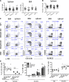
Figure 1
Figure 1
NKG2D and NKp46 are not necessary for NK-cell development. (A-B) Percentages of NK cells in the spleen and BM of WT and mutant mice are depicted for individual mice analyzed in 3 to 6 independent experiments; mean ±SD is shown for each genotype. Absolute numbers of NK cells in the spleen are shown in (B). (C-D) Representative fluorescence-activated cell sorting (FACS) profiles from a mouse of each genotype showing expression of the indicated markers (C), and percentages of cells expressing various marker combinations (n = 5 mice/genotype) (D). Data are representative of at least 4 independent experiments. (E) BM NK cells were stained for the cell-surface expression of DX5 and intracellular expression of the transcription factors T-bet and Eomes; median fluorescence intensities are indicated. Data are representative of 3 independent experiments.
Figure 1
Figure 1
NKG2D and NKp46 are not necessary for NK-cell development. (A-B) Percentages of NK cells in the spleen and BM of WT and mutant mice are depicted for individual mice analyzed in 3 to 6 independent experiments; mean ±SD is shown for each genotype. Absolute numbers of NK cells in the spleen are shown in (B). (C-D) Representative fluorescence-activated cell sorting (FACS) profiles from a mouse of each genotype showing expression of the indicated markers (C), and percentages of cells expressing various marker combinations (n = 5 mice/genotype) (D). Data are representative of at least 4 independent experiments. (E) BM NK cells were stained for the cell-surface expression of DX5 and intracellular expression of the transcription factors T-bet and Eomes; median fluorescence intensities are indicated. Data are representative of 3 independent experiments.
Figure 2
Figure 2
Differences in the NK-cell receptor repertoire in NKG2D-deficient mice. (A) Representative FACS histograms and (B) percentages of splenic NK cells expressing Ly49D, Ly49C/I, Ly49G2, Ly49F, and Ly49A are shown for 5 mice per genotype. Data are representative of at least 5 independent experiments. (C) Representative FACS histograms depicting DNAM-1 expression on splenic NK cells (CD3-gfp+), NKT cells (CD3lo, NK1.1+), and CD8+ T cells from 1 mouse of each indicated genotype (representative of at least 10 individual mice/genotype). (D) Percentages of DNAM-1–expressing NK cells in BM, spleen, and liver of 5 mice per genotype. Data are representative of at least 4 individual experiments.
Figure 3
Figure 3
Increased IFN-γ response to stimulation with IL-2, anti-Ly49D, and anti-NKp46 in NKG2D-deficient mice. (A) Percentages of IFN-γ–producing NK cells upon stimulation with IL-2 (1000 U/mL), PMA/ionomycin (upper panel), and IL-18 (5 ng/mL) + IL-12 (125 pg/mL and 250 pg/mL)(lower panel) are shown for 5 to 6 mice/genotype; data are representative of 3 individual experiments. (B) Percentages of IFN-γ–producing NK cells upon stimulation with anti-NK1.1 (25 μg/mL), anti-Ly49D (5 μg/mL), anti-NKp46 (5 μg/mL), and anti-NKG2D (25 μg/mL) antibodies (n = 3-5 mice/genotype). Data are representative of at least 3 independent experiments. (C) Percentages of IFN-γ–producing NK cells among CD27hiCD11blo (“CD27hi”), DP (“CD27+CD11b+”), and CD27loCD11bhi (“CD27lo”) subsets upon anti-Ly49D stimulation (n = 5 mice/genotype). Data are representative of 5 independent experiments.
Figure 4
Figure 4
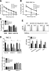
Killing activity from mutant NK cells is intact. (A-B) Killing activity of d5-activated NK cells was assessed in a 35S release assay against YAC-1 and RMA-RAE ε target cells at different effector/target ratios. (C) Killing activities of d5-sorted NK cells against RMA, RMA-S, and B16-F10 targets (E/T ratio = 10/1). Results (mean ± SD) are representative of 2 to 3 independent experiments. (D) Percentages of CD107a+ NK cells upon 5-hour incubation with YAC-1 (upper panel) and RMA and RMA-S cells (lower panel). Results are representative of 2 independent experiments. (E) Representative FACS histograms depicting Helios expression in CD11b+ NK cells from splenocytes of each genotype. Helios staining corresponds to the plain lines, whereas isotype control is depicted as the shaded histograms (upper panel). The mean fluorescence intensity of intracellular Helios staining among CD11b+ and CD11b– NK cells is shown in the lower panel (n = 3-5 mice/genotype). The data are representative of 3 experiments.
Figure 5
Figure 5
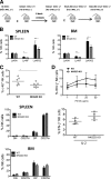
Differences in NK-receptor expression are intrinsic to mutant NK cells. (A) Schematic diagram depicting the experiment. 1:1 mixtures of fetal liver or BM cells, both gfp+, from WT mice and NKG2D-KO, NKp46-KO, or DKO mice were injected (intravenously) into lethally irradiated WT recipients (gfp-negative). FACS analysis was performed 8 to 12 weeks post transplant by gating on gfp+ NK cells. Further gating using NKG2D or NKp46 markers was used to distinguish donor NK cells of WT or mutant origin. (B-C) Percentages of NK cells expressing Ly49G2, Ly49A, DNAM-1, and c-Kit in the spleens of recipient mice reconstituted with WT/NKG2D-KO, WT/NKp46-KO, and WT/DKO precursor cell mixtures. (D) Percentages of CD11b/CD27 subsets of NK cells in recipient mice reconstituted with WT/NKG2D-KO (upper), WT/NKp46-KO (middle), and WT/DKO (lower) precursor cell mixtures. (E) Percentages of IFN-γ responding NK cells from chimeric mice stimulated with anti-NK1.1 in vitro. All data are representative of 3 independent experiments (n = 5-7 mice/genotype)
Figure 6
Figure 6
Analysis of NKG2D deficiency in BALB.B6-Cmv1r mice. (A) Schematic diagram depicting the breeding scheme used to generate NKG2D-deficient mice on a BALB.B6-Cmv1r background. C57BL/6 NKG2D KO mice were backcrossed 8 times to BALB/c mice, and the backcrossed Klrk1+/− offspring were then backcrossed once to the congenic BALB.B6-Cmv1r strain that carries B6 donor alleles from the NKC complex before intercrossing to generate BALB.B6-Cmv1r Klrk1−/− and BALB.B6-Cmv1r Klrk1+/+ littermates. (B) Percentages of splenic NK cells expressing Ly49A, Ly49F, and Ly49G2 from BALB.B6-Cmv1r NKG2D-KO and BALB.B6-Cmv1r WT littermates (gated CD3-NK1.1+). Data are mean ± SD and are representative of 2 to 3 independent experiments. (C) Percentages of c-Kit+ NK cells (upper) and CD11b/CD27 NK subsets in the spleen (middle) and BM (lower) of WT and NKG2D-KO mice. (D) Percentages of IFN-γ–producing NK cells cross-linked with ant-NK1.1 (PK136) (upper) or stimulated with IL-2 in vitro (lower). Data are expressed as mean ± SD and are representative of 3 independent experiments.
Figure 7
Figure 7
NK-cell resistance to MCMV infection is not enhanced in the absence of NKG2D. (A) B6 NKG2D-KO and WT littermates were treated with anti-NK1.1 or control IgG before infection with MCMV (1 × 10⁶ PFU intraperitoneally); virus titers were assessed in the liver, spleen, and lungs 4 days post infection. Data are representative of 3 independent experiments. (B) Groups of WT mice were treated with 250 μg of anti-NK1.1 (PK136), anti-NKG2D (MI-6), or control IgG before infection with MCMV (3 × 10⁶ PFU intraperitoneally) of MCMV; virus titers in the spleen and liver were assessed 3 days post infection. Data are representative of 2 independent experiments. All results are expressed as geometric mean ± SD of 5 mice per group. (C) Representative FACS profile of Ly49H expression on NK cells from WT and NKG2D-KO mouse. (D) Percentages of IFN-γ–producing NK cells upon stimulation with anti-Ly49H at the indicated concentrations. Data are representative of 2 of 3 independent experiments.

Similar articles

Cited by

References

    1. Biassoni R, Cantoni C, Pende D, et al. Human natural killer cell receptors and co-receptors. Immunol Rev. 2001;181:203–214. - PubMed
    1. Lanier LL. NK cell recognition. Annu Rev Immunol. 2005;23:225–274. - PubMed
    1. Kim S, Iizuka K, Kang HS, et al. In vivo developmental stages in murine natural killer cell maturation. Nat Immunol. 2002;3(6):523–528. - PubMed
    1. Carotta S, Pang SH, Nutt SL, Belz GT. Identification of the earliest NK-cell precursor in the mouse BM. Blood. 2011;117(20):5449–5452. - PubMed
    1. Fathman JW, Bhattacharya D, Inlay MA, Seita J, Karsunky H, Weissman IL. Identification of the earliest natural killer cell-committed progenitor in murine bone marrow. Blood. 2011;118(20):5439–5447. - PMC - PubMed

Publication types

MeSH terms

Substances