. 2013 Sep;28(9):1870-1884.
doi: 10.1002/jbmr.1958.

ATF4 promotes bone angiogenesis by increasing VEGF expression and release in the bone environment

Affiliations

ATF4 promotes bone angiogenesis by increasing VEGF expression and release in the bone environment

Ke Zhu et al. J Bone Miner Res. 2013 Sep.

Abstract

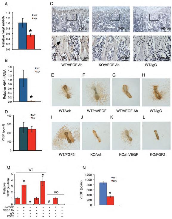
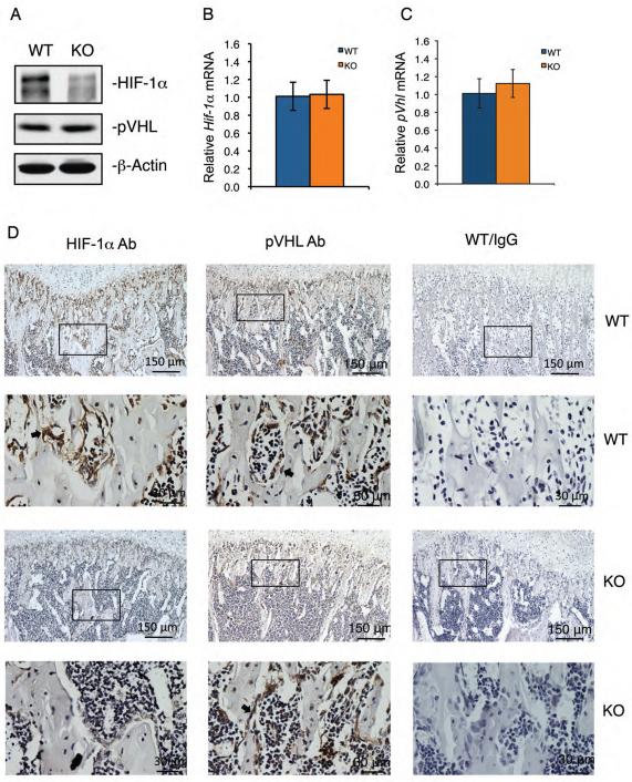
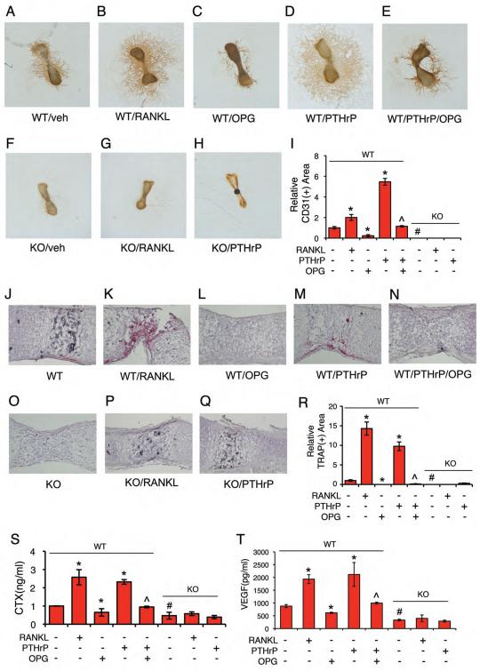
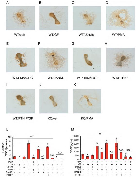
Activating transcription factor 4 (ATF4) is a critical transcription factor for bone remodeling; however, its role in bone angiogenesis has not been established. Here we show that ablation of the Atf4 gene expression in mice severely impaired skeletal vasculature and reduced microvascular density of the bone associated with dramatically decreased expression of hypoxia-inducible factor 1α (HIF-1α) and vascular endothelial growth factor (VEGF) in osteoblasts located on bone surfaces. Results from in vivo studies revealed that hypoxia/reoxygenation induction of HIF-1α and VEGF expression leading to bone angiogenesis, a key adaptive response to hypoxic conditions, was severely compromised in mice lacking the Atf4 gene. Loss of ATF4 completely prevented endothelial sprouting from embryonic metatarsals, which was restored by addition of recombinant human VEGF protein. In vitro studies revealed that ATF4 promotion of HIF-1α and VEGF expression in osteoblasts was highly dependent upon the presence of hypoxia. ATF4 interacted with HIF-1α in hypoxic osteoblasts, and loss of ATF4 increased HIF-1α ubiquitination and reduced its protein stability without affecting HIF-1α mRNA stability and protein translation. Loss of ATF4 increased the binding of HIF-1α to prolyl hydroxylases, the enzymes that hydroxylate HIF-1a protein and promote its proteasomal degradation via the pVHL pathway. Furthermore, parathyroid hormone-related protein (PTHrP) and receptor activator of NF-κB ligand (RANKL), both well-known activators of osteoclasts, increased release of VEGF from the bone matrix and promoted angiogenesis through the protein kinase C- and ATF4-dependent activation of osteoclast differentiation and bone resorption. Thus, ATF4 is a new key regulator of the HIF/VEGF axis in osteoblasts in response to hypoxia and of VEGF release from bone matrix, two critical steps for bone angiogenesis.

Keywords: ANGIOGENESIS; ATF4; BONE; OSTEOBLASTS; OSTEOCLASTS; VEGF.


Figures

Figure 1
Figure 1. Lack of ATF4 severely impairs bone vasculature during development in mice
(A) Representative photograph of calvariae from two-month-old male WT and Atf4−/− mice. (B-D) Representative μCT images of vasculature in Microfil-perfused calvariae (B) and femur (C and D) from two-month-old male WT and Atf4−/− mice. (E and F) μCT analysis of vessel volume and number within femurs from Microfil-perfused two-month-old male WT and Atf4−/− mice. n = 3, *P < 0.01 (versus WT). (G-I) IHC staining. Longitudinal tibial sections from 1-month-old male WT and Atf4−/− mice (n = 5-6) were stained with an anti-CD31 antibody or control IgG. Representative images from the metarphyseal periosteum (G, top) and diaphyseal endosteum (G, bottom) regions are shown. Original magnification, x100, arrows point to microvessels, which were stained brown.
Figure 2
Figure 2. Loss of ATF4 reduces VEGF expression in osteoblasts located on trabecular bone surfaces and abolishes endothelial sprouting from Atf4−/− metatarsals, which is restored by exogenously supplied recombinant human VEGF
(A and B) qPCR. Total RNAs were isolated from WT and Atf4−/− tibiae (n = 5-6) and analyzed by qPCR using specific primers for Vegf and Atf4 mRNAs, which were normalized to GapdhmRNA. *P < 0.01, versus WT. (C) IHC. Sections of WT and Atf4−/− tibiae (n = 5-6) were stained using specific antibodies against VEGF or normal IgG. VEGF signal was stained brown. Original magnification, x100 (top), x400 (bottom). (D) ELISA. Level of VEGF protein from WT and Atf4−/− serum was measured using an ELISA kit according to the manufacturer's instructions. n = 5-6. (E-M) Metatarsal angiogenesis assays. Metatarsals were dissected from WT and Atf4−/− E17.5 fetuses and cultured in α-MEM for 12 days, followed by IHC staining using anti-CD31 antibody as described in Materials and Methods. Representative images are shown. Microscope: Olympus SZ61. Original magnification, ×20. Endothelial sprouting from WT metatarsals (E) was significantly increased by the treatment of recombinant VEGF (10 ng/ml) (F) and blocked by an anti-VEGF neutralizing antibody (1 μg/ml) (G), but not by control IgG (1 μg/ml) (H). Endothelial sprouting from WT metatarsals was increased by FGF2 (100 ng/ml) (I). No detectable endothelial sprouting from Atf4−/− metatarsals (J). Significant endothelial sprouting from Atf4−/− metatarsals treated with VEGF (K), but not FGF2 (L). Quantitative data of each group (M). n = 6-8, *P < 0.01, versus veh, #P < 0.01, versus WT. (N)ELISA. Level of VEGF protein from WT and Atf4−/− metatarsal cultures was measured using an ELISA kit according to the manufacturer's instructions. n = 6-8, *P < 0.01, versus WT.
Figure 3
Figure 3. Inactivation of ATF4 greatly decreases the level of HIF-1α protein, but not its mRNA, in osteoblasts located on bone surfaces
(A) Western blot analysis. Protein extracts were isolated from WT and Atf4−/− tibiae (n = 5-6) and analyzed for HIF-1α and pVHL. β-Actin was used for loading control. (B and C) qPCR. Total RNAs were isolated from WT and Atf4−/− tibiae (n = 5-6) and analyzed by qPCR using specific primers for Hif-1α and pVhl mRNAs, which were normalized to Gapdh mRNA. (D)IHC. Sections of WT and Atf4−/− tibiae (n = 5-6) were stained using specific antibodies against HIF-1α, pVHL or normal IgG. Original magnification, x100 (top), x400 (bottom).
Figure 4
Figure 4. ATF4 increases HIF-1α and VEGF expression in osteoblasts in response to hypoxia and ATF4 interacts with HIF-1α and loss of ATF4 increases HIF-1α ubiquitination and decreases HIF-1α protein stability without affecting HIF-1α mRNA stability and translation
(A) Hypoxia-induced up-regulation of HIF-1α was reduced by the loss of ATF4. WT and Atf4−/− calvarial osteoblasts were treated with hypoxia (1% O2) for indicated times, followed by Western blot analysis. (B-E) ATF4 up-regulated HIF-1α and Vegf expression in a hypoxia-dependent manner. MC-4 cells were transfected with and without adenoviral vector for ATF4. Twenty-four hours later, cells were cultured in the presence and absence of hypoxia for 6h (B and C), followed by Western blot (B) and qPCR (C) analysis, or 24h (D and E), followed by qPCR analysis for Vegf (D) and Glut1 (E) mRNAs. *P < 0.01, versus AdEGFP, #P < 0.01, versus Normoxia. (F) Hif-1α mRNA stability. WT and Atf4−/− calvarial osteoblasts were treated with transcription inhibitor actinomycin D (Act D) (5 μg/ml) in the presence of 150 μM CoCl2 (i.e., to rapidly induce hypoxia (52)) for indicated times, followed by qPCR for Hif-1α mRNAs, which were normalized to Gapdh mRNAs of the 0h samples of each group. (G) In vitro translation. Translated HIF-1α proteins were subject to Western blotting for HIF-1α. Lane 1: no Hif-1α mRNA template was added, lanes 2-8: equal amounts of Hif-1α mRNA template were added in each reaction. Lane 2: nuclear extracts (NE) from primary WT calvarial osteoblasts, lane 3: NE from primary ATF4 KO calvarial osteoblasts, lane 4: NE from COS-7 transfected with ATF4 expression vector, lane 5: NE from COS-7 transfected with β-galactosidase expression vector (control vector), lane 6: purified His-ATF4 protein, lane 7: elution buffer for purifying the His-ATF4, lane 8: H2O. (H and I) HIF-1α protein stability. Primary WT and Atf4−/− calvarial osteoblasts were exposed to 150 μM cobalt chloride following addition of 10 μg/ml cycloheximide. Lysates of cells harvested at the indicated time intervals were subject to Western blot analysis of HIF-1α expression. Quantitative analysis of HIF-1α, which was normalized to β-actin, from 3 independent experiments (I). *P < 0.01 (versus KO). (J and K) co-IP assays. Whole cell extracts from hypoxic osteoblasts were immunoprecipitated by antibodies against ATF4 (J) or HIF-1α (K), followed by Western blotting for HIF-1α and ATF4. (L and M) HIF-1α ubiquitination. Whole cell lysates from hypoxic osteoblasts from WT and Atf4−/− mice were immunoprecipitated with an anti-HIF-1α antibody, followed by Western blot analysis for ubiquitin (Ub) (J). Quantitative analysis of (Ub)n-HIF-1α from 5 independent experiments (M). *P < 0.01 (versus WT).
Figure 5
Figure 5. ATF4 regulates VEGF release from bone matrix and bone angiogenesis by activating osteoclast differentiation and bone resorption
(A-I) Metatarsal angiogenesis assays. RANKL (50 ng/ml) increased (B) and OPG (300 ng/ml) decreased (C) endothelial sprouting from WT metatarsals. PTHrP (100 nM) increased endothelial sprouting in WT metatarsals (D), which was inhibited by OPG (E). Neither RANKL nor PTHrP increased endothelial sprouting from Atf4−/− metatarsals (F-H). Quantitative data of each group (I). n = 5-6. Microscope: Olympus SZ61. Original magnification, x20. (J-R) TRAP staining. WT and Atf4−/− metatarsal sections treated as indicated were stained for TRAP activity. Quantitative analysis of TRAP-positive area, which was normalized to metatarsal section area (R). n = 5-6. Original magnification, x100. (S and T) ELISA. Metatarsals were dissected from WT and Atf4−/− E17.5 fetuses and cultured in α-MEM for 14d. Culture media were harvested for CTX and VEGF assays as described in Materials and Methods. n = 5-6, *P < 0.01, versus veh, #P < 0.01, versus WT, ^P < 0.01, versus PTHrP alone.
Figure 6
Figure 6. PKC regulates VEGF release from bone matrix and bone angiogenesis through ATF4-dependent osteoclast activation and bone resorption
(A-L) Metatarsal angiogenesis assays. Endothelial sprouting in WT metatarsals (A) was completely inhibited by GF109203X (2 μM) (PKC inhibitor) (B), but not by U0126 (2 μM) (Erk1/2 inhibitor) (C). PMA (2 μM) (PKC activator) increased endothelial sprouting in WT metatarsals (D), which was suppressed by OPG (E). RANKL- and PTHrP-induced endothelial sprouting was inhibited by GF109203X (F-I). No detectable endothelial sprouting from Atf4−/− metatarsals (J). PMA failed to increase endothelial sprouting from Atf4−/− metatarsals (K). Quantitative data of each group (L). n = 5-6. Microscope: Olympus SZ61. Original magnification, x20. (M) ELISA. Levels of VEGF protein from WT and Atf4−/− metatarsal cultures treated as indicated above were measured. *P < 0.01, versus veh, #P < 0.01, versus WT, ^P < 0.01, versus PMA alone, ^^P < 0.01, versus RANKL alone, ^^^P < 0.01, versus PTHrP alone.
Figure 7
Figure 7. Hypoxia/reoxygenation-induced angiogenic response is abolished in Atf4−/− bone
(A-G) WT and Atf4−/− male mice of 1-month-old age were placed in a hypoxia chamber (8%) for 2h per day for 10d as described in Materials and Methods. Protein extracts and total RNAs were isolated from tibiae (n = 6) for Western blot analysis (A) and qPCR analysis (B-E), respectively. Longitudinal tibial sections from the chondro-osseous junction regions were stained with an anti-CD31 antibody. Representative images are shown (F). Arrows point to microvessels, which were stained brown. Microvessel density of sections from the chondro-osseous junction regions of each group was measured and compared to that of WT sections (normalized to 100%) (G). n = 6. Original magnification, x200, *P < 0.01, versus Normoxia, #P < 0.01, versus WT. (H) A working model for ATF4 promotion of bone angiogenesis.

Comment in

Similar articles

Cited by

References

    1. Schipani E, Maes C, Carmeliet G, Semenza GL. Regulation of osteogenesis-angiogenesis coupling by HIFs and VEGF. J Bone Miner Res. 2009;24:1347–1353. - PMC - PubMed
    1. Maes C, Carmeliet P, Moermans K, Stockmans I, Smets N, Collen D, Bouillon R, Carmeliet G. Impaired angiogenesis and endochondral bone formation in mice lacking the vascular endothelial growth factor isoforms VEGF164 and VEGF188. Mech Dev. 2002;111:61–73. - PubMed
    1. Wang Y, Wan C, Deng L, Liu X, Cao X, Gilbert SR, Bouxsein ML, Faugere MC, Guldberg RE, Gerstenfeld LC, et al. The hypoxia-inducible factor alpha pathway couples angiogenesis to osteogenesis during skeletal development. J Clin Invest. 2007;117:1616–1626. - PMC - PubMed
    1. Wan C, Gilbert SR, Wang Y, Cao X, Shen X, Ramaswamy G, Jacobsen KA, Alaql ZS, Eberhardt AW, Gerstenfeld LC, et al. Activation of the hypoxia-inducible factor-1alpha pathway accelerates bone regeneration. Proc Natl Acad Sci U S A. 2008;105:686–691. - PMC - PubMed
    1. Jacobsen KA, Al-Aql ZS, Wan C, Fitch JL, Stapleton SN, Mason ZD, Cole RM, Gilbert SR, Clemens TL, Morgan EF, et al. Bone formation during distraction osteogenesis is dependent on both VEGFR1 and VEGFR2 signaling. J Bone Miner Res. 2008;23:596–609. - PMC - PubMed

Publication types

MeSH terms

Substances