. 2013 Jun 21;288(25):18353-65.
doi: 10.1074/jbc.M113.461988. Epub 2013 May 6.

Identification of a secondary promoter within the human B cell receptor component gene hCD79b

Affiliations

Identification of a secondary promoter within the human B cell receptor component gene hCD79b

Eung Jae Yoo et al. J Biol Chem. .

Abstract

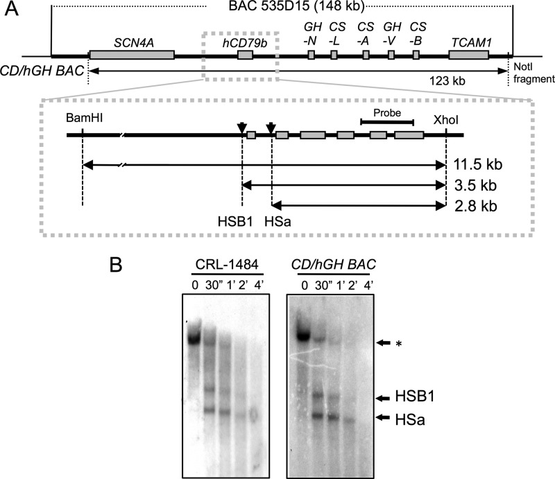
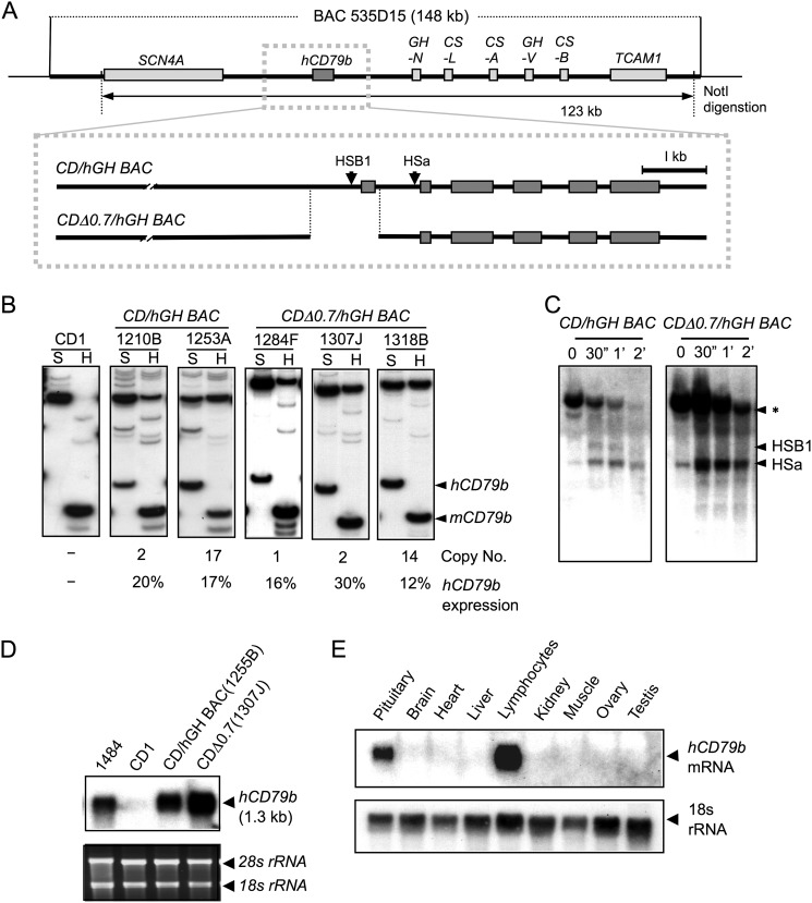
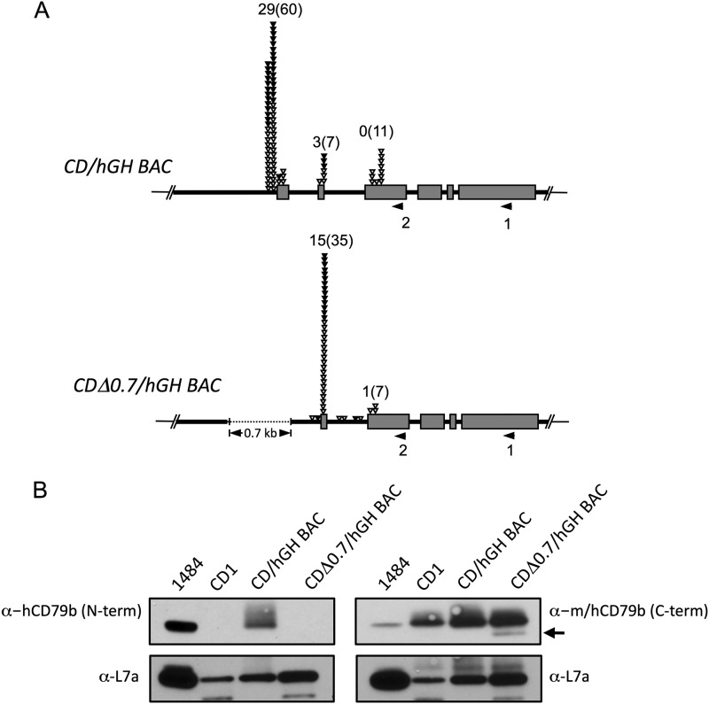
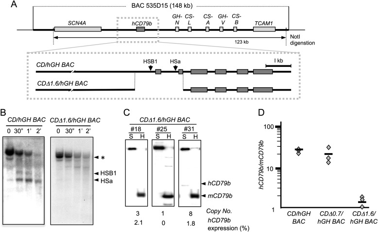
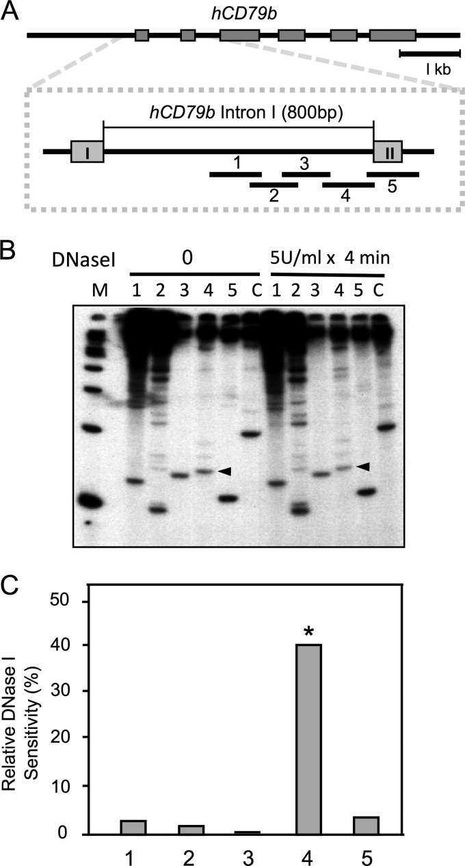
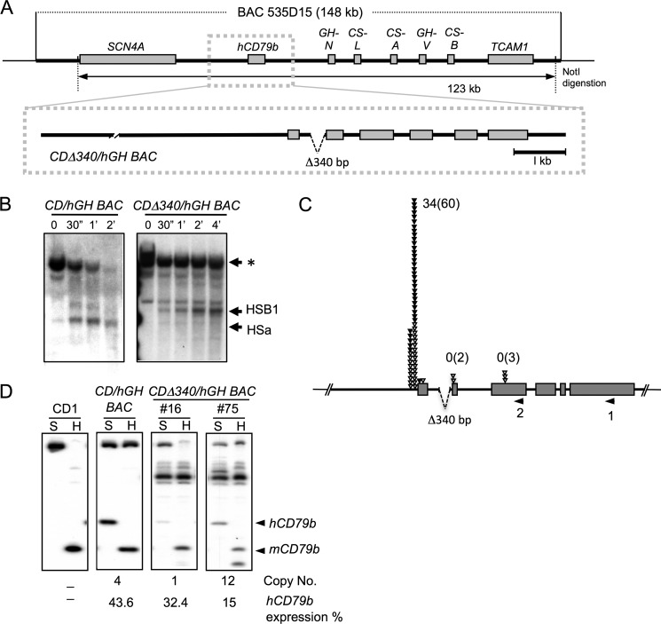
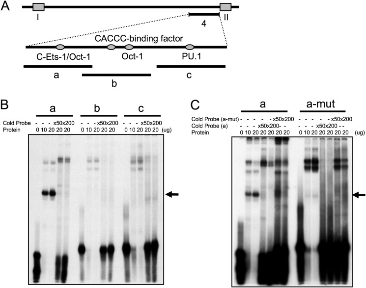
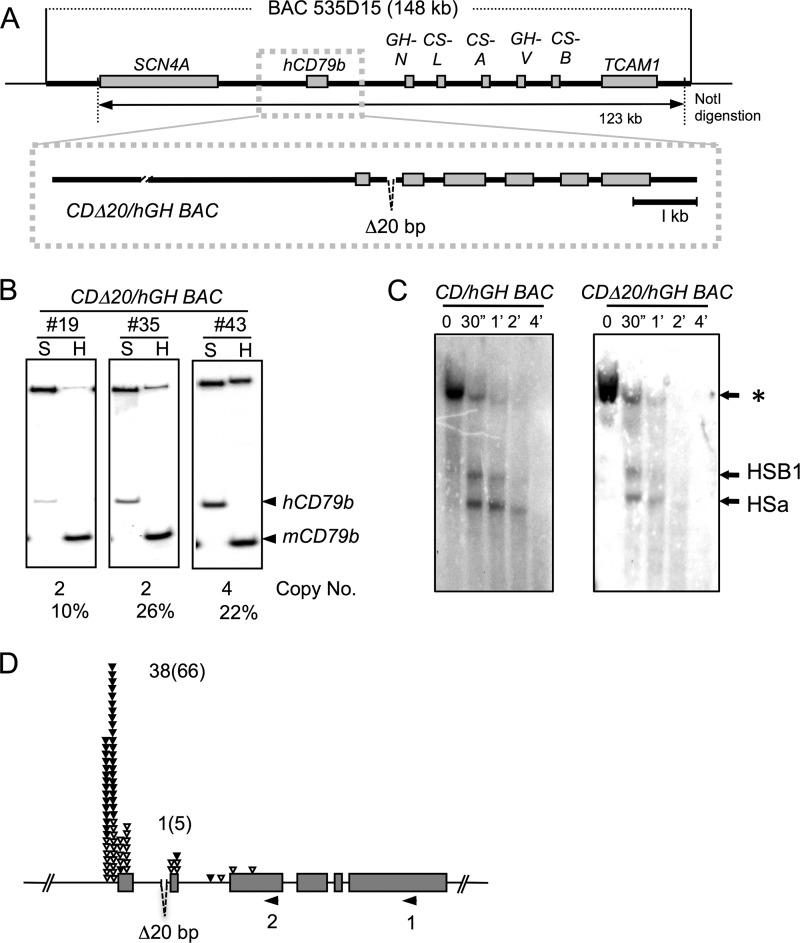
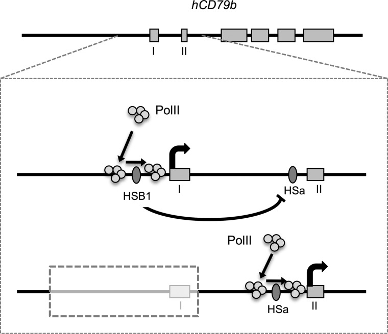
The human B cell-specific protein, CD79b (also known as Igβ and B29) constitutes an essential signal transduction component of the B cell receptor. Although its function is central to the triggering of B cell terminal differentiation in response to antigen stimulation, the transcriptional determinants that control CD79b gene expression remain poorly defined. In the present study, we explored these determinants using a series of hCD79b transgenic mouse models. Remarkably, we observed that the previously described hCD79b promoter along with its associated enhancer elements and first exon could be deleted without appreciable loss of hCD79b transcriptional activity or tissue specificity. In this deletion setting, a secondary promoter located within exon 2 maintained full levels and specificity of hCD79b transcription. Of note, this secondary promoter was also active, albeit at lower levels, in the wild-type hCD79b locus. The activity of the secondary promoter was dependent on the action(s) of a conserved sequence element mapping to a chromatin DNase I hypersensitive site located within intron 1. mRNA generated from this secondary promoter is predicted to encode an Igβ protein lacking a signal sequence and thus unable to serve normal B cell receptor function. Although the physiologic role of the hCD79b secondary promoter and its encoded protein remain unclear, the current data suggest that it has the capacity to play a role in normal as well as pathologic states in B cell proliferation and function.

Keywords: B Cell; DNase I Hypersensitive Site; Gene Expression; Immunology; Secondary Promoter; Transcription Enhancers; Transcription Promoter; Transgenic Mice; hCD79b.


Figures

FIGURE 1.
FIGURE 1.
Map of the hCD79b/hGH locus. A, a 123-kb NotI-digested DNA fragment (CD/hGH BAC transgene), isolated from a 148-kb human BAC clone (BAC 535D15), was used to generate a set of transgenic mouse lines. The map of the locus is shown at the top with the respective genes labeled. An expanded view of the hCD79b gene region is shown below this map, with the six exons represented as rectangles. The diagram also summarizes the DNase I HS mapping strategy. After partial digestion of chromatin samples with DNase I, the extracted DNA was digested with BamHI and XhoI. The 3.5- and 2.8-kb sub-bands, released by the partial DNase I-digestion were visualized by Southern blot analysis (B) using the indicated 0.9-kb NdeI/NcoI fragment as a hybridization probe (Probe). B, DNase I mapping and Southern blot analysis. DNase I HS mapping was carried out on nuclei from a human B cell line (CRL-1484) and primary lymphocytes isolated from a CD/hGH BAC transgenic mouse. Purified nuclei were treated with 30 units of DNase I for the indicated times. *, position of the originating 11.5-kb BamHI/XhoI fragment and the two DNase I-generated sub-bands corresponding to cleavage at each of the two DNase I HS sites, HSB1 and HSa. These are indicated and migrated at 3.5 and 2.8 kb, respectively.
FIGURE 2.
FIGURE 2.
Deletion of the hCD79b promoter and adjacent exon 1 failed to alter levels of hCD79b mRNA expression. A, map of the unmodified wild-type CD/hGH BAC transgene (as in Fig. 1A) and of the derivative transgene lacking a 0.7-kb 5′ segment encompassing the hCD79b promoter and exon 1 (CDΔ0.7/hGH BAC). The 122-kb CDΔ0.7/hGH BAC fragment was released by NotI digestion and used to generate a set of transgenic mouse lines (1284F, 1307J, and 1318B). B, quantification of mouse and human CD79b mRNAs. Human and mouse CD79b mRNA from the B cells of the indicated lines were co-amplified by RT-PCR, and cDNAs were distinguished by restriction enzyme digestions. SfcI (S) digestion of the 32P-5′-end-labeled hCD79b cDNA generated a 95-bp product corresponding to the hCD79b cDNA. Digestion with HinfI (H) exclusively generates a 63-bp mCD79b cDNA product. The transgene copy numbers indicated below each panel were determined by Southern blot analysis. hCD79b expression per transgene copy was normalized to endogenous mCD79b expression, and values were indicated as percentages below each respective lane. The mean expression level was minimally changed by the deletion. C, DNase I hypersensitive site mapping confirms the deletion of HSB1. HS mapping was carried out as described (Fig. 1 and “Experimental Procedures”). The wild-type CD/hGH BAC transgene locus has both HSs intact, whereas the CDΔ0.7/hGH BAC transgene locus assembles HSa but lacks HSB1. D, Northern blot analysis of B cell mRNA from the wild-type CD/hGH BAC and the CDΔ0.7/hGH BAC transgenes revealed no change in hCD79b mRNA size despite the deletion. Total RNAs were extracted from lymphocytes of each sample and separated on a denaturing agarose gel and hybridized with a probe specific to human CD79b mRNA. A human plasma cell line (CRL-1484) was used as a positive control and B cells from a non-transgenic CD1 mouse served as a negative (species specificity) control. Ethidium bromide-stained 28 and 18 S rRNAs were used for loading controls. E, tissue-specific expression of hCD79b was maintained in the absence of the promoter and exon I. Northern blots of RNAs isolated from the tissues of a CDΔ0.7/hGH BAC mouse were analyzed for hCD79b expression. Expression was detected only in lymphocytes and pituitary as observed with the non-deleted transgene (9). 18 S rRNA was used as a loading control.
FIGURE 3.
FIGURE 3.
A secondary promoter for the hCD79b is markedly enhanced subsequent to deletion of the primary promoter, and its mRNA encodes an N-terminally deleted CD79b protein. A, 5′-RACE assay analyses of B cell mRNA from the wild-type CD/hGH BAC revealed two sites of polymerase II transcription initiation. Each RNA sample was reverse transcribed using hCD79b-specific exon 6 primer (1), and the 3′-ends of these cDNAs were extended with poly(G). Poly(G)-tailed cDNAs were then amplified with exon 3 primer (2) and poly(C) adaptor primer and cloned into pGEM-T vector for sequencing. Each triangle indicates a transcription start site from each sequenced clone in the 50-bp window. The total numbers of cDNAs containing an additional non-templated terminal C (corresponding to the 5′-cap structure) are shown, and the total numbers of cDNAs mapping to the indicated position (with and without the additional C) are included in parentheses. The position of the 0.7-kb deletion that removes the primary promoter and exon 1 is indicated by the dotted line. B, hCD79b proteins were detected by human-specific rabbit monoclonal antibody that recognizes the N terminus of the protein (left; ab134103) and a rabbit monoclonal antibody that detects the C terminus of both the mouse and human CD79b (right; ab134147). The inability to detect an hCD79b protein in lymphocytes from the CDΔ0.7/hGH BAC transgene with the N terminus-specific antibody (left) was consistent with the exon 1-truncated structure of the mRNA initiated from the secondary promoter. The antibody recognizing the C terminus of both mouse and human CD79b revealed a smaller hCD79b (∼30 kDa) in the protein extract from the CDΔ0.7/hGH BAC lymphocytes (arrow), supporting the presence of a protein encoded by the mRNA originating from the secondary promoter. Ribosomal protein, L7a (30 kDa), detected using rabbit polyclonal antibody, served as an internal loading control.
FIGURE 4.
FIGURE 4.
A deletion of the hCD79b gene that encompasses the primary and secondary promoters ablates gene expression. A, map of the CDΔ1.6/hGH BAC transgene. A 1.6-kb segment of the hCD79b gene was removed from the CD/hGH BAC transgene by homologous recombination. This deleted region extended the 5′-flanking region through exon 2. The modified 121-kb transgene, released by NotI digestion, was purified and used to generate a set of transgenic mouse lines (#18, #25, and #31). B, DNase I HS mapping of chromatin from CDΔ1.6/hGH BAC lymphocytes confirmed elimination of HSB1 and HSa. The origins of the chromatin for the two assays are indicated above the Southern blot images, and the migration of HS fragments is shown to the right. C, expression of hCD79b mRNA was dramatically diminished by loss of the primary and secondary transcription start sites in the CDΔ1.6/hGH BAC lymphocytes. hCD79b mRNA levels were normalized to endogenous mCD79b expression and to transgene copy number. These normalized values are indicated as percentages of mCD79b mRNA expression below each respective lane. Three independent lines with unique transgene insertion sites were analyzed. The transgene copy numbers were determined by Southern blot analysis. D, summary of hCD79b mRNA expression levels, from intact and deleted constructs, in B cells of the transgenic lines. Each symbol represents the normalized level of hCD79b mRNA in a separate transgenic mouse line. The loss of the primary promoter (Δ0.7) had no significant effect on mRNA levels, whereas extension of the deletion to encompass the secondary promoter (Δ1.6) resulted in a dramatic loss of gene expression.
FIGURE 5.
FIGURE 5.
The specific location of HSa was mapped with a tiled amplimer array. A, chromatin from CD/hGH BAC lymphocytes was subjected to partial DNase I digestion followed by amplification of the purified DNA with five partially overlapping amplimer sets. The positioning of the amplimer sets is shown in the expanded view of the 5′-end of the hCD79b gene. B, amplicons were incorporated with [32P] α-dCTP and separated on a 6% polyacrylamide gel. Correctly sized bands (amplimer 1, 120 bp; amplimer 2, 100 bp; amplimer 3, 127 bp; amplimer 4, 129 bp; amplimer 5, 113 bp; mGAPDH, 236 bp) were quantified. The arrowheads indicate the amplicons (4) that show the most sensitivity to DNase I treatment. C, the products of each amplimer set from DNase I-digested samples were normalized to each signal from non-digested control sample (see “Experimental Procedures”). The decrease in the amplicon signal due to DNase I digestion was calculated and plotted as a percentage of relative sensitivity. *, amplimer with the greatest level of DNase I sensitivity and thus the location of HSa. mGAPDH was used for the normalization of PCR amplifications.
FIGURE 6.
FIGURE 6.
Deletion of HSa results in a selective loss of transcription initiation from the hCD79b secondary promoter. A, deletion of HSa from the CD/hGH BAC transgene. A 340-bp segment of intron 1 encompassing HSa was deleted from the CD/hGH locus, and the modified human gene was released from its BAC 535D15 vector. After purification, the fragment was injected into mouse embryos to generate a set of CDΔ340/hGH BAC transgenic mice (#16 and #75). B, DNase I HS mapping of CDΔ340/hGH BAC lymphocyte chromatin confirmed selective elimination of HSa. Nuclei from the indicated wild type and 340 bp-deleted human transgenic mouse lymphocytes were analyzed as in Fig. 1B. HSB1 remained intact despite the loss of the HSa determinant. C, hCD79b transcription initiation from exon 2 was selectively repressed in the HSa-deleted transgene. hCD79b TSSs in the CDΔ340/hGH BAC were mapped and quantitiated by 5′-RACE. Each inverted triangle indicates a transcription start site from each sequenced clone in a 50-bp window. The filled triangles indicate individual cDNA clones generated by the 5′-RACE that contain an additional non-templated C corresponding to the 5′-7meG cap. The numbers of C-containing clones are shown for each site, and the total numbers of clones are noted in parentheses. D, the 340-bp deletion encompassing HSa fails to alter the net expression from the hCD79b locus. Co-RT-PCR was performed to examine hCD79b expression in CDΔ340/hGH BAC. The origin of each lymphocyte RNA sample is indicated above the respective panel; CD1, nontransgenic mouse; BAC17, CD/hGH BAC; #16 and #75, CDΔ340/hGH BAC.
FIGURE 7.
FIGURE 7.
Ets family protein binds to the DNase I hypersensitive region of hCD79b. A, diagram of the HSa region in hCD79b intron 1 (see also Fig. 5). Three probes (a–c) encompassing the HSa (region 4; see Fig. 5) were used for gel shift analyses. Transcription factor binding sites predicted by informatics analyses of the corresponding sequence are indicated. B, band shift analysis. Nuclear extract from the human plasma cell line CRL-1484 was incubated with each 32P-labeled probe (a–c) and resolved on a native 7.5% polyacrylamide gel. Non-labeled probes were added at the indicated excess to the labeled probe (×50 and ×200) for self-competition studies. The arrow to the right indicates the position of the putative Ets protein-DNA complex. C, band shift analysis with probe “a” containing a mutation at the Ets binding site (a-mut) (GAAGTA → AGAGTA). The mutant probe was used to confirm the specific binding of Ets family proteins. The arrow indicates the specific band shift with Ets protein using probe “a” and its selective loss when the Ets site is mutated (probe “a-mut”).
FIGURE 8.
FIGURE 8.
Deletion of the Ets protein binding site in hCD79b intron 1 selectively represses use of the secondary promoter. A, a 20-bp segment of intron I encompassing the c-Ets-1 binding site (GAAGTA) was deleted from the CD/hGH BAC human transgene. The resulting NotI digestion released a 123-kb insert that was used to establish a set of CDΔ20/hGH BAC transgenic mice (#19, #35, and #43). B, hCD79b mRNA expression in mouse lymphocytes of transgenic mouse lines. Human and mouse CD79b mRNA were co-amplified by RT-PCR, and cDNAs were distinguished by restriction enzyme digestion. Expression per copy number is indicated below each gel segment and is relatively unchanged. C, deletion of the c-Ets-1 site in intron I fails to inhibit formation of HSa. Shown is DNase I HS mapping of purified lymphocytes isolated from the wild-type CD/hGH BAC and the derived transgene with deletion of the c-Ets-1 site in intron I (CDΔ20/hGH BAC). *, position of the originating 11.5-kb BamHI/XhoI fragment and the two DNase I-generated sub-bands corresponding to cleavage at each of the two DNase I HS sites, HSB1 and HSa. These are indicated and migrated at 3.5 and 2.8 kb, respectively. D, 5′-RACE analysis of CDΔ20/hGH BAC transcription initiation. The analysis reveals a ratio of 38:1 for verified (presence of a non-templated 3′-terminal C) TSSs in the hCD79b transcription start sites in the CDΔ20/hGH BAC. All explanations and designations used in this figure are same as in Fig. 3A and Fig. 6C.
FIGURE 9.
FIGURE 9.
Proposed model of interactions between the primary and secondary promoters at the hCD79b locus. HSB1 recruits chromatin remodeling factors and B cell-specific transcriptional factors to the primary promoter. Transcriptional machineries, including RNA polymerase II, recognize the −0.5 kb to −0.2 kb region and track through open chromatin in the promoter region and start transcription. Activity of the primary promoter inhibits the activity of HSa and the secondary promoter. When the primary promoter is deleted or inactivated, HSa and associated cis-acting determinants are able to mediate full levels of B cell-specific transcription from the adjacent secondary promoter.

Similar articles

Cited by

References

    1. Gold M. R., Matsuuchi L., Kelly R. B., DeFranco A. L. (1991) Tyrosine phosphorylation of components of the B-cell antigen receptors following receptor crosslinking. Proc. Natl. Acad. Sci. 88, 3436–3440 - PMC - PubMed
    1. Benschop R. J., Cambier J. C. (1999) B cell development. Signal transduction by antigen receptors and their surrogates. Curr. Opin. Immunol. 11, 143–151 - PubMed
    1. Calame K. L., Lin K. I., Tunyaplin C. (2003) Regulatory mechanisms that determine the development and function of plasma cells. Annu. Rev. Immunol. 21, 205–230 - PubMed
    1. Huang X., Takata K., Sato Y., Tanaka T., Ichimura K., Tamura M., Oka T., Yoshino T. (2011) Downregulation of the B-cell receptor signaling component CD79b in plasma cell myeloma. A possible post-transcriptional regulation. Pathol. Int. 61, 122–129 - PubMed
    1. Dobbs A. K., Yang T., Farmer D., Kager L., Parolini O., Conley M. E. (2007) Cutting edge. A hypomorphic mutation in Igβ. J. Immunol. 179, 2055–2059 - PubMed

Publication types

MeSH terms

LinkOut - more resources