. 2013 Sep;6(5):1133-45.
doi: 10.1242/dmm.011916. Epub 2013 May 2.

Conditional deletion of neurogenin-3 using Nkx2.1iCre results in a mouse model for the central control of feeding, activity and obesity

Affiliations

Conditional deletion of neurogenin-3 using Nkx2.1iCre results in a mouse model for the central control of feeding, activity and obesity

Neal Anthwal et al. Dis Model Mech. 2013 Sep.

Abstract

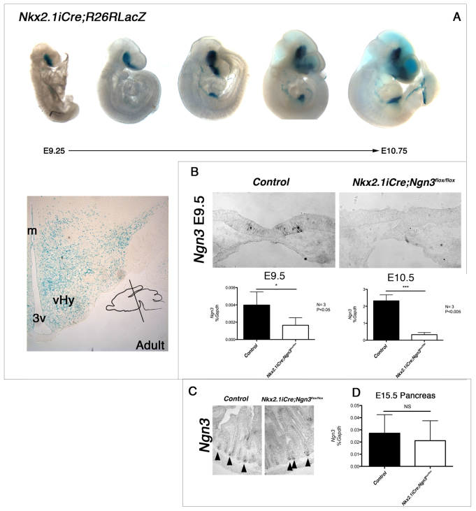
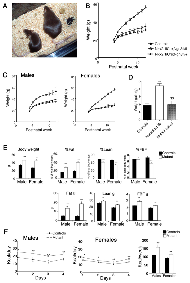
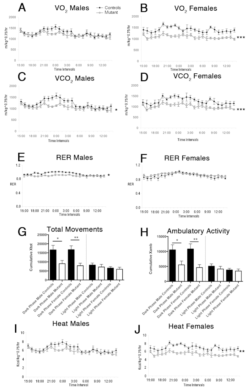
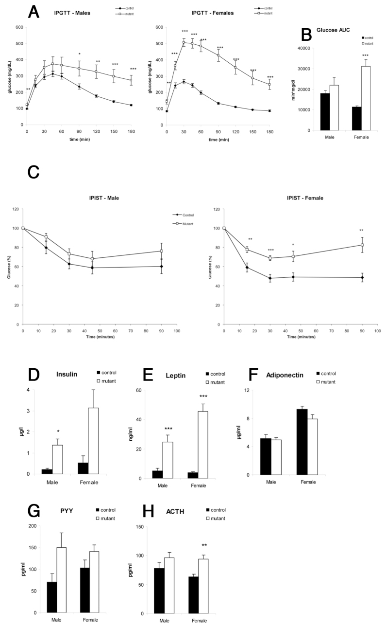
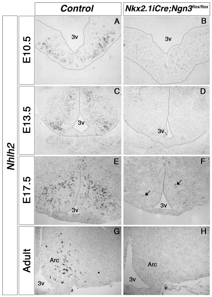
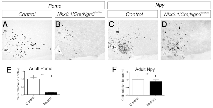
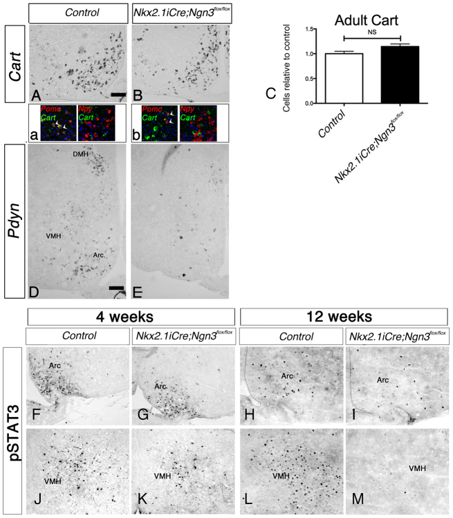
The ventral hypothalamus acts to integrate visceral and systemic information to control energy balance. The basic helix-loop-helix transcription factor neurogenin-3 (Ngn3) is required for pancreatic β-cell development and has been implicated in neuronal development in the hypothalamus. Here, we demonstrate that early embryonic hypothalamic inactivation of Ngn3 (also known as Neurog3) in mice results in rapid post-weaning obesity that is associated with hyperphagia and reduced energy expenditure. This obesity is caused by loss of expression of Pomc in Pomc- and Cart-expressing (Pomc/Cart) neurons in the arcuate nucleus, indicating an incomplete specification of anorexigenic first order neurons. Furthermore, following the onset of obesity, both the arcuate and ventromedial hypothalamic nuclei become insensitive to peripheral leptin treatment. This conditional mouse mutant therefore represents a novel model system for obesity that is associated with hyperphagia and underactivity, and sheds new light upon the roles of Ngn3 in the specification of hypothalamic neurons controlling energy balance.


Figures

Fig. 1.
Fig. 1.
Nkx2.1iCre drives Ngn3 deletion in the ventral forebrain. (A) Expression of Nkx2.1iCre in the developing mouse embryo. R26RLacZ reporter females were crossed with Nkx2.1iCre males, and resulting embryos stained with X-Gal to reveal lacZ reporter expression. β-gal-positive cells are observed in the ventral forebrain as early as E9.25 and throughout the adult hypothalamus. β-gal-positive cells are also observed in endodermal-derived tissues from around E10.0. Insert represents angle of section throughout study. m, midline; 3v, third ventricle; vHy, ventral hypothalamus. (B) Ngn3 was conditionally deleted in Nkx2.1-expressing cells by crossing Nkx2.1iCre males with Ngn3flox/flox females, resulting in Ngn3 conditional mutant mice (Nkx2.1iCre/+;Ngn3flox/flox). Following deletion, Ngn3 expression was not observable by in situ hybridization at E9.5. However, qRT-PCR reveals low levels of Ngn3 in the E9.5 ventral forebrain, whereas only residual levels are seen at E10.5. Student’s t-test: *P<0.05 and ***P<0.005. (C,D) Despite iCre-positive cells being observed in the developing endoderm, no change was seen in the expression of Ngn3 by in situ hybridization in the adult intestinal crypt cells (C), nor by qRT-PCR in the developing pancreas (D). Arrowheads in C indicate Ngn3-positive crypt cells. NS, not significant.
Fig. 2.
Fig. 2.
Nkx2.1iCre/+;Ngn3flox/flox mice become viscerally obese and are hyperphagic. (A) Examples of control (left) and conditional mutant (right) 8-week-old female littermates. (B,C) Mutant Nkx2.1iCre/+;Ngn3flox/flox mice become significantly heavier than control and Nkx2.1iCre/+;Ngn3flox/+ littermates from postnatal week 5 (around 1 week after weaning), and continue to gain mass well into mature adult stages. The rate of weight gain is more rapid in females than males (C). (D) Ad libitum-fed Nkx2.1iCre/+;Ngn3flox/flox mice gain more weight per week between 4 and 6 weeks of age than do Nkx2.1iCre/+;Ngn3flox/flox mutants pair-fed to control littermates. (E) Body weight, fat mass, lean mass and free body fluids (FBF) presented as percentage body weight (%) and as total mass (g) in 14-week-old control and mutant male and female mice. Body weight and proportion of body mass made up of fat tissue is significantly increased in mutant animals, whereas the percentage lean mass is reduced. The percentage free body fluid is significantly reduced in female mutant mice compared with controls, but not in males. (F) Both male and female Nkx2.1iCre/+;Ngn3flox/flox mice show a significant increase in daily and cumulative weekly food intake. Student’s t-test: *P<0.05, **P<0.01, ***P<0.005; NS, not significant.
Fig. 3.
Fig. 3.
Nkx2.1iCre/+;Ngn3flox/flox mutant mice exhibit decreased energy expenditure at 17 weeks of age compared with controls. (A,B) Oxygen consumption in male and female mice. There is a trend for decreased oxygen consumption in mutant male mice compared with control mice particularly in the night-time dark phase (A), whereas, in female mutant mice, oxygen consumption if significantly reduced compared with controls (B). (C,D) CO2 production is significantly reduced in both male (C) and female (D) mutant mice compared with control littermates. (E,F) Respiratory exchange ration (RER) is significantly lower in male mutant mice compared with controls (E); no difference is observed in females (F). (G,H) Movement, measured by total movement (G) and ambulatory activity (H), is significantly reduced in mutant mice during the dark phase but not during the light phase. (I,J) There is a reduction in heat production in mutant mice. Male mice show a trend of reduced heat production compared with controls, particularly in the dark phase (I), whereas heat production is consistently reduced in female mutant mice (J). *P<0.05, **P<0.01, ***P<0.005.
Fig. 4.
Fig. 4.
Nkx2.1iCre/+;Ngn3flox/flox mice display reduced glucose tolerance and insulin sensitivity, and show changes in blood hormone levels compared with controls. (A,B) Intraperitoneal glucose tolerance test (IPGTT). White squares, mutants; black squares, controls. Female mutant mice showed significantly higher glucose levels at all time points of the test, whereas, in male mutants, levels were not significantly higher immediately following injection; however, the glucose decrease following glycemic peak is slower (A). Female mutant mice showed significantly higher area under glucose curve (AUC) than controls (B). (C) Insulin sensitivity test (IPIST). Female mutant mice are significantly insulin resistant, whereas male mice do not reach significance. (D–H) Blood insulin, leptin, Pyy and Acth levels are increased in mutant mice (D,E,G,H), whereas adiponectin levels are unaffected (F). *P<0.05, **P<0.01, ***P<0.005.
Fig. 5.
Fig. 5.
Expression of Nhlh2 in the hypothalamus of developing and adult Nkx2.1iCre/+;Ngn3flox/flox mice. In situ hybridization reveals that Nhlh2 expression is absent in the early embryonic hypothalamus of mutant mice (A–D). In late development, a few Nhlh2-positive cells are observed per section (E,F; arrows); however, no expression is observed in the adult mutant ARC (G,H). 3v, third ventricle; Arc, arcuate nucleus.
Fig. 6.
Fig. 6.
Expression by in situ hybridization of Pomc and Npy in the hypothalamus of Nkx2.1iCre/+;Ngn3flox/flox and control mice. (A–D) Anorexigenic Pomc expression compared with the expression in control mice is reduced in the ARC (A,B), whereas orexigenic Npy expression is unchanged (C,D). (E,F) Quantification of the number of Pomc- and Npy-expressing neurons in the ARC of Nkx2.1iCre/+;Ngn3flox/flox mice compared with control mice. The number of Pomc+ cells is reduced by around 80% in Nkx2.1iCre/+;Ngn3flox/flox mutant mice (E), whereas the number of Npy+ cells is unchanged (F). Student’s t-test: NS, not significant; **P<0.01.
Fig. 7.
Fig. 7.
Analysis of Cart and Pdyn expression by in situ hybridization, and of pSTAT3 by immunohistochemistry, in Nkx2.1iCre/+;Ngn3flox/flox and control mice. (A–E) Hypothalamic expression analyzed by in situ hybridization of Cart and Pdyn in adult Nkx2.1iCre/+;Ngn3flox/flox mice. (A,B) Cart expression is largely unchanged in mutant mice compared with controls. Double-fluorescence in situ hybridization (a,b) shows that Cart is always coexpressed with Pomc in the ARC of control mice, whereas both Cart/Pomc double-labeled cells (arrowheads) and Cart single-labeled cells are observed in the mutant condition. Cart is never coexpressed with Npy in control or mutant mice. (C) Quantification by cell counting shows that the number of Cart+ cells in adult mutant mice is unaffected. (D,E) Expression of Pdyn, the gene encoding the opioid dynorphin, is reduced in the VMH, DMH and ARC, where it is coexpressed by a subset of POMC+ neurons, in the conditional mutant mice compared with controls. (F–M) Immunohistochemical analysis of the expression of pSTAT3 following leptin challenge in 4-week- and 12-week-old Nkx2.1iCre/+;Ngn3flox/flox mutant mice. pSTAT3 is a intracellular effector of LepR. (F,G,J,K) Expression is unaffected in the ARC (F,G) and VMH (J,K) of pre-obese mutant mice at 4 weeks of age. (H,I,L,M) Obese 12-week-old mutant mice display reduced pSTAT3 expression in both the ARC (H,I) and VMH (L,M), demonstrating the onset of leptin insensitivity. DMH, dorsomedial hypothalamic nucleus; VMH, ventromedial hypothalamic nucleus; Arc, arcuate nucleus.

Similar articles

Cited by

References

    1. Bingham N. C., Anderson K. K., Reuter A. L., Stallings N. R., Parker K. L. (2008). Selective loss of leptin receptors in the ventromedial hypothalamic nucleus results in increased adiposity and a metabolic syndrome. Endocrinology 149, 2138–2148 - PMC - PubMed
    1. Bumaschny V. F., Yamashita M., Casas-Cordero R., Otero-Corchón V., de Souza F. S., Rubinstein M., Low M. J. (2012). Obesity-programmed mice are rescued by early genetic intervention. J. Clin. Invest. 122, 4203–4212 - PMC - PubMed
    1. Chen H., Simar D., Lambert K., Mercier J., Morris M. J. (2008). Maternal and postnatal overnutrition differentially impact appetite regulators and fuel metabolism. Endocrinology 149, 5348–5356 - PubMed
    1. Chen H., Simar D., Morris M. J. (2009). Hypothalamic neuroendocrine circuitry is programmed by maternal obesity: interaction with postnatal nutritional environment. PLoS ONE 4, e6259. - PMC - PubMed
    1. Cowley M. A., Smart J. L., Rubinstein M., Cerdán M. G., Diano S., Horvath T. L., Cone R. D., Low M. J. (2001). Leptin activates anorexigenic POMC neurons through a neural network in the arcuate nucleus. Nature 411, 480–484 - PubMed

Publication types

MeSH terms