

Functional analysis of a de novo ACTB mutation in a patient with atypical Baraitser-Winter syndrome
- PMID: 23649928
- PMCID: PMC3745514
- DOI: 10.1002/humu.22350
Functional analysis of a de novo ACTB mutation in a patient with atypical Baraitser-Winter syndrome
Abstract
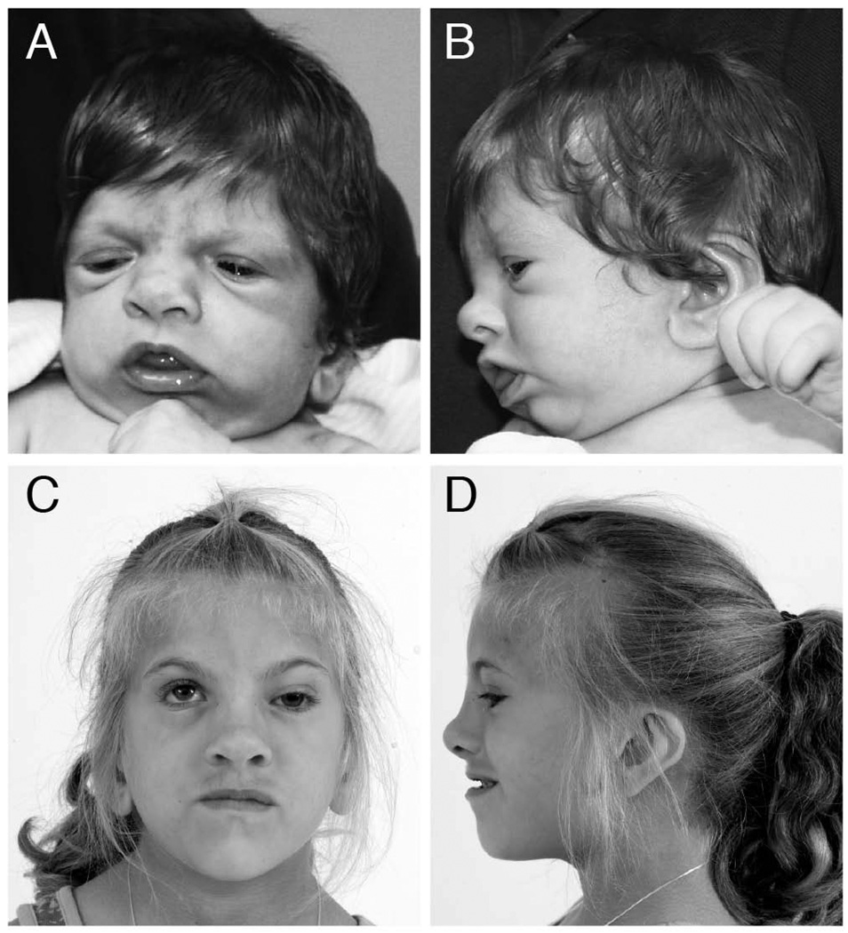
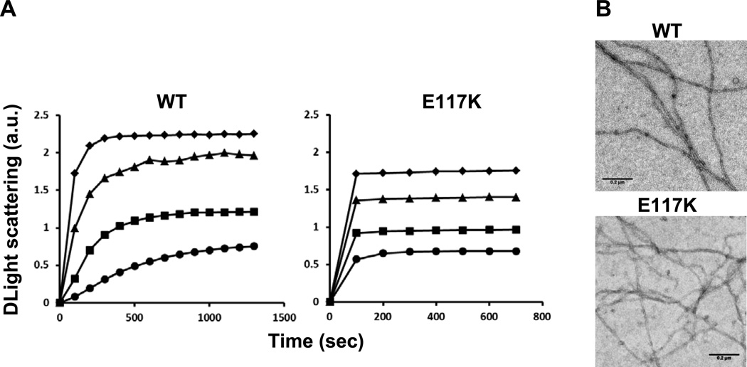
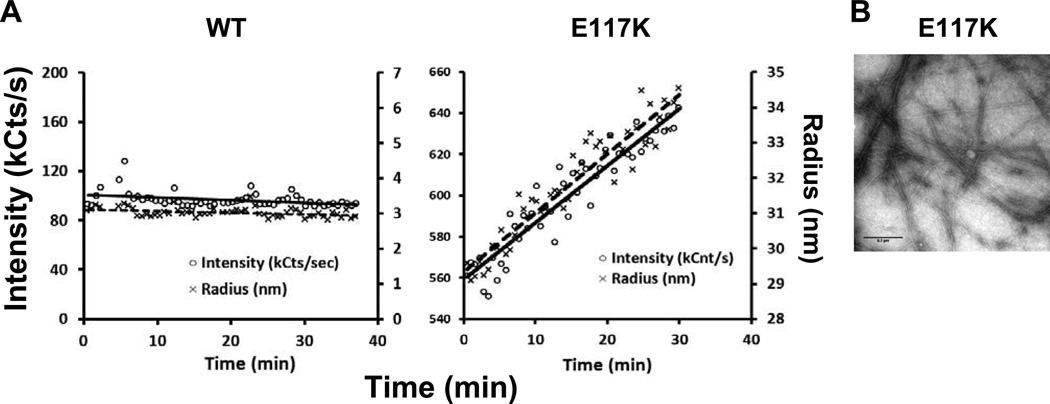
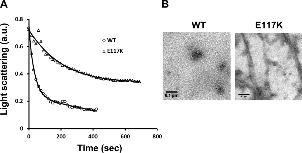
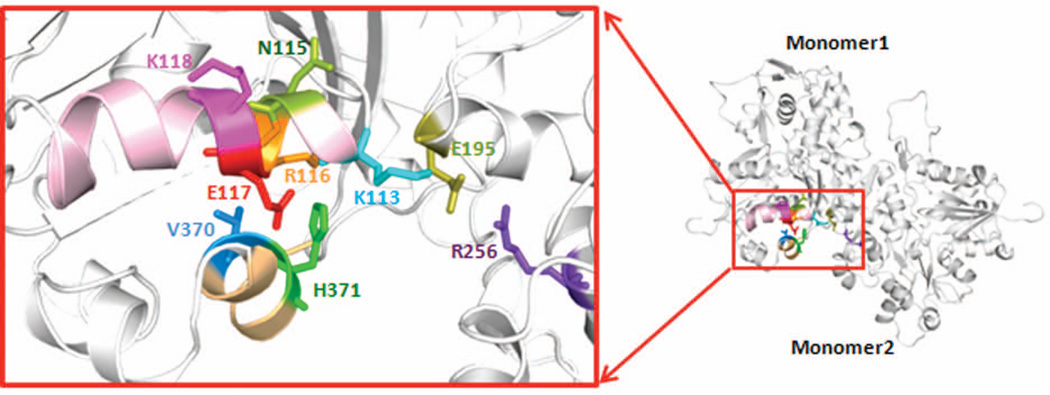
Exome sequence analysis can be instrumental in identifying the genetic etiology behind atypical disease. We report a patient presenting with microcephaly, dysmorphic features, and intellectual disability with a tentative diagnosis of Dubowitz syndrome. Exome analysis was performed on the patient and both parents. A de novo missense variant was identified in ACTB, c.349G>A, p.E117K. Recent work in Baraitser-Winter syndrome has identified ACTB and ACTG1 mutations in a cohort of individuals, and we rediagnosed the patient with atypical Baraitser-Winter syndrome. We performed functional characterization of the variant actin and show that it alters cell adhesion and polymer formation supporting its role in disease. We present the clinical findings in the patient, comparison of this patient to other patients with ACTB/ACTG1 mutations, and results from actin functional studies that demonstrate novel functional attributes of this mutant protein.
Keywords: ACTB; Baraitser-Winter syndrome; Dubowitz; actin.
© 2013 WILEY PERIODICALS, INC.
Conflict of interest statement
All other authors declare no conflict of interest.
Figures
Similar articles
-
De novo mutations in the actin genes ACTB and ACTG1 cause Baraitser-Winter syndrome.Nat Genet. 2012 Feb 26;44(4):440-4, S1-2. doi: 10.1038/ng.1091. Nat Genet. 2012. PMID: 22366783 Free PMC article.
-
A likely pathogenic ACTG1 variant in a child showing partial phenotypic overlap with Baraitser-Winter syndrome.Am J Med Genet A. 2023 Jun;191(6):1565-1569. doi: 10.1002/ajmg.a.63157. Epub 2023 Feb 21. Am J Med Genet A. 2023. PMID: 36810952
-
The First Korean Case of Baraitser-Winter Cerebro-Fronto-Facial Syndrome with a Novel Mutation in ACTB Diagnosed Via Targeted Gene Panel Sequencing and Literature Review.Ann Clin Lab Sci. 2020 Nov;50(6):818-824. Ann Clin Lab Sci. 2020. PMID: 33334799
-
Baraitser-Winter cerebrofrontofacial syndrome.Clin Genet. 2017 Jul;92(1):3-9. doi: 10.1111/cge.12864. Epub 2016 Nov 30. Clin Genet. 2017. PMID: 27625340 Review.
-
Trio exome sequencing identified a novel de novo WASF1 missense variant leading to recurrent site substitution in a Chinese patient with developmental delay, microcephaly, and early-onset seizures: A mutational hotspot p.Trp161 and literature review.Clin Chim Acta. 2021 Dec;523:10-18. doi: 10.1016/j.cca.2021.08.030. Epub 2021 Aug 31. Clin Chim Acta. 2021. PMID: 34478686 Review.
Cited by
-
Multilineage ACTB mutation in a patient with fibro-osseous maxillary lesion and pilocytic astrocytoma.Am J Med Genet A. 2018 Sep;176(9):2037-2040. doi: 10.1002/ajmg.a.40475. Epub 2018 Aug 27. Am J Med Genet A. 2018. PMID: 30152002 Free PMC article. No abstract available.
-
Baraitser and Winter syndrome with growth hormone deficiency.J Pediatr Neurosci. 2014 Sep-Dec;9(3):257-9. doi: 10.4103/1817-1745.147583. J Pediatr Neurosci. 2014. PMID: 25624931 Free PMC article.
-
Comprehensive genotype-phenotype correlation in lissencephaly.Quant Imaging Med Surg. 2018 Aug;8(7):673-693. doi: 10.21037/qims.2018.08.08. Quant Imaging Med Surg. 2018. PMID: 30211035 Free PMC article. Review.
-
Essential nucleotide- and protein-dependent functions of Actb/β-actin.Proc Natl Acad Sci U S A. 2018 Jul 31;115(31):7973-7978. doi: 10.1073/pnas.1807895115. Epub 2018 Jul 16. Proc Natl Acad Sci U S A. 2018. PMID: 30012594 Free PMC article.
-
Prevalence of Cytoplasmic Actin Mutations in Diffuse Large B-Cell Lymphoma and Multiple Myeloma: A Functional Assessment Based on Actin Three-Dimensional Structures.Int J Mol Sci. 2020 Apr 27;21(9):3093. doi: 10.3390/ijms21093093. Int J Mol Sci. 2020. PMID: 32349449 Free PMC article.
References
-
- Bryan KE, Wen KK, Zhu M, Rendtorff ND, Feldkamp M, Tranebjaerg L, Friderici KH, Rubenstein PA. Effects of human deafness gamma-actin mutations (DFNA20/26) on actin function. J Biol Chem. 2006;281:20129–20139. - PubMed
-
- Furness DN, Katori Y, Mahendrasingam S, Hackney CM. Differential distribution of beta- and gamma-actin in guinea-pig cochlear sensory and supporting cells. Hear Res. 2005;207:22–34. - PubMed
Publication types
MeSH terms
Substances
Grants and funding
LinkOut - more resources
Full Text Sources
Other Literature Sources
Molecular Biology Databases
Miscellaneous
