. 2013 Jul;61(7):1018-28.
doi: 10.1002/glia.22483. Epub 2013 May 7.

Evidence for miR-181 involvement in neuroinflammatory responses of astrocytes

Affiliations

Evidence for miR-181 involvement in neuroinflammatory responses of astrocytes

Emmette R Hutchison et al. Glia. 2013 Jul.

Abstract

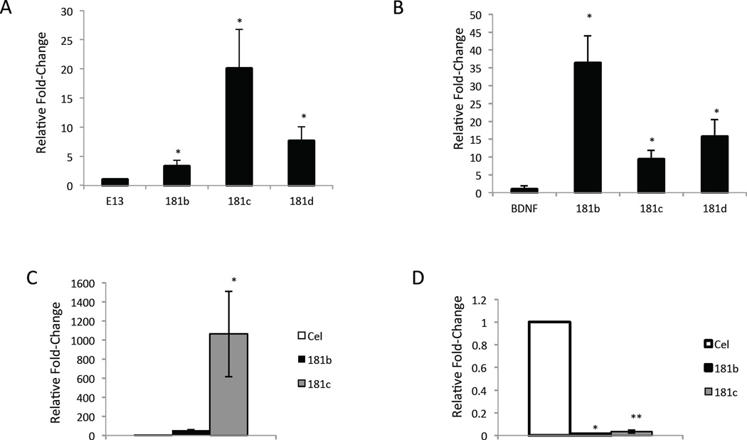
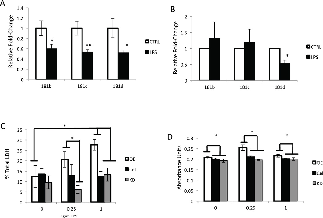
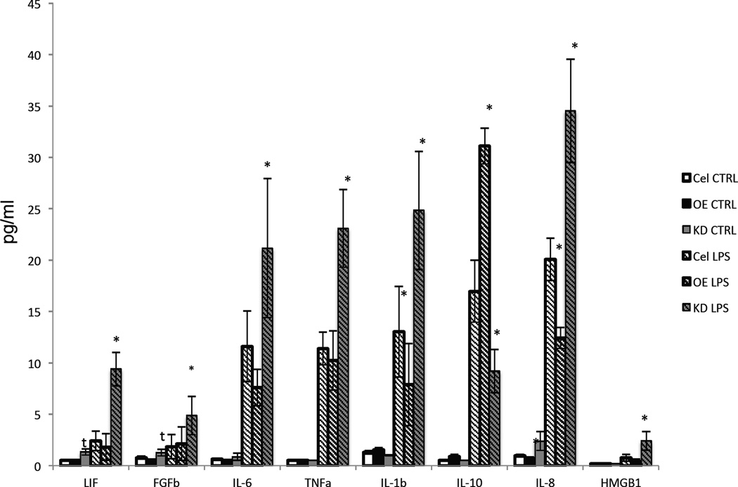
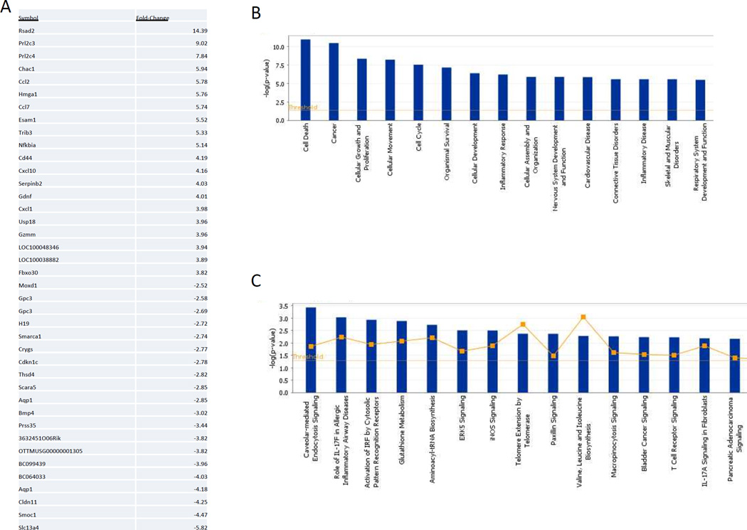
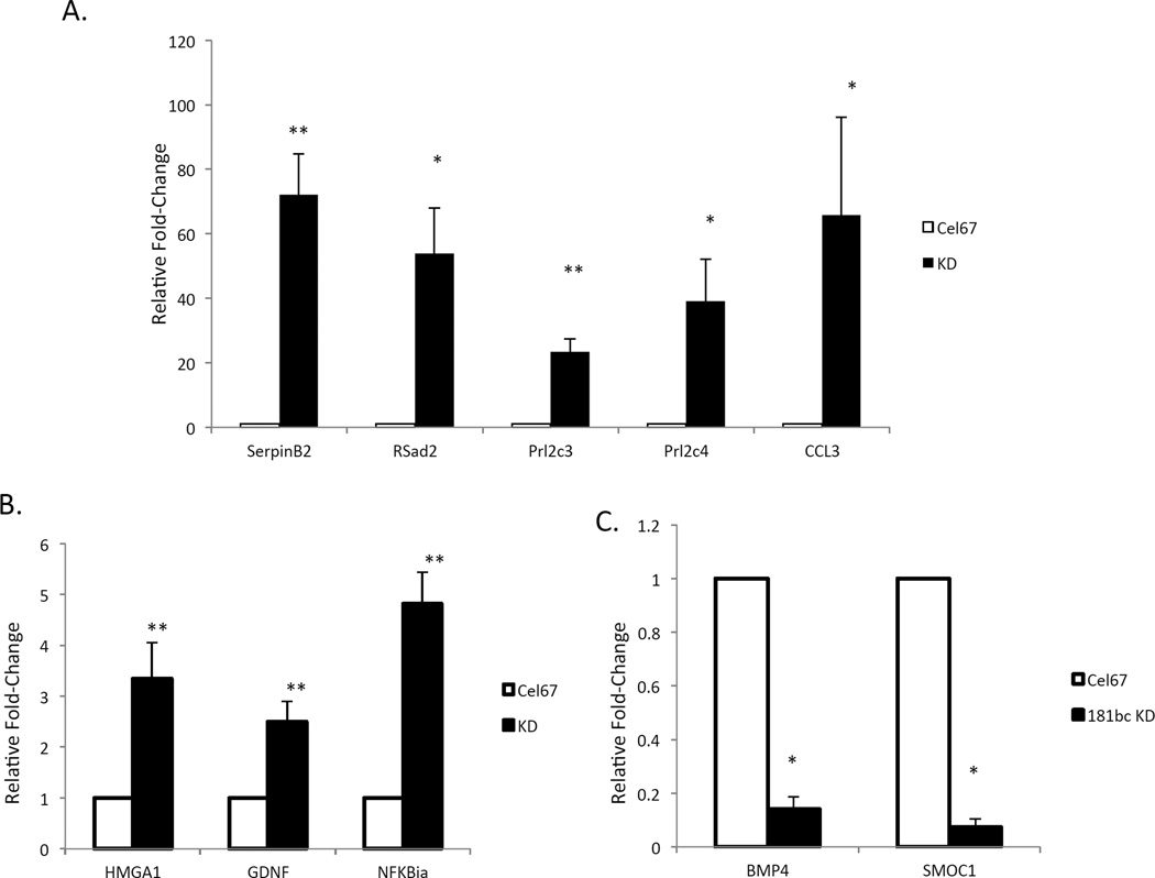
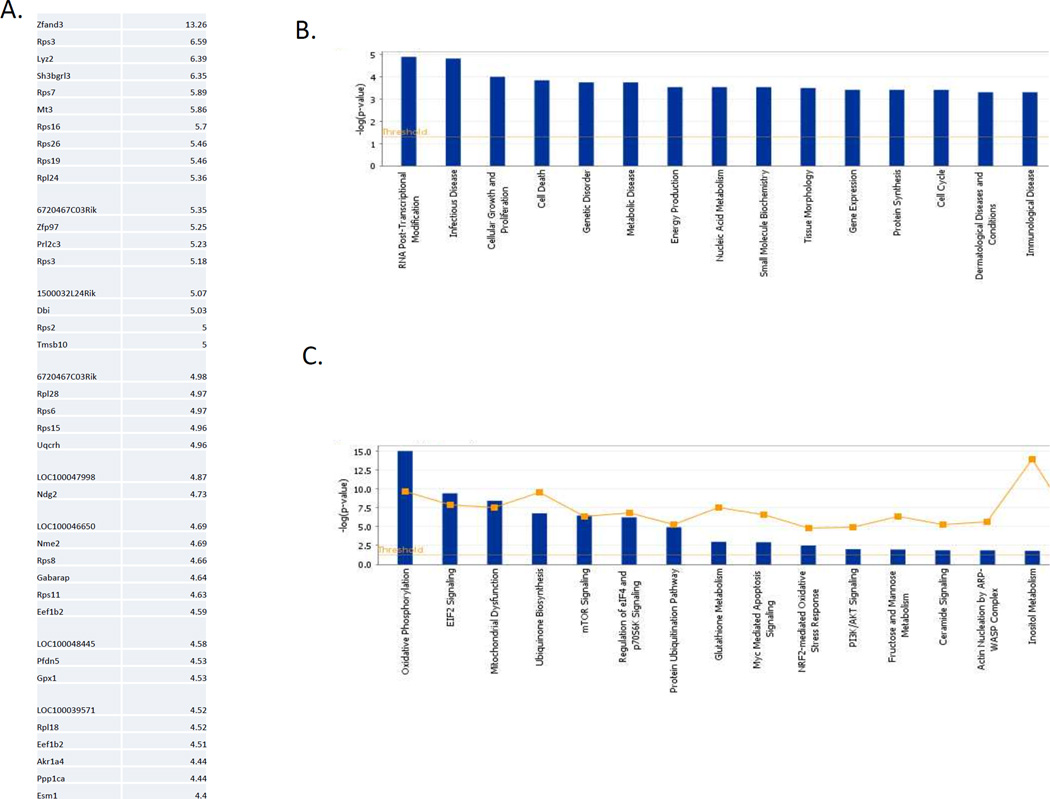
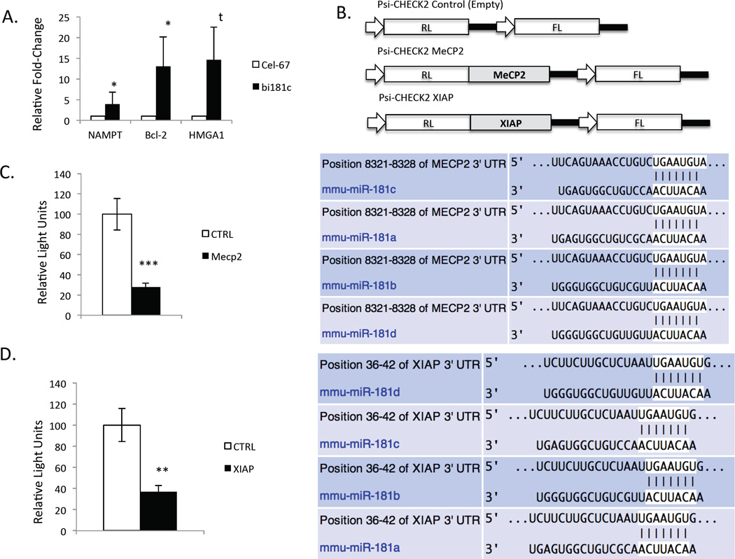
Inflammation is a common component of acute injuries of the central nervous system (CNS) such as ischemia, and degenerative disorders such as Alzheimer's disease. Glial cells play important roles in local CNS inflammation, and an understanding of the roles for microRNAs in glial reactivity in injury and disease settings may therefore lead to the development of novel therapeutic interventions. Here, we show that the miR-181 family is developmentally regulated and present in high amounts in astrocytes compared to neurons. Overexpression of miR-181c in cultured astrocytes results in increased cell death when exposed to lipopolysaccharide (LPS). We show that miR-181 expression is altered by exposure to LPS, a model of inflammation, in both wild-type and transgenic mice lacking both receptors for the inflammatory cytokine TNF-α. Knockdown of miR-181 enhanced LPS-induced production of pro-inflammatory cytokines (TNF-α, IL-6, IL-1β, IL-8) and HMGB1, while overexpression of miR-181 resulted in a significant increase in the expression of the anti-inflammatory cytokine IL-10. To assess the effects of miR-181 on the astrocyte transcriptome, we performed gene array and pathway analysis on astrocytes with reduced levels of miR-181b/c. To examine the pool of potential miR-181 targets, we employed a biotin pull-down of miR-181c and gene array analysis. We validated the mRNAs encoding MeCP2 and X-linked inhibitor of apoptosis as targets of miR-181. These findings suggest that miR-181 plays important roles in the molecular responses of astrocytes in inflammatory settings. Further understanding of the role of miR-181 in inflammatory events and CNS injury could lead to novel approaches for the treatment of CNS disorders with an inflammatory component.


Figures

Figure 1
Figure 1. miR-181s are enriched in astrocytes, modify cell proliferation and vulnerability to degeneration in a model of neuroinflammation
A. Expression levels of miR-181b-d in E13 telencephalon versus adult cortex (181b p<0.04, 181c p<0.02, 181d p<0.03, n=4). B. Expression of miR-181b-d in cultured astrocytes compared to BDNF-differentiated neurons (181b p<0.03, 181c p<0.01, 181d p<0.03, n=4). C. Levels of miR-181 b and c in cultured astrocytes 24 hours after transfection with a mirR-181c mimic (181c p<0.04, n=3). D. Levels of miR-181b and miR-181c in cultured astrocytes 48 hours after transfection with miR-181c and miR-181b microRNA hairpin inhibitors (181b p<0.04, 181c p<0.00002, n=4).
Figure 2
Figure 2. Decline in R-181 levels following exposure to LPS in the cerebral cortex of mice in vivo and in cultured astrocytes
A. miR-181b-d expression in cerebral cortex from LPS-treated wild-type mice versus vehicle-treated control mice, 4 h after LPS administration (181b p<0.02, 181c p<0.007, 181d p<0.02, n=4). B. miR-181b-d expression in cortex from LPS treated TNFα receptor knockout mice, 4 h after IP injection of LPS (181d p<0.04, n=3). C. Effects of miR-181 overexpression (OE) or knockdown (KD) on the vulnerability of cultured astrocytes to cell death caused by exposure to LPS, as measured by LDH levels in the culture medium 48 h after transfection and 6 h after LPS treatment (1 μg/ml LPS: OE p<0.03, OEvKD p<0.05; 1 μg vs. Basal: OE p<0.04, n=6; 250 ng/ml LPS: OEvKD p<0.02, n=4). D. In cultured astrocytes 48 h after miR-181 OE or miR-181 KD, followed by a 6 h LPS treatment, the relative levels of cellular metabolic activity were measured using an MTS reduction assay (1 μg/ml: n=8, OEvCTRL p<0.02; OEvKD p<0.01, OEVNoLPSOE p<0.04; 250 ng/ml: OEvCTRL p<0.03, KDvsCTRL p<0.01, OEvKD p<0.007, OEvNoLPSOE p<0.01, CTRLVSNoLPSCTRL p<0.04, n=4; 0 mg LPS: OEvCTRL p<0.003, OEvKD p<0.05, n=4).
Figure 3
Figure 3. Reduction of miR-181 levels alters both basal and LPS-induced cytokine and FGF2 production
A. Multiplex immunoassay was used to detect levels of LIF (basal: KD p<0.055, OEvKD p<0.055; LPS: KD p<0.02, LPS KDvOE p<0.002; LPS vs. Basal: KD p<0.01), FGF2 (Basal: KD p<0.066, KDvOE p<0.07; LPS: KD p<0.02, KDvOE p<0.004), IL-6 (LPS: KD p<0.02, KDvOE p<0.004), TNF-α (LPS: KD p<0.02, KDvOE p<0.02; LPS vs Basal: OE p<0.04, CTRL p<0.01, KD p<0.01), IL-1β (LPS: OE p<0.03, KD p<0.006, KDvOE p<0.01; LPS vs Basal: CTRL p<0.057, KD p<0.02), IL-10 (LPS: OE p<0.02, KD p<0.007, OEvKD p<0.007; LPS vs. Basal: OE p<0.002, CTRL p<0.01, KD p<0.03), IL-8 (LPS: OE p<0.02, KD p<0.05, KDvOE p<0.02; Basal OE p<0.005; LPS vs Basal: OE p<0.003, CTRL p<0.004, KD p<0.01) and HMGB1 (LPS: KDvOE p<0.05, KDvCTRL p<0.06) in media from cultured astrocytes that had been treated with either 1 µg/ml LPS or vehicle (n = 3).
Figure 4
Figure 4. Reduction of miR-181 expression alters levels of transcripts involved in cell death and inflammation responses
A. A list of the twenty transcripts that increased by the greatest magnitude, and the twenty transcripts that decreased by the greatest magnitude in response to simultaneous knockdown of miR-181b and miR-181c. B. Pathway analysis of transcriptional changes demonstrated that alterations related to cell death, cell cycle regulation, cell differentiation, cancer and inflammatory responses, and related signaling pathways were affected. C. Signaling pathways of interest altered by miR-181b and c knock-down included iNOS signaling.
Figure 5
Figure 5. Evidence that miR-181 regulates diverse signaling pathways in astrocytes
A. The mRNAs encoding SerpinB2 (p<0.007, n=4), RSad2 (p<0.02, n=4), Prl2c3 (p<0.007, n=4), Prl2c4 (p<0.03, n=4) and CCL3 (p<0.04, n=4) increased significantly after reducing miR-181b and c expression in cultured astrocytes. B. Increased levels of mRNAs encoding HMGA1 (p<0.007, n=4), GDNF (p<0.004, n=3) and NFKBia (p<0.008, n=4) after silencing miR-181b/c. C. Lower levels of BMP4 (p<0.05, n=4) and SMOC1 (p<0.04, n=4) mRNAs in astrocytes in which miR-181b/c levels were reduced.
Figure 6
Figure 6. Direct interaction of miR-181 with transcriptome
A. Top 40 most highly enriched transcripts after pull-down of biotinylated miR-181c. B. Pathway analysis indicating that transcripts that interact with miR-181c encode proteins implicated controlling cell death. C. Putative miR-181c targets include proteins implicated in EIF2 signaling, mTOR signaling and mitochondrial dysfunction.
Figure 7
Figure 7. MeCP2 and XIAP mRNAs are direct targets of miR-181
A. NAMPT, Bcl-2 and HMGA1 mRNAs are all enriched in biotin pull-down of miR-181c (HMGA1 p<0.08, n=3, Bcl-2 p<0.02, n=4, NAMPT p<0.07, n=4). B. Schematic representation of Psi-CHECK2 reporter constructs generated to test putative miR-181 targets MeCP2 and XIAP mRNAs. C. Analysis of Psi-CHECK2 MeCP2 reporter activity 48 h after miR-181 OE (p<0.0004, n=6). D. Analysis of Psi-CHECK2 XIAP reporter activity 48 h after miR-181 OE (p<0.001, n=6).

References

    1. Ballas N, Lioy DT, Grunseich C, Mandel G. Non-cell autonomous influence of MeCP2-deficient glia on neuronal dendritic morphology. Nat Neurosci. 2009;12:311–317. - PMC - PubMed
    1. Baune BT, Wiede F, Braun A, Golledge J, Arolt V, Koerner H. Cognitive dysfunction in mice deficient for TNF-α and its receptors. Am J Med Genet B Neuropsychiatr Genet. 2008;147B:1056–1064. - PubMed
    1. Bethea JR, Castro M, Keane RW, Lee TT, Dietrich WD, Yezierski RP. Traumatic spinal cord injury induces nuclear factor-kappaB activation. J. Neurosci. 1998;18:3251–3260. - PMC - PubMed
    1. Boldin MP, Baltimore D. MicroRNAs, new effectors and regulators of NF-κB. Immunol Rev. 2012;246:205–220. - PubMed
    1. Bruce AJ, Boling W, Kindy MS, Peschon J, Kraemer PJ, Carpenter MK, Holtsberg FW, Mattson MP. Altered neuronal and microglial responses to excitotoxic and ischemic brain injury in mice lacking TNF receptors. Nat Med. 1996;2:788–794. - PubMed

Publication types

MeSH terms

LinkOut - more resources