. 2013 Jun;161A(6):1238-63.
doi: 10.1002/ajmg.a.35934. Epub 2013 May 6.

Elements of morphology: standard terminology for the external genitalia

Affiliations

Elements of morphology: standard terminology for the external genitalia

Raoul C M Hennekam et al. Am J Med Genet A. 2013 Jun.

Abstract

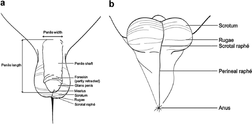
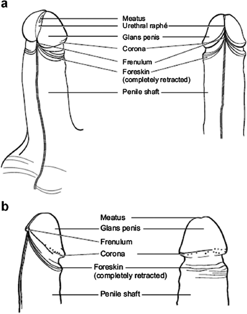
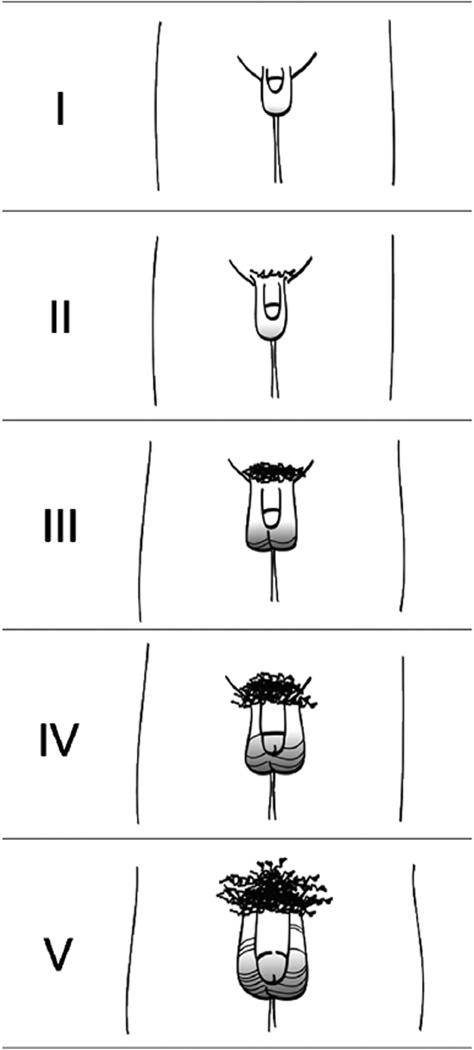
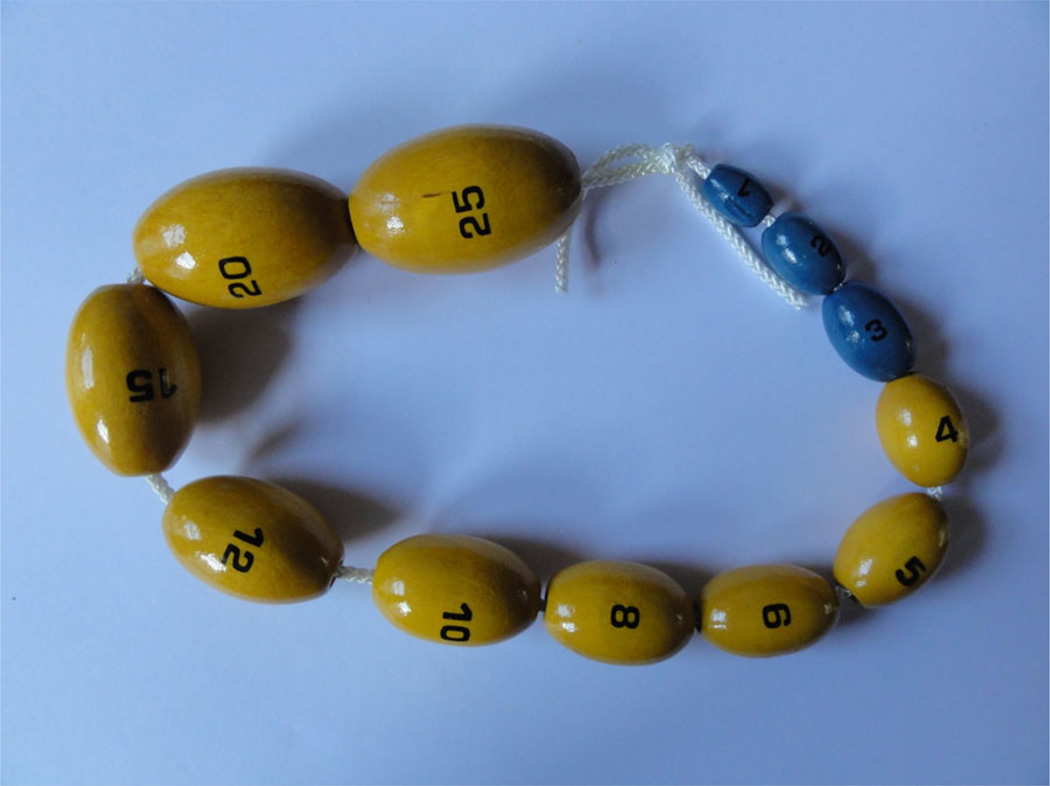
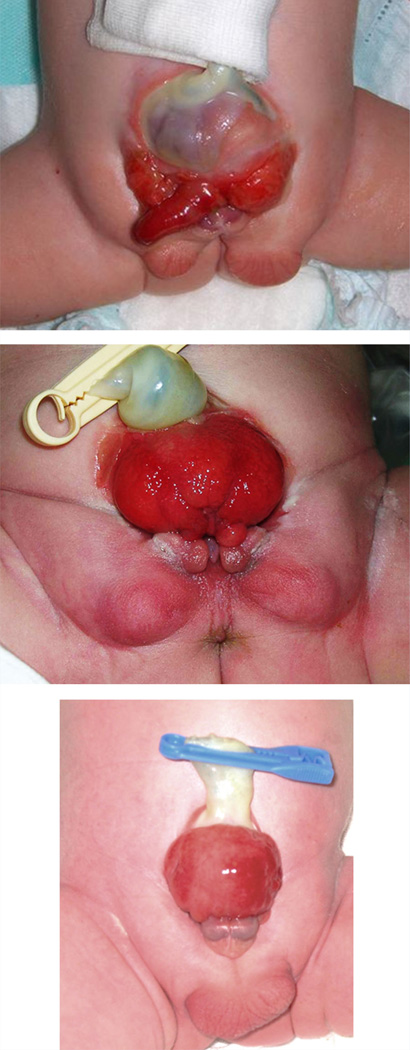
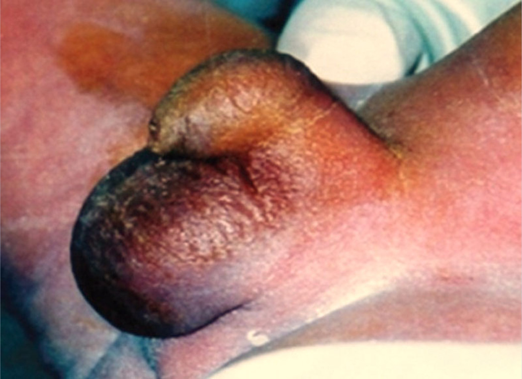
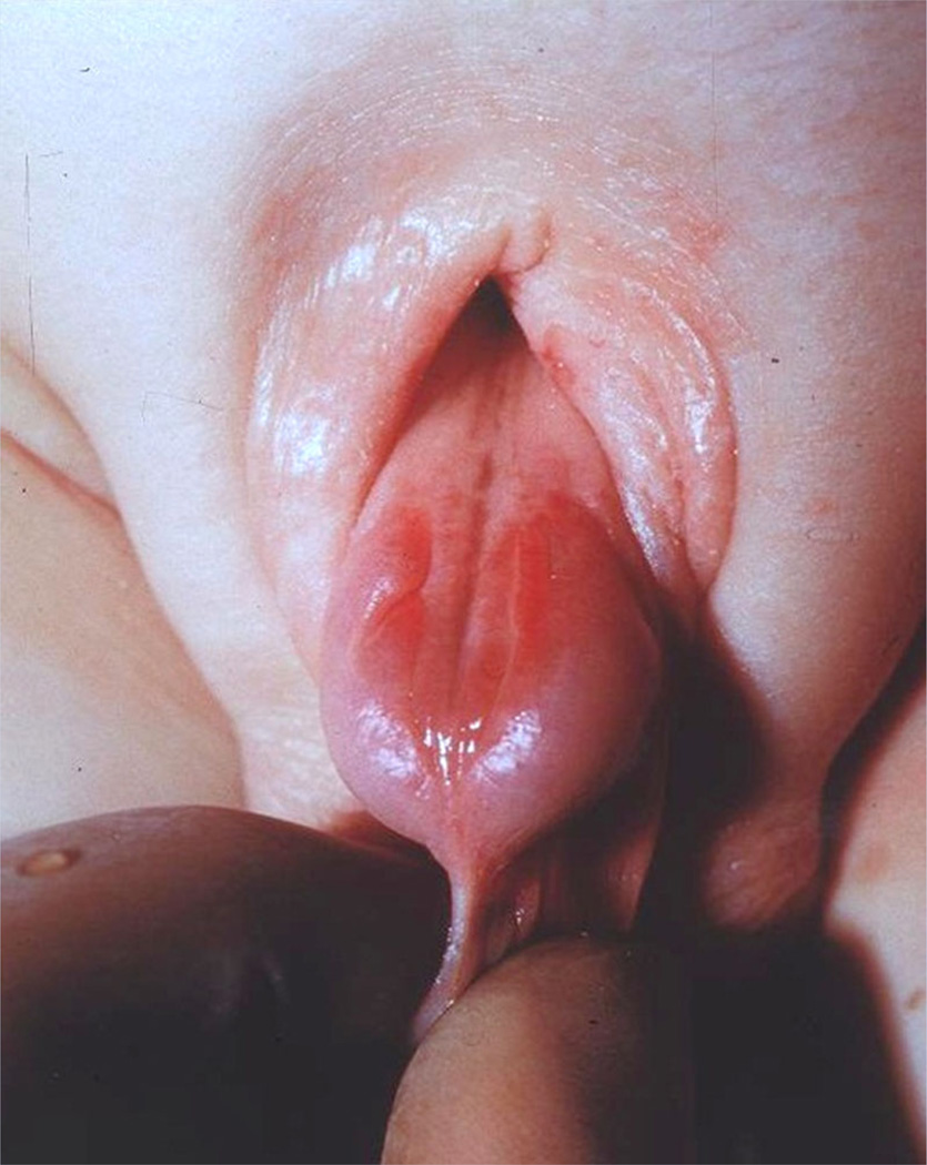
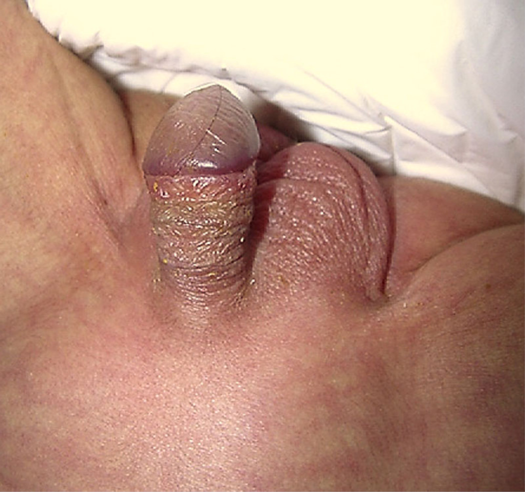
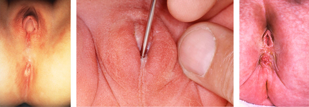
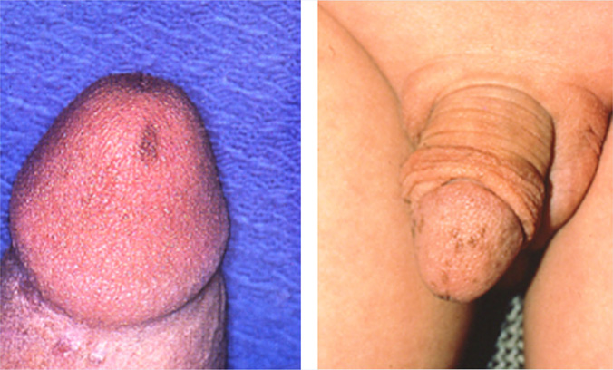
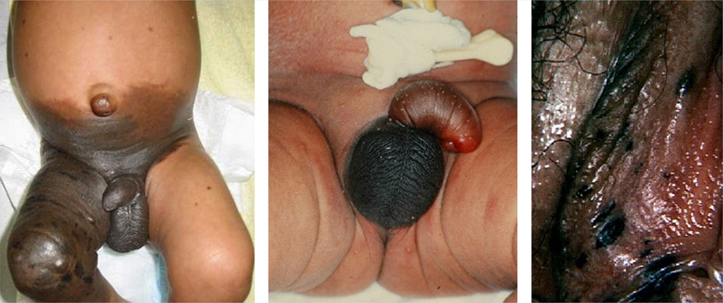
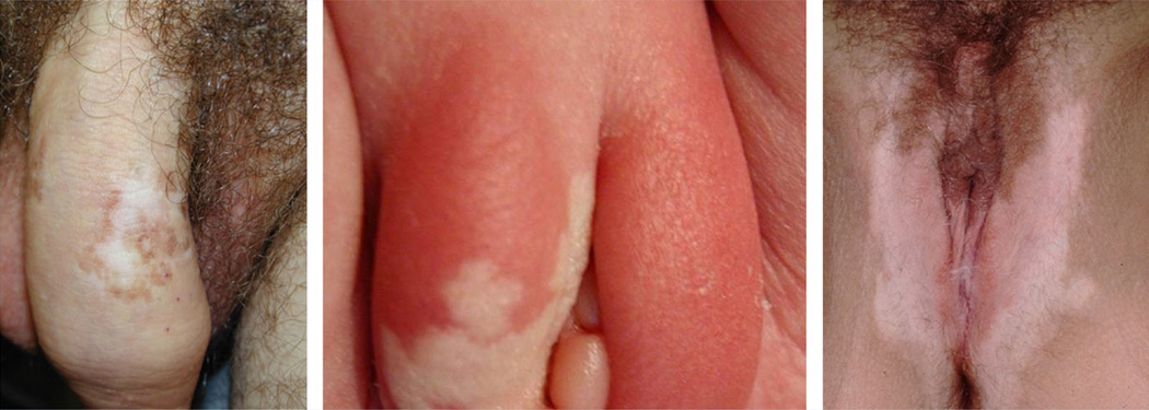
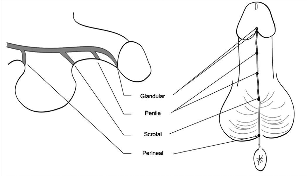
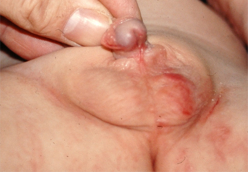
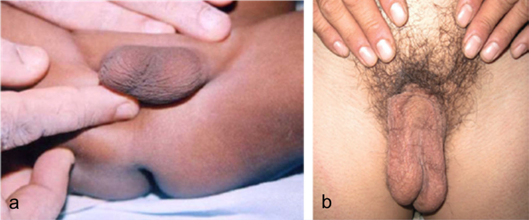
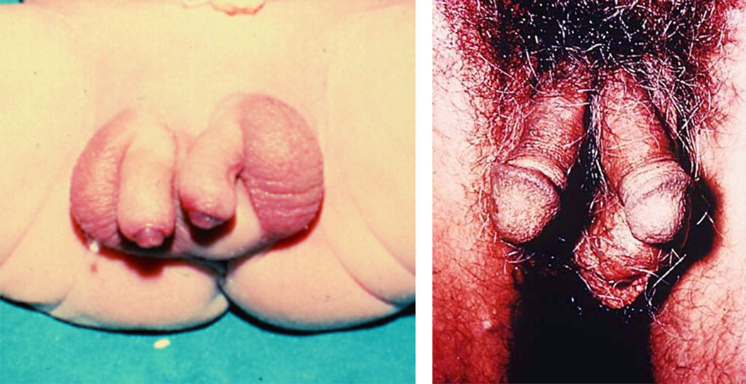
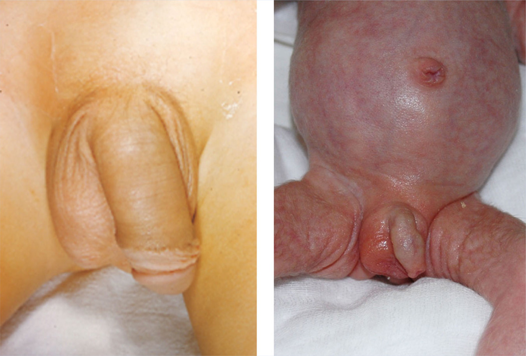
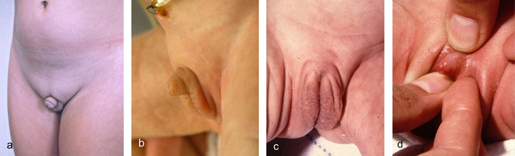
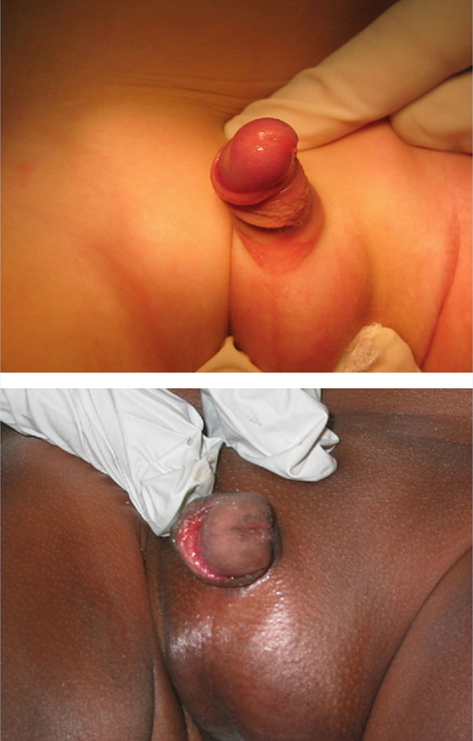
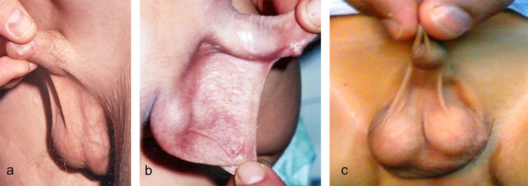
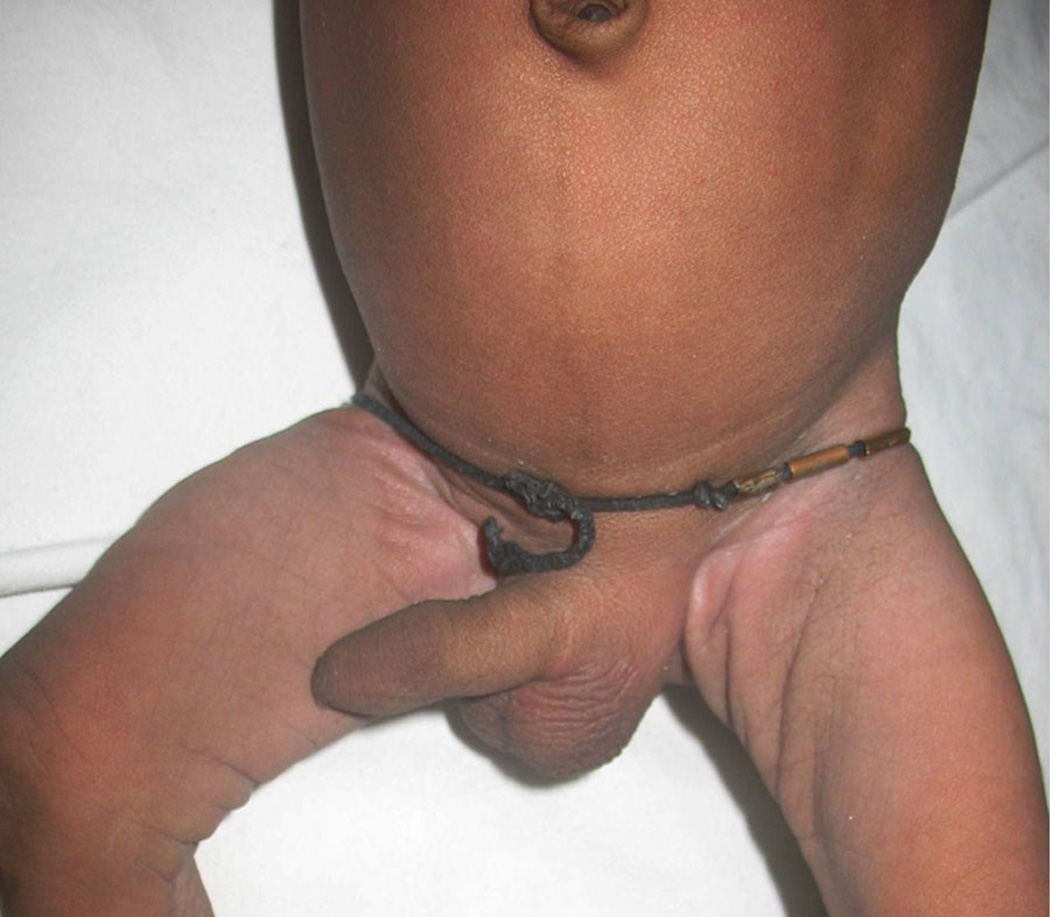
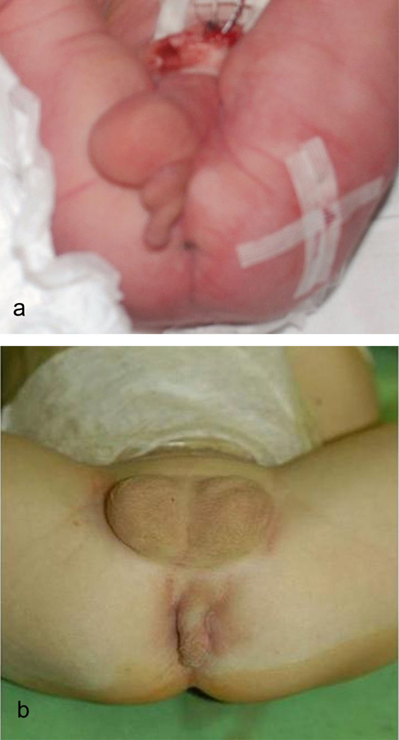
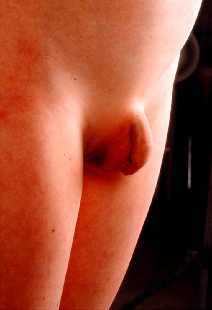
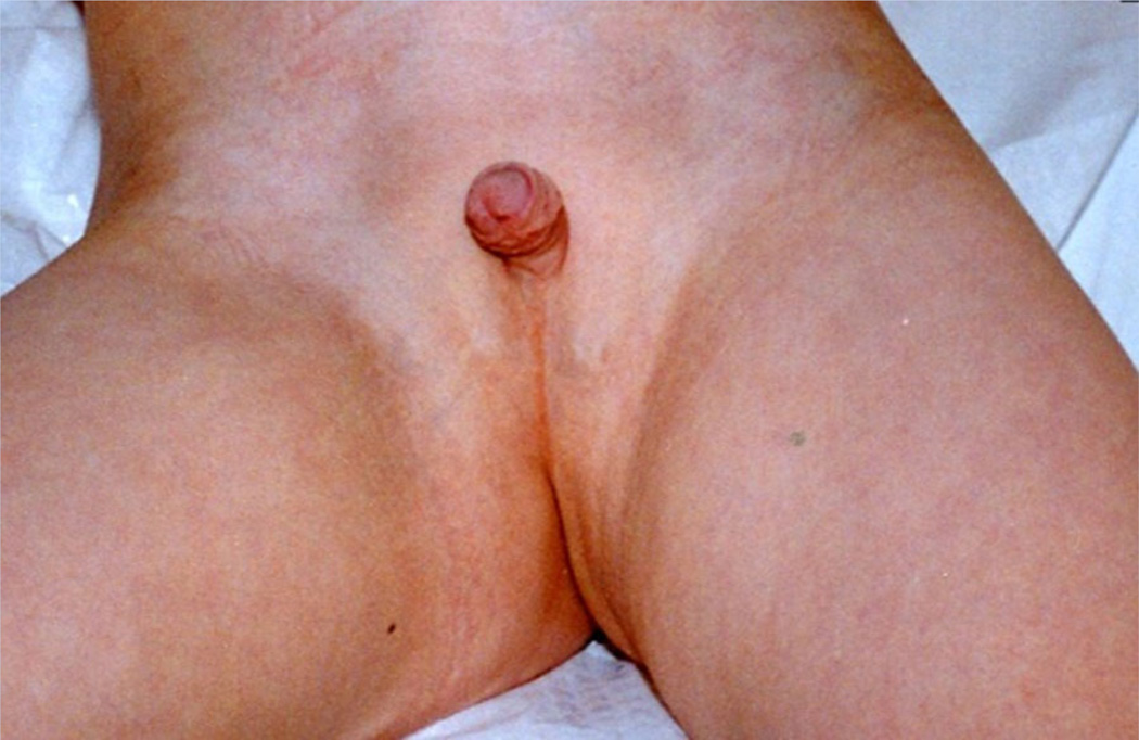
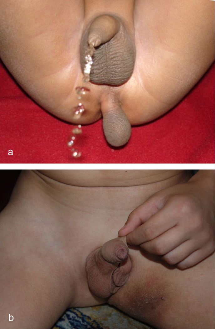
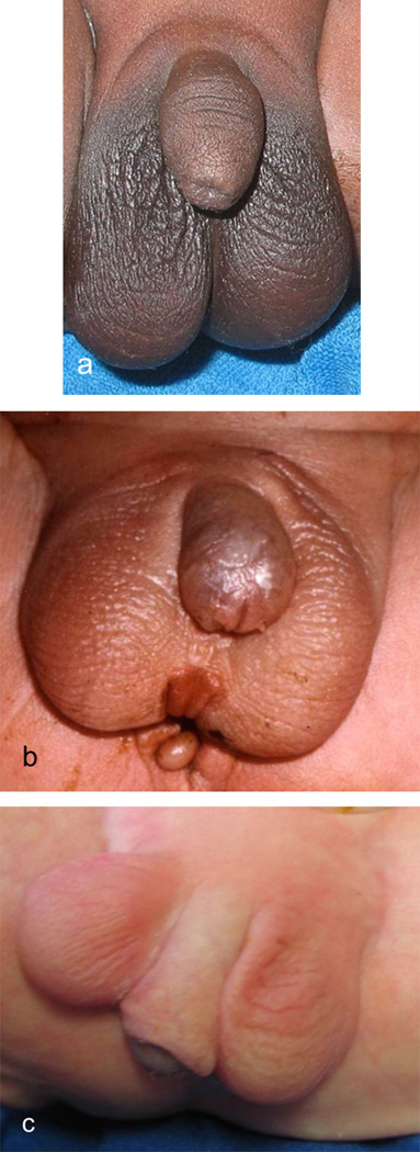
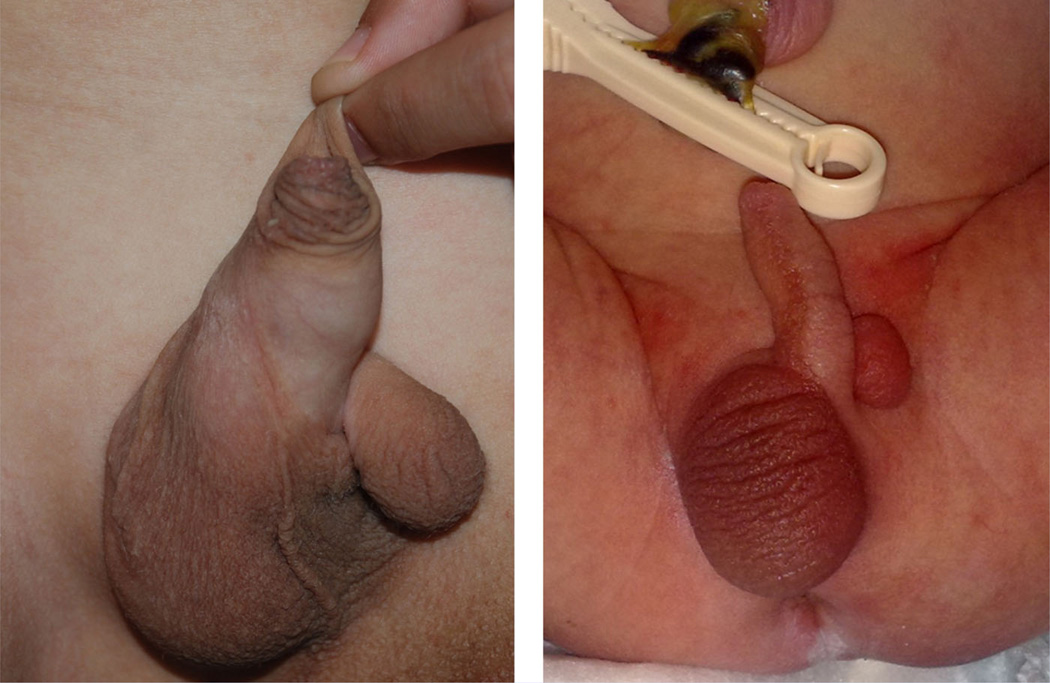
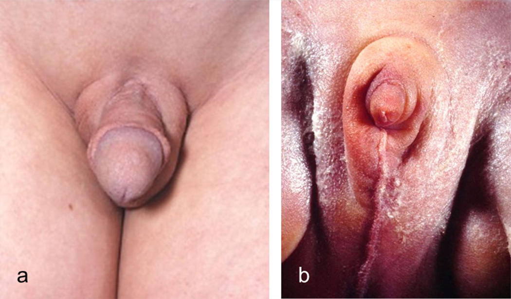
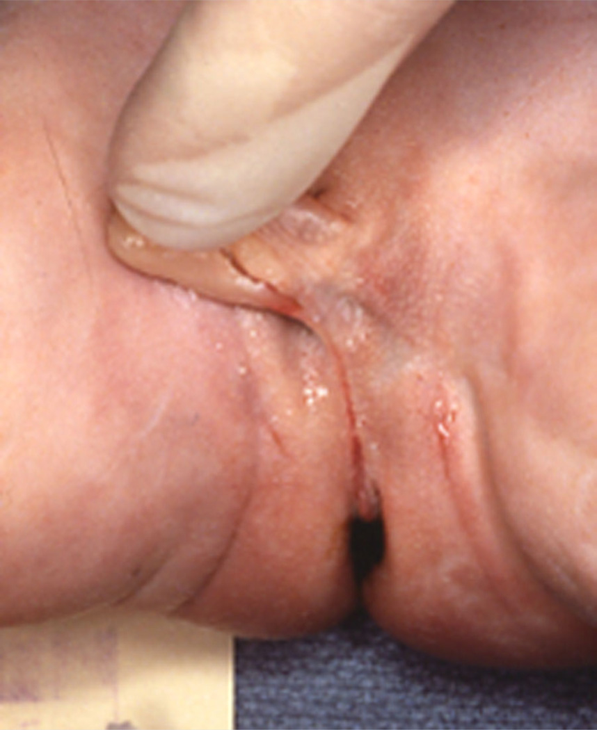
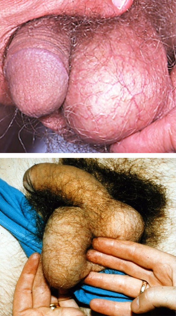
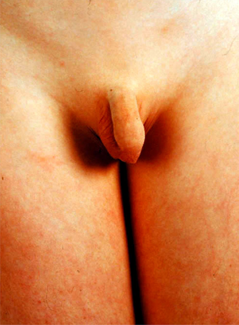
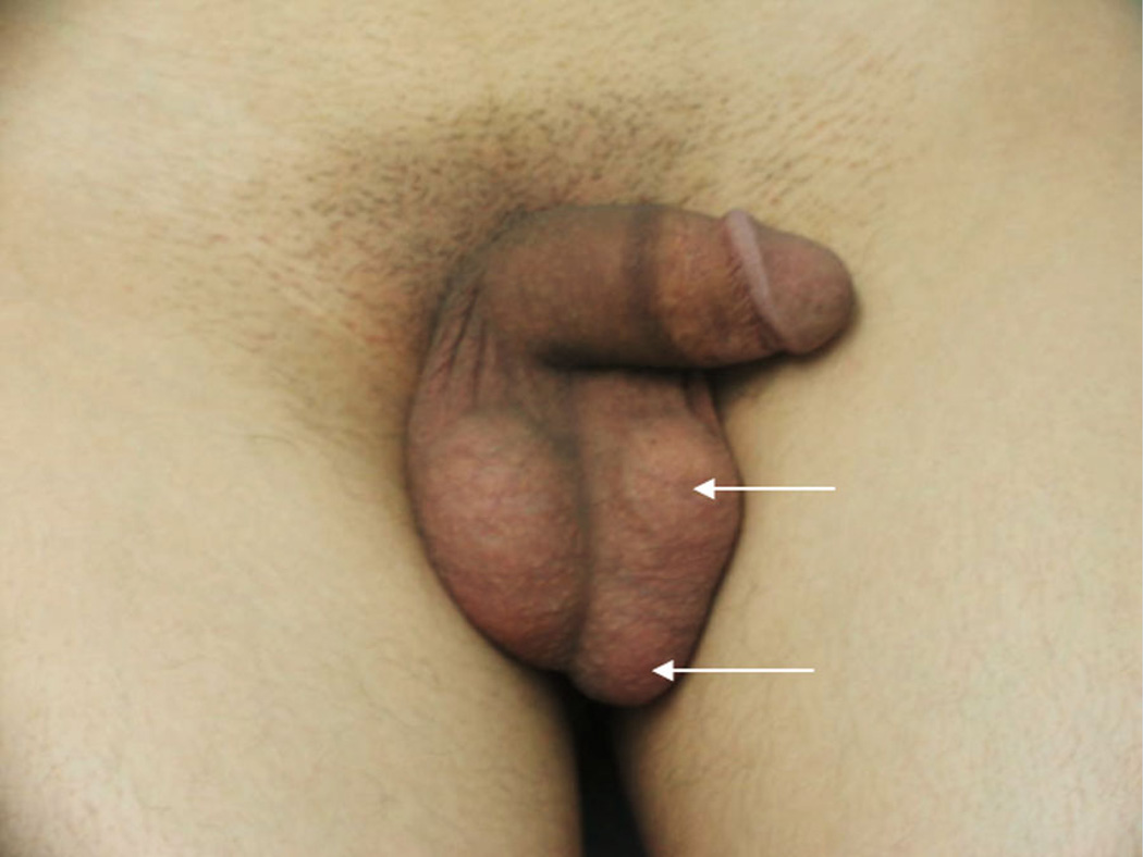
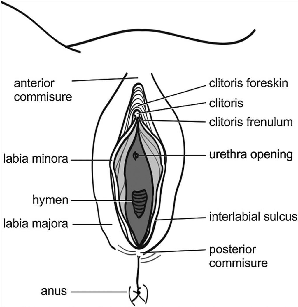
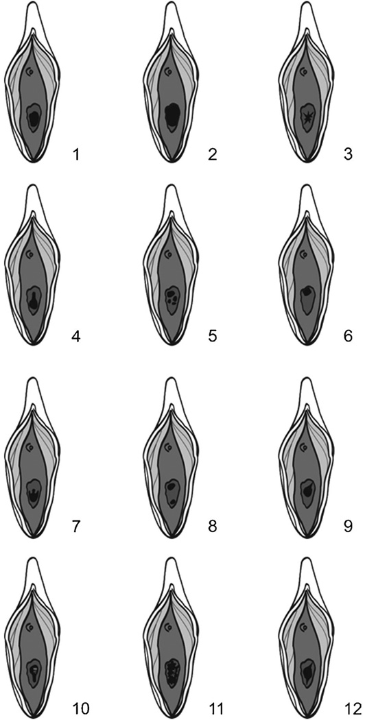
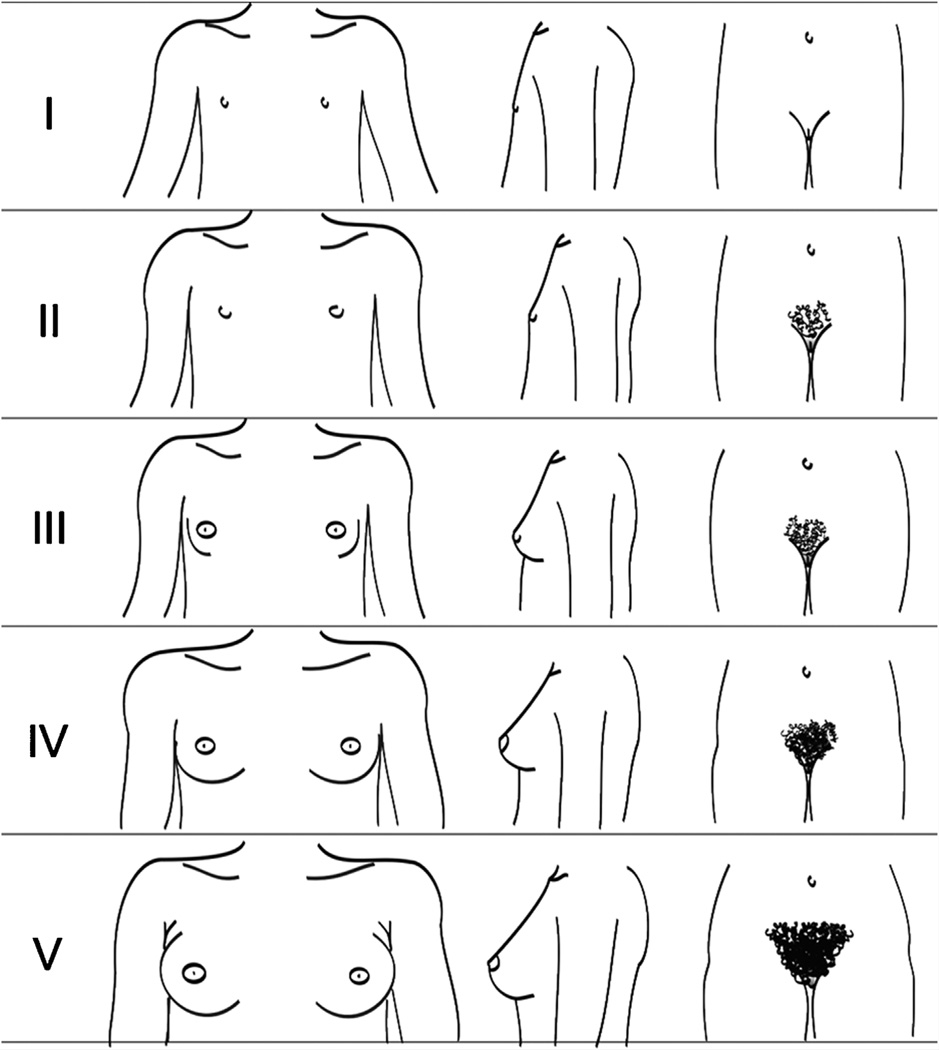
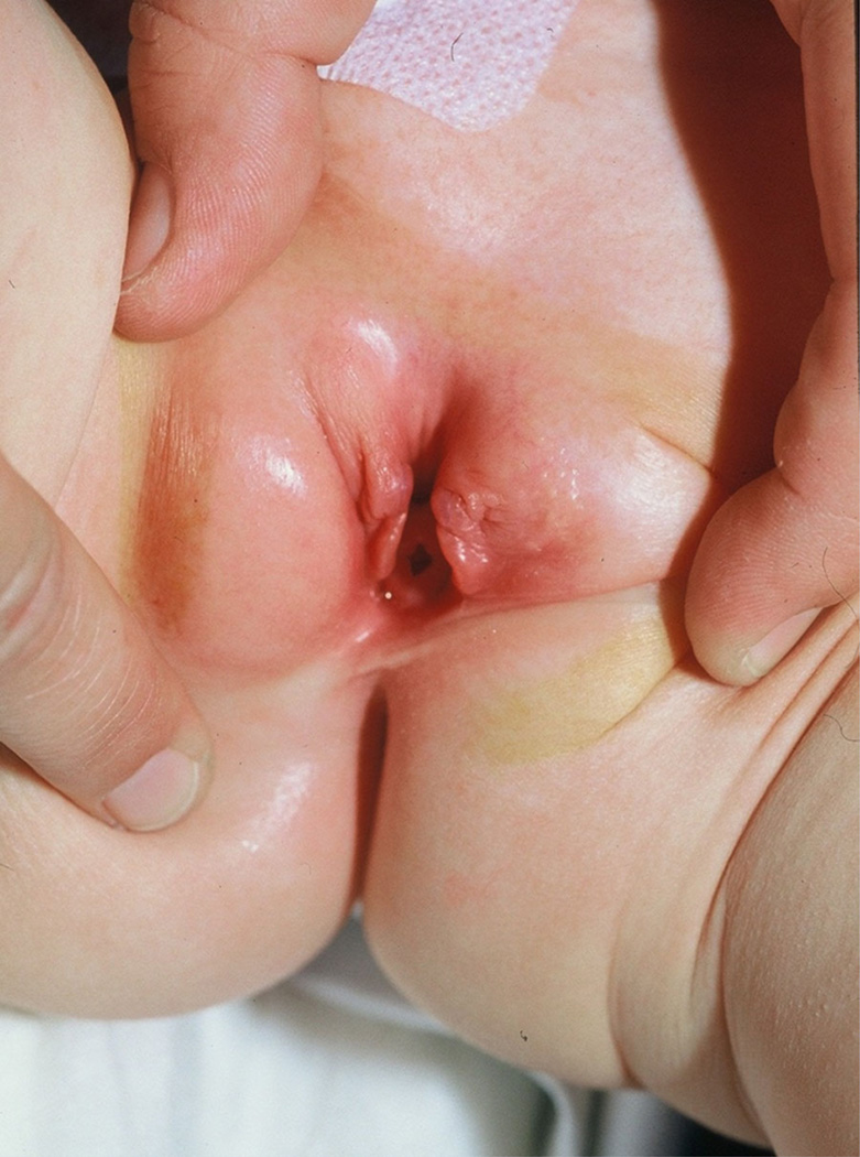
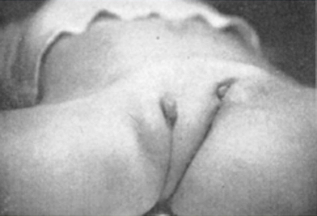
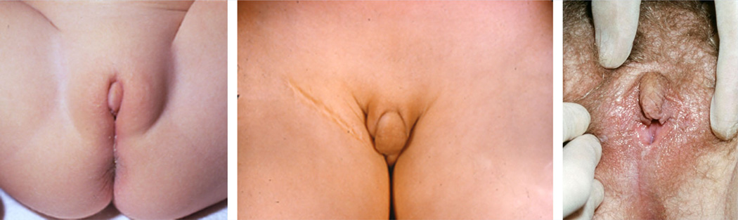
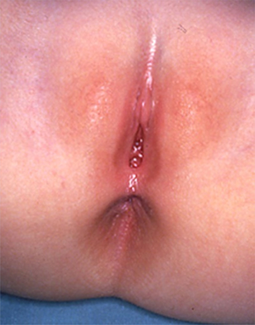
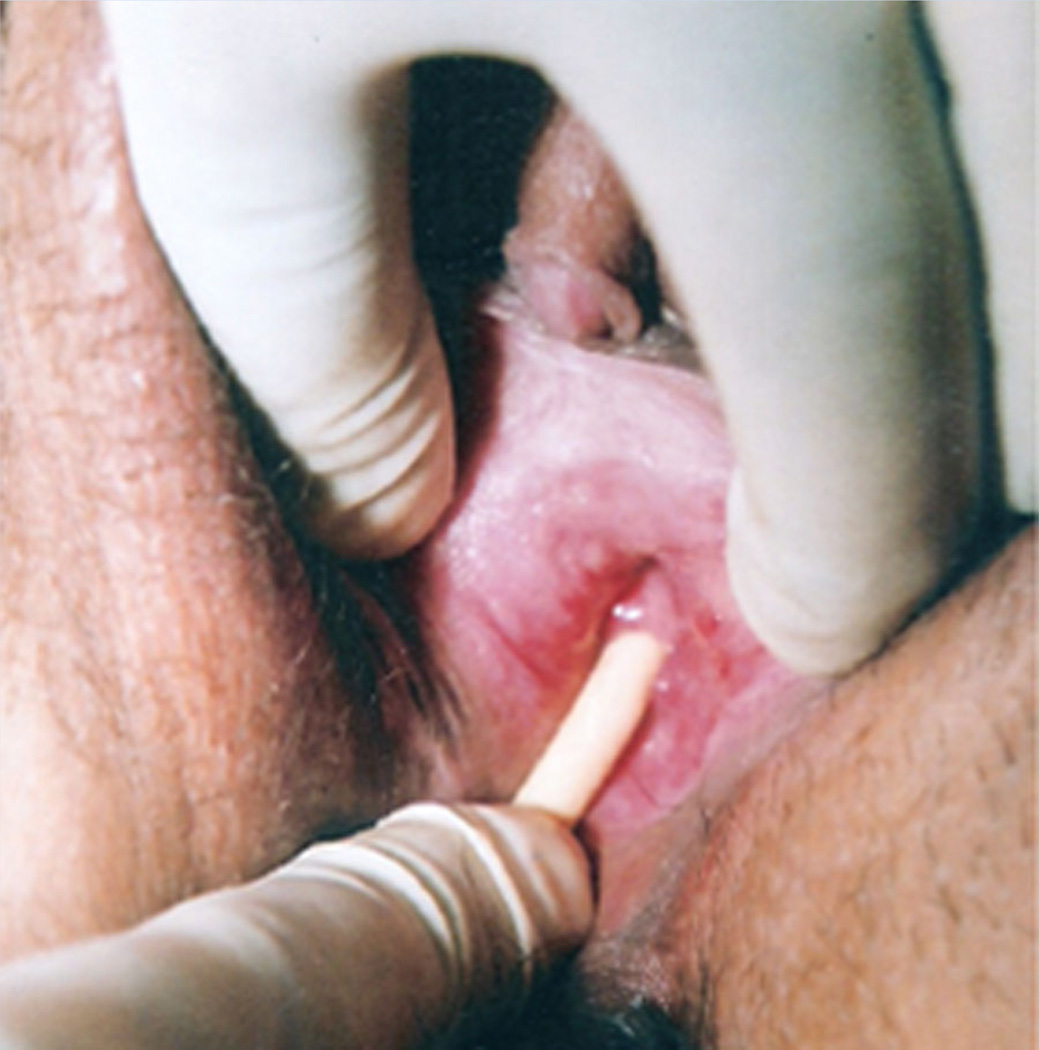
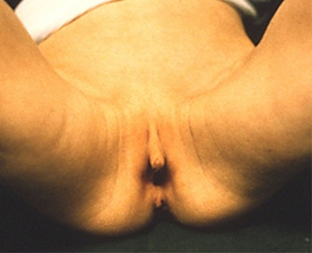
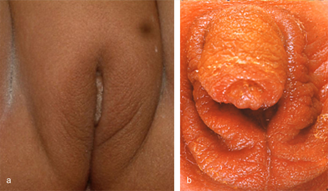
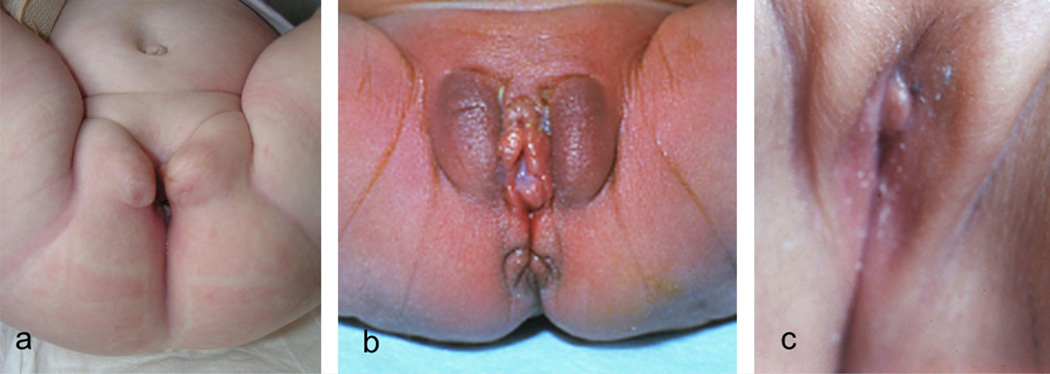
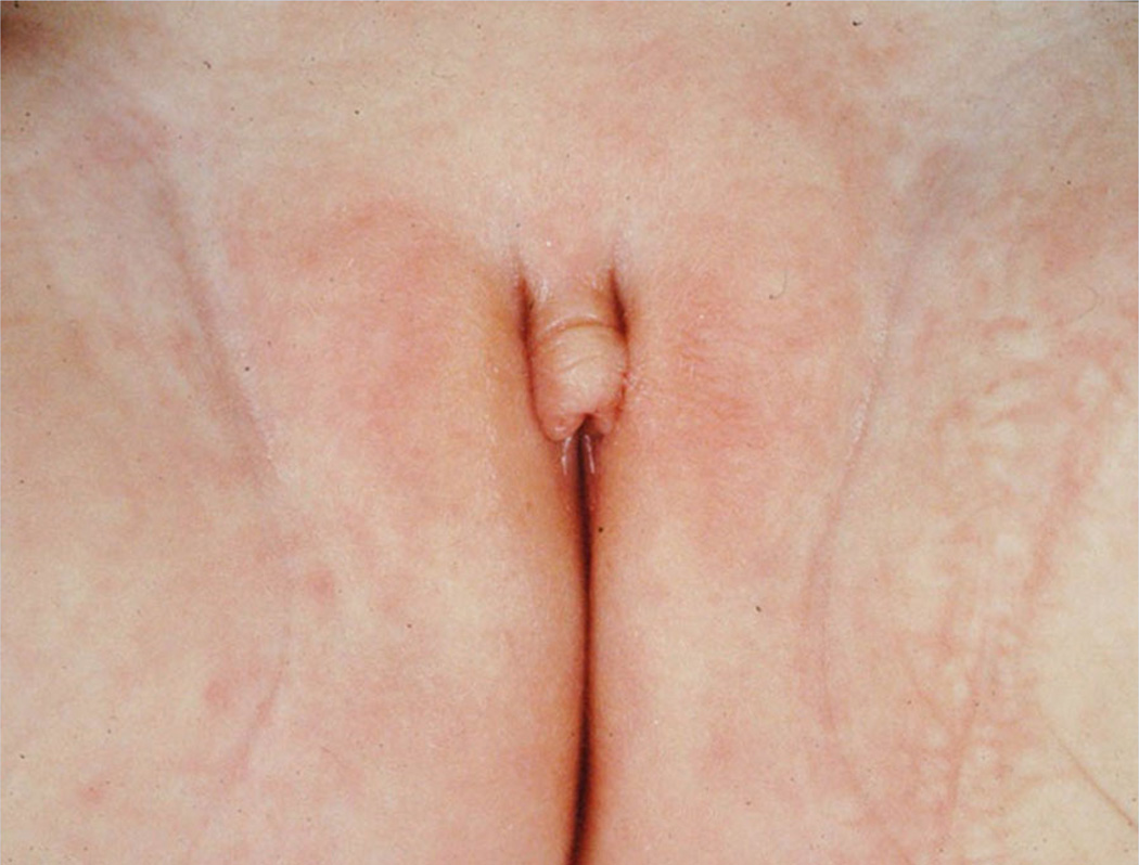
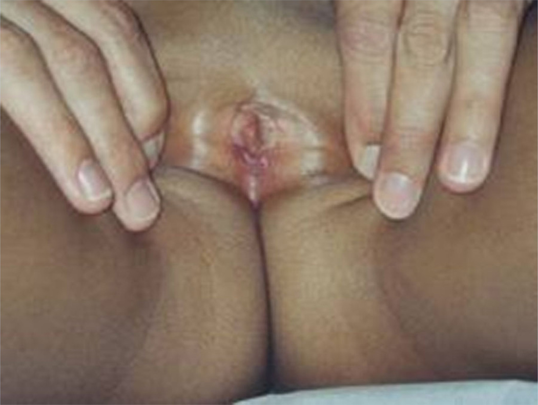
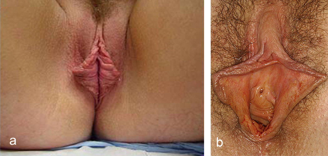
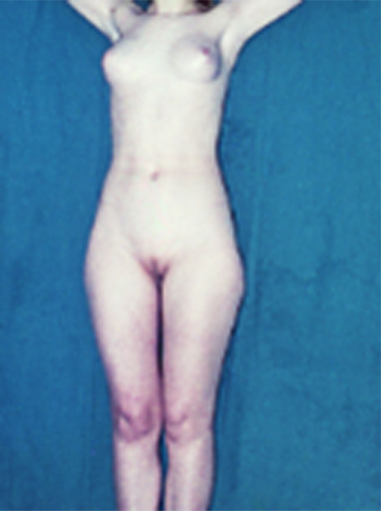
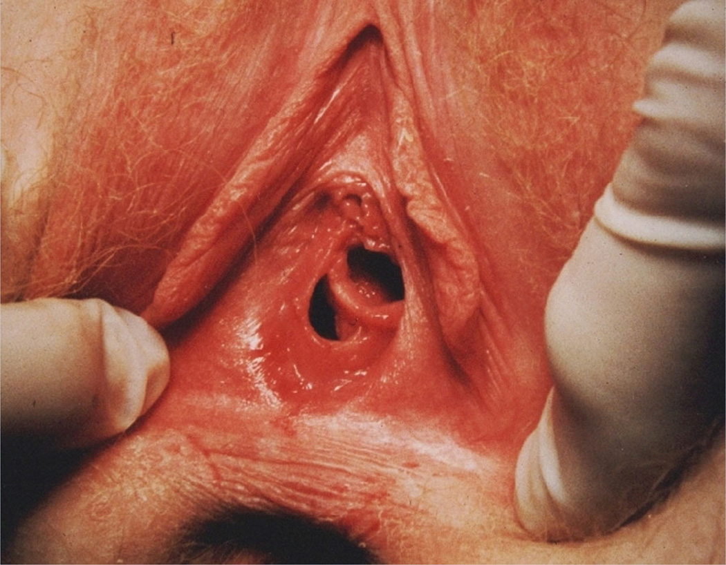
An international group of clinicians working in the field of dysmorphology has initiated the standardization of terms used to describe human morphology. The goals are to standardize these terms and reach consensus regarding their definitions. In this way, we will increase the utility of descriptions of the human phenotype and facilitate reliable comparisons of findings among patients. Discussions with other workers in dysmorphology and related fields, such as developmental biology and molecular genetics, will become more precise. Here we introduce the anatomy of the male and female genitalia, and define and illustrate the terms that describe the major characteristics of these body regions. Published 2013. This article is a U.S. Government work and is in the public domain in the USA.


Figures

FIG. 1
FIG. 1
Anatomy of external male genitalia. a: Anterior overview. b: Inferior overview.
FIG. 2
FIG. 2
Anatomy of external male genitalia. a: Anterior and side view of the penis with the preputium removed. b: Detailed side and posterior view of the penis with the preputium removed.
FIG. 3
FIG. 3
Puberty stages males according to Tanner [1986].
FIG. 4
FIG. 4
Prader orchidometer.
FIG. 5
FIG. 5
Bladder exstrophia. Courtesy of Dr. Anne-Karoline Ebert. Reprinted with permission from Ebert et al. [2009].
FIG. 6
FIG. 6
Chordee. Courtesy of Dr. Montasser El-Koutby. Reprinted with permission from El-Koutby and El Gohary [2010].
FIG. 7
FIG. 7
Epispadias. Courtesy of Dr. Anne-Karoline Ebert. Reprinted with permission from Ebert et al. [2009].
FIG. 8
FIG. 8
Foreskin, absence. Courtesy of Dr. S. Sadeghipour and Dr. N. Esmailzadehha. Reprinted with permission from Sadeghipour and Esmailzadehha [2010].
FIG. 9
FIG. 9
Ambiguous genitalia.
FIG. 10
FIG. 10
Freckled genitalia.
FIG. 11
FIG. 11
Hyperpigmented genitalia in males (left and middle) and female (right).
FIG. 12
FIG. 12
Hypopigmented genitalia in male (left) and females (middle and right).
FIG. 13
FIG. 13
Hydrocele.
FIG. 14
FIG. 14
Various forms of hypospadias. There are various classification systems (see text).
FIG. 15
FIG. 15
Microphallus.
FIG. 16
FIG. 16
Absent penis. Courtesy of Dr. Ruchi Gupta (panel A) and Dr. Li Fubiao (panel B). Panel A reprinted with permission from Rattan et al. [2010]. Panel B reprinted with permission from Wang et al. [2011].
FIG. 17
FIG. 17
Bifid penis.
FIG. 18
FIG. 18
Long penis (in newborn children).
FIG. 19
FIG. 19
Short penis. Please note that on panel C the penis seems to be absent, but can be made visible in the same patient in panel D by pushing surrounding tissues backward.
FIG. 20
FIG. 20
Torsion of the penis. Courtesy of Dr. Barry Kogan. Reprinted with permission from Bauer and Kogan [2009].
FIG. 21
FIG. 21
Webbed penis. Panels B and C courtesy of Dr. Montasser El-Koutby. Reprinted with permission from El-Koutby and El Gohary [2010].
FIG. 22
FIG. 22
Wide penis (newborn child). Courtesy of Dr. Sheela Nampoothiri.
FIG. 23
FIG. 23
Penoscrotal transposition. Courtesy of Dr. Trine Prescott and Dr. Kathrine Bj?rgo (panel A) and Dr Khaled Fathi (panel B). Panel B reprinted with permission from Fathi et al. [2010].
FIG. 24
FIG. 24
Sparse pubic hair (in a pubertal male).
FIG. 25
FIG. 25
Absent scrotum.
FIG. 26
FIG. 26
Accessory scrotum. Courtesy of Dr. Paval Ganesan (panel A) and Dr. Zoran Gucev (panel B). Panel B reprinted with permission from Gucev et al. [2010].
FIG. 27
FIG. 27
Bifid scrotum. Note that in panels A and B the scrotum shows only a mild indentation and in panel C the scrotum is completely bifid.
FIG. 28
FIG. 28
Ectopic scrotum (panel a courtesy of Dr. Zoran Gucev, panel b courtesy of Dr Stefanos Gardikis). Panel A reprinted with permission from Gucev et al. [2010]. Panel B reprinted with permission from Gardikis et al. [2012].
FIG. 29
FIG. 29
Shawl scrotum. Note only partly overriding scrotum in panel A and complete overriding in panel B.
FIG. 30
FIG. 30
Small scrotum.
FIG. 31
FIG. 31
Large testis.
FIG. 32
FIG. 32
Small testis (in a pubertal boy).
FIG. 33
FIG. 33
Supernumerary testis (two testes in left hemiscrotum indicated by arrows). Courtesy of Dr. Ali Feyzullah S?ahin. Reprinted with permission from Yalcinkaya et al. [2011].
FIG. 34
FIG. 34
Anatomy of female external genitalia.
FIG. 35
FIG. 35
Variation in opening of virginal hymen in newborns.
FIG. 36
FIG. 36
Puberty stages females according to Tanner [1986].
FIG. 37
FIG. 37
Bifid clitoris. Note the co-occurrence of epispadias. Courtesy of Dr. Anne-Karoline Ebert. Reprinted with permission from Ebert et al. [2009].
FIG. 38
FIG. 38
Duplicated clitoris. Reprinted with permission from Kurth [1958].
FIG. 39
FIG. 39
Large clitoris.
FIG. 40
FIG. 40
Small clitoris.
FIG. 41
FIG. 41
Hypospadias (a catheter being in hypospadiac opening high in the anterior vaginal wall). Courtesy of Dr. Amilal Bhat. Reprinted with permission from Bhat et al. [2010].
FIG. 42
FIG. 42
Absent labia majora.
FIG. 43
FIG. 43
Large labia majora. Courtesy of Dr. Ariachery Ammini. Reprinted with permission from Kulshreshtha et al. [2010].
FIG. 44
FIG. 44
Labia majora with mild (a) and more marked (b) rugae formation. Note enlargement of clitoris as well in panel B. Panel a courtesy of Dr. Ariachery Ammini. Panel a reprinted with permission from Atilgan et al. [2009].
FIG. 45
FIG. 45
Small upper part (panels a and b) and lower part (panel C) of labia majora.
FIG. 46
FIG. 46
Absent labia minora. Note this causes a seemingly large clitoris which is in fact of normal size.
FIG. 47
FIG. 47
Fused labia minora. Courtesy of Dr. AKC Leung.
FIG. 48
FIG. 48
Prominent labia minora which is unilateral on the right panel. Panel a courtesy of Dr. Warren Ellsworth, panel b courtesy of Dr. Charles Malata. Panel a reprinted with permission from Ellsworth et al. [2010]. Panel B reprinted with permission from Solanki et al. [2010].
FIG. 49
FIG. 49
Sparse pubic hair (in a pubertal female).
FIG. 50
FIG. 50
Atretic opening of the vagina.
FIG. 51
FIG. 51
Septated vagina.

References

    1. Adams MV. The multicultural imagination: Race, color, and the unconscious. London: Routledge; 1996. p. 164.
    1. Ajmani ML, Jain SP, Saxena SK. Anthropometric study of male external genitalia of 320 healthy Nigerian adults. Anthropol Anz. 1985;43:179–186. - PubMed
    1. Akbiyik F, Kutlu AO. External genital proportions in prepubertal girls:Amorphometric reference for female genitoplasty. J Urol. 2010;184:1476–1481. - PubMed
    1. Allanson JE, Biesecker LG, Carey JC, Hennekam RCM. Elements of morphology: Introduction. Am J Med Genet Part A. 2009a;149A:2–5. - PMC - PubMed
    1. Allanson JE, Cunniff C, Hoyme HE, McGaughan J, Muenke M, Neri G. Elements of morphology: Standard terminology for the head and face. Am J Med Genet Part A. 2009b;149A:6–28. - PMC - PubMed

Publication types

LinkOut - more resources