. 2013 May 21;110(21):8662-7.
doi: 10.1073/pnas.1307152110. Epub 2013 May 6.

Transcytosis and brain uptake of transferrin-containing nanoparticles by tuning avidity to transferrin receptor

Affiliations

Transcytosis and brain uptake of transferrin-containing nanoparticles by tuning avidity to transferrin receptor

Devin T Wiley et al. Proc Natl Acad Sci U S A. .

Abstract

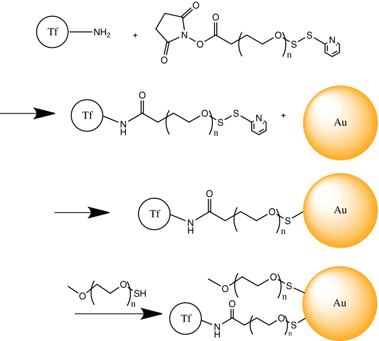
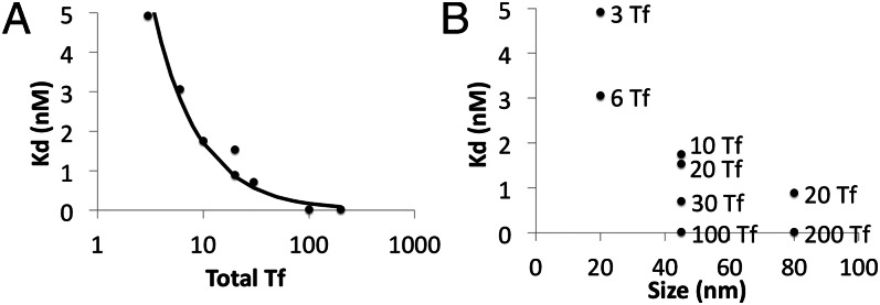
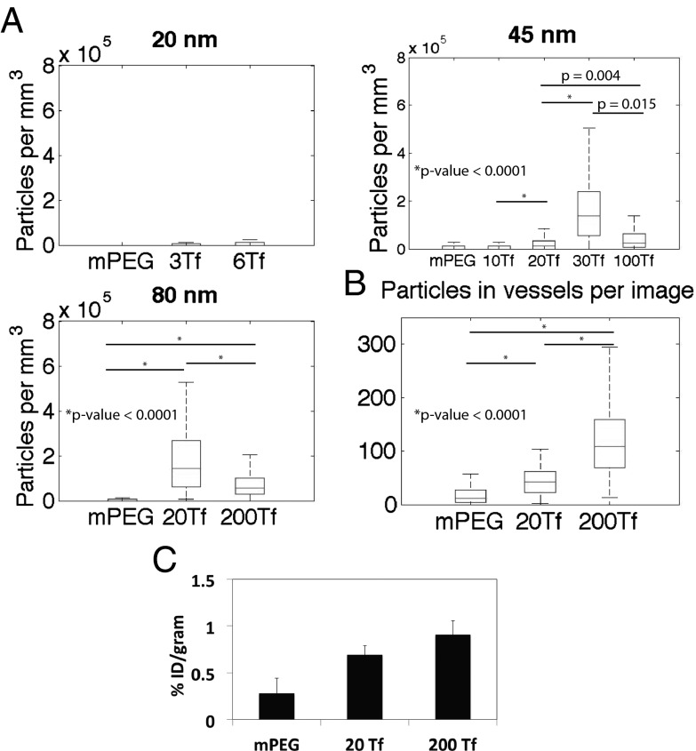
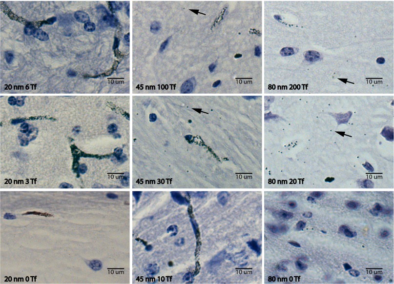
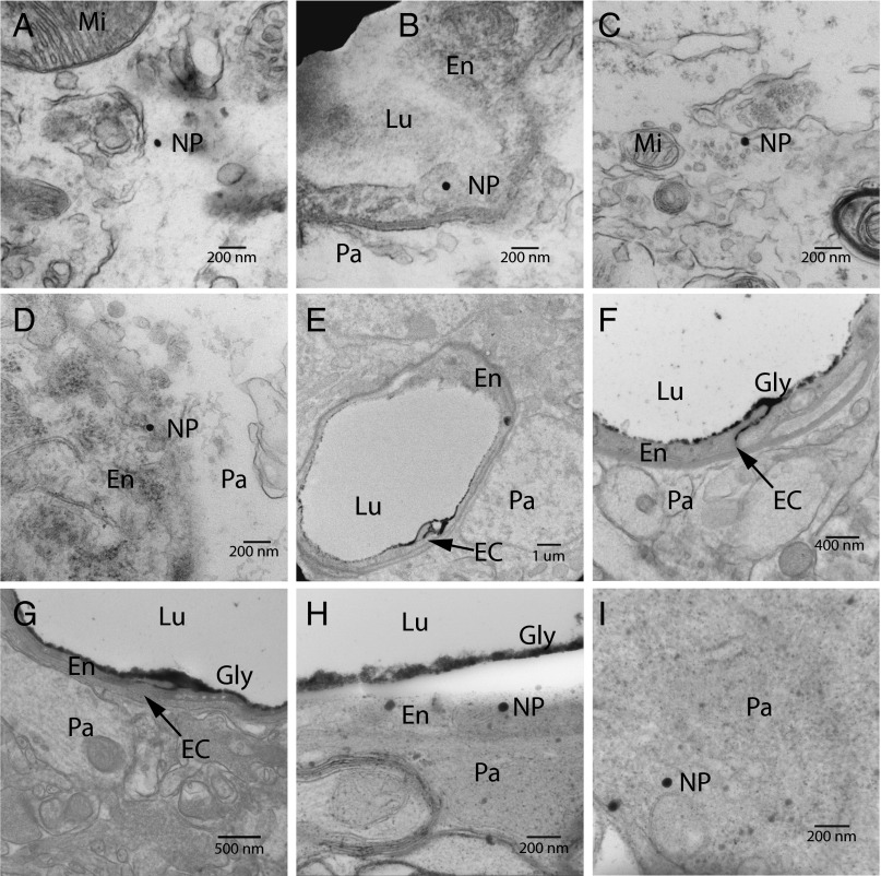
Receptor-mediated transcytosis across the blood-brain barrier (BBB) may be a useful way to transport therapeutics into the brain. Here we report that transferrin (Tf)-containing gold nanoparticles can reach the brain parenchyma from systemic administration in mice through a receptor-mediated transcytosis pathway. This transport is aided by tuning the nanoparticle avidity to Tf receptor (TfR), which is correlated with nanoparticle size and total amount of Tf decorating the nanoparticle surface. Nanoparticles of both 45 nm and 80 nm diameter reach the brain parenchyma, and their accumulation there (visualized by silver enhancement light microscopy in combination with transmission electron microscopy imaging) is observed to be dependent on Tf content (avidity); nanoparticles with large amounts of Tf remain strongly attached to brain endothelial cells, whereas those with less Tf are capable of both interacting with TfR on the luminal side of the BBB and detaching from TfR on the brain side of the BBB. The requirement of proper avidity for nanoparticles to reach the brain parenchyma is consistent with recent behavior observed with transcytosing antibodies that bind to TfR.


Conflict of interest statement

The authors declare no conflict of interest.

Figures

Fig. 1.
Fig. 1.
Representation of the targeted nanoparticle assembly process (n ∼120; PEG molecular mass, 5,000 Da).
Fig. 2.
Fig. 2.
Binding data of gold nanoparticle formulations on Neuro2A cells. (A) Effects of nanoparticle Tf content on Kd of the nanoparticle to Neuro2A TfRs. (B) Effects of nanoparticle size and Tf content on Kd of the nanoparticle to Neuro2A TfRs.
Fig. 3.
Fig. 3.
Quantitative studies of gold nanoparticles in the brain. (A) Quantitation of the nanoparticles observed in the brain parenchyma by silver enhancement after tail vein injection (n = 3 for 45-nm and 80-nm formulations; n = 1 for 20-nm formulations). (B) Quantitation of 80-nm formulations in the blood vessels by silver enhancement. P values of A and B were calculated from nonnormal distributions using the Mann–Whitney U test (Wilcoxon rank-sum test) of compiled data from all mice investigated in each nanoparticle formulation. (C) ICP-MS data of bulk brain gold content from the 80-nm formulations. mPEG: n = 2; 20 Tf and 200 Tf: n = 3. Error bars indicate SE.
Fig. 4.
Fig. 4.
Sample images from hematoxylin-stained and silver-enhanced brain sections. Shown are images from a range of nanoparticle formulations injected systemically with brains resected and processed 8 h later. Black arrows accentuate clearly visible nanoparticles. (Left) 20-nm nanoparticles with 6 Tf, 3 Tf, and 0 Tf (from top to bottom). (Center) 45-nm nanoparticles with 100 Tf, 30 Tf, and 10 Tf. (Right) 80-nm nanoparticles with 200 Tf, 20 Tf, and 0 Tf.
Fig. 5.
Fig. 5.
TEM images of gold nanoparticles in the brain. Mi, mitochondria; Lu, lumen; En, endothelial cell; Pa, parenchyma; EC, endothelial cleft; NP, nanoparticle; Gly, glycocalyx. (A) 80- nm, 20-Tf nanoparticle in the parenchyma. (B) 80-nm, 20-Tf nanoparticle inside a vesicle of a BBB endothelial cell. (C) 80-nm, 200-Tf nanoparticles in the parenchyma. (D) 80-nm, 200-Tf nanoparticle near the basal surface of an endothelial cell. (E) Perfusion fixation with lanthanum nitrate showing lanthanum penetrating the interendothelial cleft with no subendothelial staining. (F and G) Injection of 80-nm particles, followed by perfusion fixation at 8 h postinjection. Neither the 20-Tf/Au formulation (F) nor the 200-Tf/Au formulation (G) degrades the BBB tight junctions to lanthanum nitrate; the same interendothelial cleft penetration with no subendothelial staining is seen. (H) 80-nm, 200-Tf particle inside and near the apical surface of the endothelial cell. (Note that the lanthanum nitrate-stained glycocalyx separated from the cell surface owing to the electron beam during imaging.) (I) 80-nm, 200-Tf particle found within the brain parenchyma after perfusion fixation.

References

    1. Neuwelt E, et al. Strategies to advance translational research into brain barriers. Lancet Neurol. 2008;7(1):84–96. - PubMed
    1. Yu YJ, et al. Boosting brain uptake of a therapeutic antibody by reducing its affinity for a transcytosis target. Sci Transl Med. 2011;3(84):84ra44. - PubMed
    1. Friden PM, Olson TS, Obar R, Walus LR, Putney SD. Characterization, receptor mapping and blood-brain barrier transcytosis of antibodies to the human transferrin receptor. J Pharmacol Exp Ther. 1996;278(3):1491–1498. - PubMed
    1. Davis ME, Chen ZG, Shin DM. Nanoparticle therapeutics: An emerging treatment modality for cancer. Nat Rev Drug Discov. 2008;7(9):771–782. - PubMed
    1. Kamaly N, Xiao Z, Valencia PM, Radovic-Moreno AF, Farokhzad OC. Targeted polymeric therapeutic nanoparticles: Design, development and clinical translation. Chem Soc Rev. 2012;41(7):2971–3010. - PMC - PubMed

Publication types