. 2013 Jun 4;110(23):9451-6.
doi: 10.1073/pnas.1219997110. Epub 2013 May 6.

Endothelial TLR4 activation impairs intestinal microcirculatory perfusion in necrotizing enterocolitis via eNOS-NO-nitrite signaling

Affiliations

Endothelial TLR4 activation impairs intestinal microcirculatory perfusion in necrotizing enterocolitis via eNOS-NO-nitrite signaling

Ibrahim Yazji et al. Proc Natl Acad Sci U S A. .

Abstract

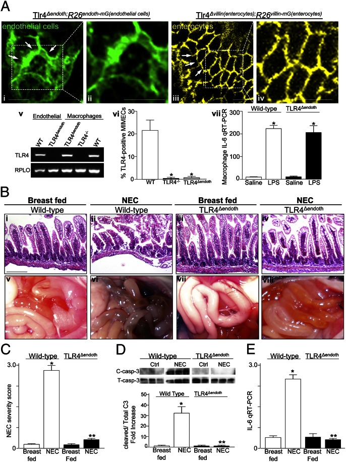
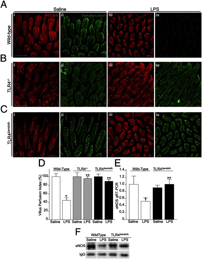
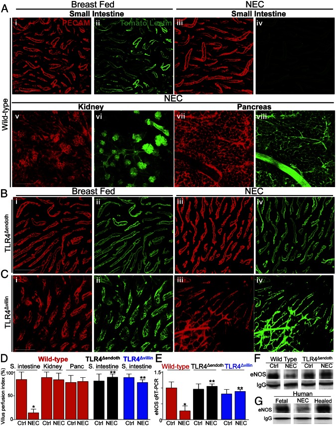
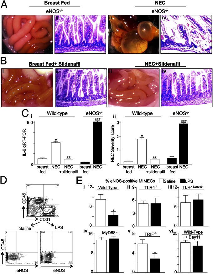
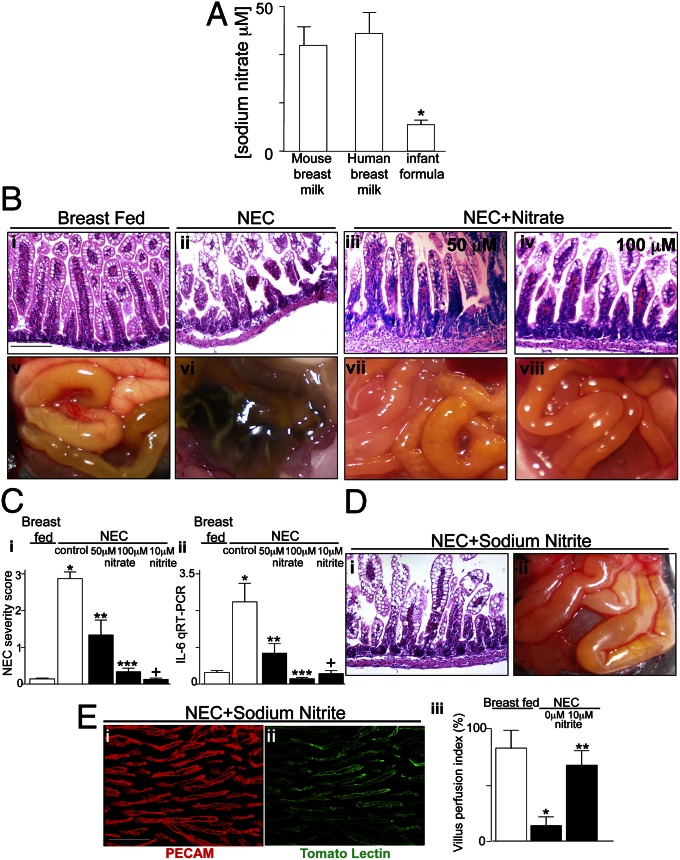
Necrotizing enterocolitis (NEC) is a devastating disease of premature infants characterized by severe intestinal necrosis and for which breast milk represents the most effective protective strategy. Previous studies have revealed a critical role for the lipopolysaccharide receptor toll-like receptor 4 (TLR4) in NEC development through its induction of mucosal injury, yet the reasons for which intestinal ischemia in NEC occurs in the first place remain unknown. We hypothesize that TLR4 signaling within the endothelium plays an essential role in NEC development by regulating perfusion to the small intestine via the vasodilatory molecule endothelial nitric oxide synthase (eNOS). Using a unique mouse system in which we selectively deleted TLR4 from the endothelium, we now show that endothelial TLR4 activation is required for NEC development and that endothelial TLR4 activation impairs intestinal perfusion without effects on other organs and reduces eNOS expression via activation of myeloid differentiation primary response gene 88. NEC severity was significantly increased in eNOS(-/-) mice and decreased upon administration of the phosphodiesterase inhibitor sildenafil, which augments eNOS function. Strikingly, compared with formula, human and mouse breast milk were enriched in sodium nitrate--a precursor for enteral generation of nitrite and nitric oxide--and repletion of formula with sodium nitrate/nitrite restored intestinal perfusion, reversed the deleterious effects of endothelial TLR4 signaling, and reduced NEC severity. These data identify that endothelial TLR4 critically regulates intestinal perfusion leading to NEC and reveal that the protective properties of breast milk involve enhanced intestinal microcirculatory integrity via augmentation of nitrate-nitrite-NO signaling.

Keywords: infant formula; neonatal inflammation; neonatal nutrition; prematurity; sepsis.


Conflict of interest statement

Conflict of interest statement: M.T.G. holds a patent for the use of nitrite salts in cardiovascular diseases and consults with Aires Pharmaceuticals.

Figures

Fig. 1.
Fig. 1.
Generation of mice lacking TLR4 within the intestinal endothelium and demonstration that TLR4 activation in the endothelium is required in the pathogenesis of NEC. (A, iiv) Representative confocal micrographs showing expression of GFP in the ilea of Tlr4Δendoth;R26endoth-mG(endothelial cells) (i and ii) and Tlr4Δvillin(enterocytes);R26villin-mG(enterocytes) (iii and iv) mice. Green expression reveals endothelial cells (arrows, i and ii), and yellow indicates enterocytes (arrows, iii and iv) in which TLR4 has been deleted according to the breeding strategy described in Materials and Methods. Shown in ii and iv are magnifications corresponding to the regions outlined in i and iii, respectively. (Scale bars: 100 μm.) (v) RT-PCR showing expression of TLR4 in endothelial cells (MIMECs) and peritoneal macrophages from the indicated strain. (vi) Percent of MIMECs from the indicated strain that express TLR4 as assessed by flow cytometry. *P < 0.05 vs. wild-type (WT). (vii) qRT-PCR expression of IL-6 in peritoneal macrophages from the indicated strain after treatment with saline or LPS. *P < 0.05 vs. saline. (B) Representative histomicrographs of the terminal ileum (iiv) and photographs (vviii) of the intestines after fresh autopsy of newborn mice that were either breast-fed (i, iii, v, and vii) or induced to develop NEC (ii, iv, vi, and viii). (C) NEC severity score (mean ± SEM) in the strain and group indicated. (D) Measurement of apoptosis as the ratio of expression of cleaved caspase-3 to total caspase-3 by SDS/PAGE. (E) Expression of IL-6 by qRT-PCR (mean ± SEM) in wild-type or TLR4Δendoth mice that were either breast-fed or subjected to experimental NEC as indicated. *P < 0.05 vs. wild-type breast-fed controls; **P < 0.05 NEC in TLR4δendoth vs. wild-type mice. Results are representative of three separate experiments.
Fig. 2.
Fig. 2.
TLR4 activation in the endothelium leads to reduced perfusion of the intestinal microcirculation and reduced expression of eNOS. (AC) Representative confocal micrographs from whole-mount sections of terminal ileum from wild-type (A), TLR4−/− (B), or TLR4Δendoth mice (C) that were either treated with saline (i and ii) or LPS (iii and iv) 6 h before intracardiac injection with the fluorescent tracer tomato lectin. Whole mounts were immunostained with antibodies to PECAM-1 to assess the microvasculature of the intestinal mucosa (red images); the corresponding blood flow (Lucifer yellow) appears in green. (D) Villus perfusion index (VPI) as described in Materials and Methods (mean ± SEM). *P < 0.05 LPS vs. saline; **P < 0.005 LPS treated wild-type vs. LPS-treated TLR4−/− or TLR4Δendoth mice. (E) qRT-PCR (mean ± SEM) showing the expression of eNOS in wild-type or TLR4Δendoth mice treated with saline or LPS as indicated. *P < 0.05 LPS vs. saline; **P < 0.005 LPS-treated wild-type vs. TLR4Δendoth mice. (F) SDS/PAGE for eNOS expression in immunoprecipitates that had been pulled down with antibodies to eNOS from the terminal ileum of wild-type or TLR4Δendoth mice that were treated with saline or LPS; IgG is shown as a loading control. (Scale bars: 100 μm.)
Fig. 3.
Fig. 3.
TLR4 signaling in the endothelium leads to impaired perfusion of the intestinal microcirculation in the pathogenesis of NEC and reduced expression of eNOS. (AC) Representative confocal micrographs from whole mount sections of terminal ileum (A, iiv; B; and C), kidney (A, v and vi), or pancreas (A, vii and viii), from wild-type (A), TLR4Δendoth (B), or TLR4Δvillin mice that were either breast fed (i and ii) or induced to develop NEC (A, iiiviii; B, iii and iv; and C, iii and iv). Mice were then subjected to intracardiac injection with the fluorescent tracer tomato lectin 5 min before they were killed. Whole mounts were immunostained with antibodies to PECAM-1 to assess the microvasculature of the indicated organ(red images); the corresponding blood flow (Lucifer yellow) appears in green. (D) VPI (mean ± SEM) as described in Materials and Methods. *P < 0.05 NEC vs. control; **P < 0.005 NEC wild-type vs. NEC TLR4Δendoth or TLR4Δvillin mice. (E) qRT-PCR (mean ± SEM) showing the expression of eNOS in wild-type, TLR4Δendoth, or TLR4Δvillin mice that were either breast-fed controls or induced to develop NEC as indicated. *P < 0.05 NEC vs. control; **P < 0.005 NEC in wild-type vs. TLR4Δendoth or TLR4Δvillin mice. (F and G) SDS/PAGE of immunoprecipitates of eNOS that had first been pulled down with antibodies to eNOS from intestinal MIMECs obtained from either the terminal ileum of wild-type or TLR4Δendoth mice that were either breast-fed controls or subjected to experimental NEC (F) or from the resected ileum from a fetus, from an infant with NEC, and a neonate 6 wk after the resolution of NEC at the time of stoma reversal (G). IgG is shown as a loading control. Results are representative of three separate experiments. (Scale bars: 100 μm.)
Fig. 4.
Fig. 4.
The severity of experimental NEC is increased in eNOS−/− mice and reduced after administration of sildenafil, and TLR4 reduces the expression of eNOS in isolated MIMECs in a MyD88-dependent manner. (A and B) Photographs of the intestines and histomicrographs obtained from eNOS−/− (A) or wild-type mice that had been administered sildenafil as described in Materials and Methods (B) that were either breast fed (i and ii) or induced to develop NEC (iii and iv). (C) qRT-PCR showing expression of IL-6 within the intestinal mucosa (i) or NEC severity score (ii) in wild-type or eNOS−/− mice under the condition indicated. *P < 0.05 wild-type NEC vs. breast fed; **P < 0.05 NEC plus sildenafil vs. NEC; ***NEC in eNOS−/− vs. NEC in wild-type mice. Results are representative of three separate experiments with at least 10 mice per experiment. (D and E) Mouse MIMECs were isolated from the ileum of mice based upon their staining for CD31+ CD45−. Mice had been treated 6 h prior with saline or LPS and were wild type, TLR4−/−, TLR4Δendoth, MyD88−/−, or TRIF−/− or had been pretreated with the NF-κB inhibitor BAY 11 as indicated. (D) Representative plot of sorted MIMECs in the presence of saline or LPS as shown. (E) Percent eNOS-positive MIMECs from the indicated strain treated with saline or LPS as indicated. *P < 0.05. Results are representative of three separate experiments. All values are mean ± SEM.
Fig. 5.
Fig. 5.
Sodium nitrate is enriched in breast milk and reduces the severity of experimental NEC. (A) Concentration (mean ± SEM) of sodium nitrate in mouse and human breast milk and the infant formula. (B) Representative histomicrographs (iiv) of the terminal ileum and photographs of the intestines of newborn mice that were breast fed (i and v), induced to develop NEC (ii and vi), or induced to develop NEC after daily oral gavage with sodium nitrate at the doses indicated (iii, iv, vii, and viii). (C) NEC severity score (i) and qRT-PCR showing the expression of IL-6 in the intestinal mucosa (ii) of the indicated group. *P < 0.05, breast fed vs. NEC-control; **P < 0.001 NEC vs. NEC plus sodium nitrate (50 μM); ***P < 0.001 NEC vs. NEC plus sodium nitrate (100 μM); +P < 0.05 NEC vs. NEC plus sodium nitrite (10 μM). (D and E) Representative histomicrograph (D, i) and representative photograph of intestines at time of death (D, ii) of mice induced to develop NEC with daily administration of sodium nitrite (10 μM); whole-mount confocal images of terminal ileum of mice showing expression of PECAM-1 (E, i, red) and distribution of the fluorescent tracer tomato lectin (E, ii, green) to reveal intestinal perfusion quantified through the VPI (E, iii) as in Fig. 2. *P < 0.05 vs. breast fed; **P < 0.05 vs. NEC-0 μM nitrite. (Scale bars: 100 μm.)

References

    1. Neu J, Walker WA. Necrotizing enterocolitis. N Engl J Med. 2011;364(3):255–264. - PMC - PubMed
    1. Dominguez KM, Moss RL. Necrotizing enterocolitis. Clin Perinatol. 2012;39(2):387–401. - PubMed
    1. Gephart SM, McGrath JM, Effken JA, Halpern MD. Necrotizing enterocolitis risk: State of the science. Adv Neonatal Care. 2012;12(2):77–87. - PMC - PubMed
    1. Jilling T, et al. The roles of bacteria and TLR4 in rat and murine models of necrotizing enterocolitis. J Immunol. 2006;177(5):3273–3282. - PMC - PubMed
    1. Leaphart CL, et al. A critical role for TLR4 in the pathogenesis of necrotizing enterocolitis by modulating intestinal injury and repair. J Immunol. 2007;179(7):4808–4820. - PubMed

Publication types

MeSH terms