. 2013 May 28;110(22):9112-7.
doi: 10.1073/pnas.1217508110. Epub 2013 May 6.

Impaired learning resulting from respiratory syncytial virus infection

Affiliations

Impaired learning resulting from respiratory syncytial virus infection

Janyra A Espinoza et al. Proc Natl Acad Sci U S A. .

Abstract

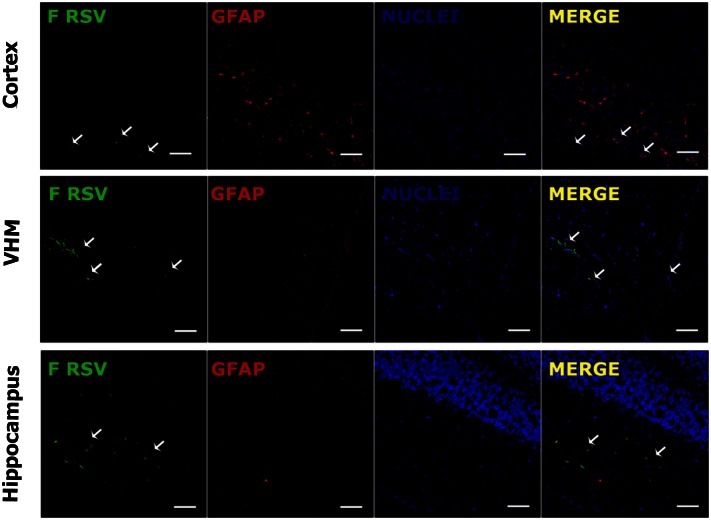
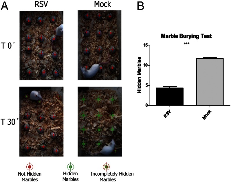
Respiratory syncytial virus (RSV) is the major cause of respiratory illness in infants worldwide. Neurologic alterations, such as seizures and ataxia, have been associated with RSV infection. We demonstrate the presence of RSV proteins and RNA in zones of the brain--such as the hippocampus, ventromedial hypothalamic nucleus, and brainstem--of infected mice. One month after disease resolution, rodents showed behavioral and cognitive impairment in marble burying (MB) and Morris water maze (MWM) tests. Our data indicate that the learning impairment caused by RSV is a result of a deficient induction of long-term potentiation in the hippocampus of infected animals. In addition, immunization with recombinant bacillus Calmette-Guérin (BCG) expressing RSV nucleoprotein prevented behavioral disorders, corroborating the specific effect of RSV infection over the central nervous system. Our findings provide evidence that RSV can spread from the airways to the central nervous system and cause functional alterations to the brain, both of which can be prevented by proper immunization against RSV.

Keywords: LTD; LTP; behavior; cognition; inflammation.


Conflict of interest statement

The authors declare no conflict of interest.

Figures

Fig. 1.
Fig. 1.
Detection of RSV transcripts in the brain and blood of infected BALB/c mice. Data in the graph show the infection kinetics of RSV in the lung, blood, and CNS of infected mice at different times postinfection. Copy numbers for N transcripts normalized by 5,000 copies of a β-actin as housekeeping gene are shown. The peak of viral charge in brain was observed at day 3 postinfection, and the virus was able to spread to the brainstem in the following days.
Fig. 2.
Fig. 2.
RSV proteins were detected in lung and brain of infected mice. Confocal photomicrograph sections of lung and brain tissue. Viral proteins were observed by immunofluorescence (green fluorescence) in a confocal microscope using an anti-F-RSV or anti–N-RSV antibody. The nuclei were stained with thiazole orange-oligonucleotide conjugates (TOPRO-3) (blue). (A) Images of infected lung tissues 3 d after RSV infection. (Upper) Staining for F protein. (Lower) Staining for N protein. (B) Pictures of infected brain cortex 3 d after RSV infection. (Upper) Staining for F protein. (Lower) Staining for RSV Nucleoprotein. (C) Images of infected olfactory bulb tissues 1 d after RSV infection. (Upper) Staining for the F protein. (Lower) Staining for the nucleoprotein. (D) Images of infected choroid plexus after 3 d of RSV infection. (Upper) Staining for F protein. (Lower) Staining for N protein. (Scale bar, 150 µm.) (E) Quantification of total F-RSV channel fluorescence intensity per 40× field by pixel analysis of immunofluorescence for whole-brain and lung. Mouse α-F RSV, primary antibody; goat α-mouse, secondary antibody. More than 10 fields were analyzed per treatment.
Fig. 3.
Fig. 3.
Detection of F-RSV protein in specific brain regions. Confocal analyses were performed using tissue sections obtained from RSV-infected mice. Tissue was stained for nuclei (TOPRO-3; blue), GFAP (astrocyte marker; red), and F protein (RSV; green). Staining of brain tissue sections obtained at day 7 postinfection shows presence of viral protein adjacent to astrocytes zones. The white arrows show the clusters of RSV protein expression in the cortex, the VMH, and the hippocampus. (Scale bar, 150 µm.)
Fig. 4.
Fig. 4.
Treatment with CD49d decreased CNS dissemination of RSV. BALB/c mice were infected intranasally with RSV and received a single dose of 70 µg anti-CD49d blocking antibody intravenously. Three days postinfection, mice were killed and the tissues prepared for RNA extraction. Data in the graph show the number of copies of RSV nucleoprotein transcripts in the brains of controls animals and animals treated with anti-CD49d antibody. Statistical analysis, one-way ANOVA; **P < 0.05, n = 5 per treatment.
Fig. 5.
Fig. 5.
RSV infection caused behavioral alterations. (A) Six-week-old BALB/c mice were challenged intranasally with RSV and 30 d later were subjected to MB tests to measure their cognitive capacity. The panels show representative images of MB tests for RSV-infected and control mice. (Upper) Initial position of marbles for RSV-infected and mock mice. (Lower) Position of the marbles 30 min after beginning the experiment. (B) Data in the graph show the quantification of hidden marbles for mock and RSV-infected mice. Statistical analysis, Student t test; ***P < 0.0001, n = 10 per treatment.
Fig. 6.
Fig. 6.
RSV infection impaired learning and memory. Nine 3-wk-old Sprague-Dawley rats were i.n. challenged with RSV or vehicle; 30 d after the infections, rats were subjected to MWM tests to evaluate their cognitive capacity. (A) Data in the graph show the mean escape latency recorded from MWM test. Infected (●) and control (■) rats were subjected for 5 d to a MWM test. The day of each trial is indicated on the X-axis, and the mean ± SE escape latency is indicated on the Y-axis. The mean escape latency is the average of four trials performed on the day of the same experimental group. Statistical analysis (Mann–Whitney U test) showed significant differences on days 1, 2, and 3 with P ≤ 0.05 (n = 9). (B) Mean escape latency from four trials for infected and control groups. Statistical analysis (unpaired t test) showed no significant differences between the control and infected animals.
Fig. 7.
Fig. 7.
Impaired synaptic plasticity resulting from RSV infection. (A) Rat hippocampal slice preparation and typical electrode placements for studying synaptic plasticity at SC synapses onto CA1, showing the regions CA1, CA3, and the DG. DG, dentate gyrus; MF, mossy fiber; SC, Schaffer collateral; Stim, stimulating electrode; Rec, recording electrode. (B) Illustration of LTP and LTD in the CA1 region of the hippocampus. (C) LTP in the stratum radiatum of the CA1 region of hippocampus. Synaptic strength, defined as the initial slope of the fEPSP (normalized to baseline), is plotted as a function of time. Data in the graph show the LTP induction elicited by high-frequency tetanic stimulation (100 Hz stimulation for 1 s) for RSV-infected (■) or mock control (formula image) rats. (D) LTD in the CA1 region of the hippocampus. Data in graph show the LTD elicited by low-frequency stimulation (5 Hz stimulation for 3 min given twice with a 3-min interval) for RSV-infected (■) or mock control (formula image) rats. (Scale bar, 0.5 mV, 10 ms.) Statistical analysis, unpaired t test; ***P < 0.0001, n = 7 slides, 4 rats for each group.
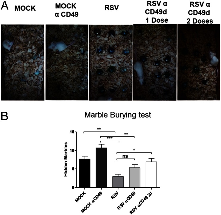
Fig. 8.
Fig. 8.
CD49d blockade prevents the cognition impairment caused by RSV infection. Six-week-old BALB/c mice were treated with one or two doses of 70 μg anti- CD49d and i.n. inoculated with RSV or mock control. (A) Representative images of MB tests for each treatment. Mice receiving two doses of anti-CD49d showed no altered performance in MB tests after RSV infection in relation to control mice. (B) Data in the graph show the quantification of hidden marbles for all experimental groups. Statistical analysis, unpaired t test; ***P < 0.0001, n = 5.
Fig. 9.
Fig. 9.
Immunization with rBCG-N prevents the cognition impairment caused by RSV infection. Six-wk-old BALB/c mice were immunized with either rBCG-N or BCG-WT and intranasally inoculated with RSV or mock control. (A) Data in the graph show the viral load in lungs (gray bars) and brains (white bars) for unimmunized, rBCG-N-, and BCG-WT-immunized and uninfected mice. (B) Quantification of MB test for rBCG-N-, BCG-WT-immunized, and uninfected mice. Statistical analysis unpaired t test; ***P < 0.0001, n = 10.

References

    1. Bueno SM, et al. Protective T cell immunity against respiratory syncytial virus is efficiently induced by recombinant BCG. Proc Natl Acad Sci USA. 2008;105(52):20822–20827. - PMC - PubMed
    1. Bueno SM, et al. Host immunity during RSV pathogenesis. Int Immunopharmacol. 2008;8(10):1320–1329. - PubMed
    1. González PA, Bueno SM, Riedel CA, Kalergis AM. Impairment of T cell immunity by the respiratory syncytial virus: Targeting virulence mechanisms for therapy and prophylaxis. Curr Med Chem. 2009;16(34):4609–4625. - PubMed
    1. González PA, et al. Respiratory syncytial virus impairs T cell activation by preventing synapse assembly with dendritic cells. Proc Natl Acad Sci USA. 2008;105(39):14999–15004. - PMC - PubMed
    1. Sweetman LL, Ng YT, Butler IJ, Bodensteiner JB. Neurologic complications associated with respiratory syncytial virus. Pediatr Neurol. 2005;32(5):307–310. - PubMed

Publication types

MeSH terms

LinkOut - more resources