. 2013 May 1;8(5):e62869.
doi: 10.1371/journal.pone.0062869. Print 2013.

LPLUNC1 inhibits nasopharyngeal carcinoma cell growth via down-regulation of the MAP kinase and cyclin D1/E2F pathways

Affiliations

LPLUNC1 inhibits nasopharyngeal carcinoma cell growth via down-regulation of the MAP kinase and cyclin D1/E2F pathways

Yixin Yang et al. PLoS One. .

Erratum in

  • PLoS One. 2013;8(8). doi:10.1371/annotation/e120c2f3-efb3-4bac-931d-b53751136785

Abstract

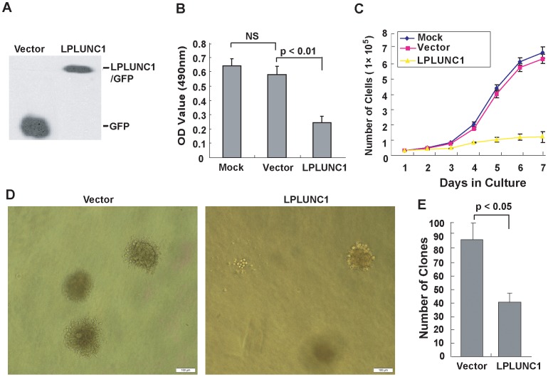
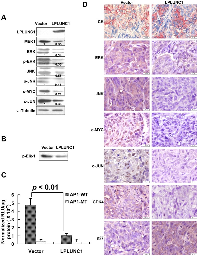
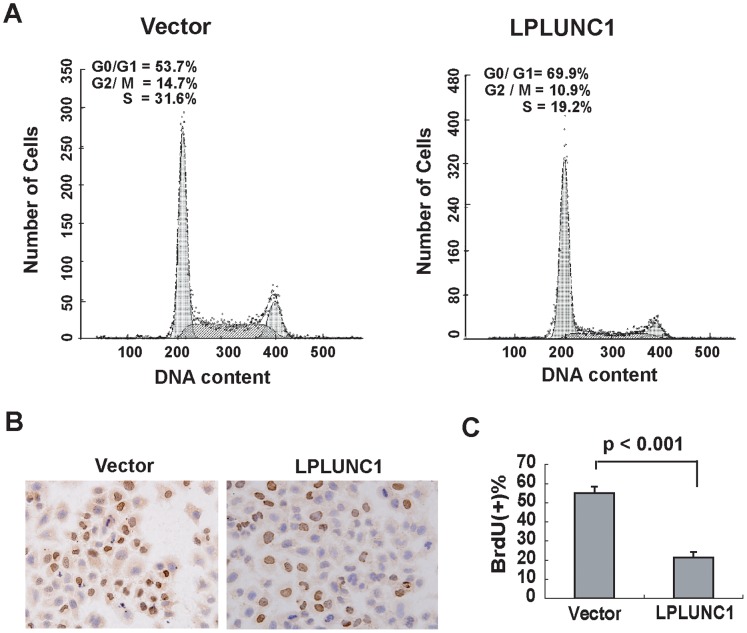
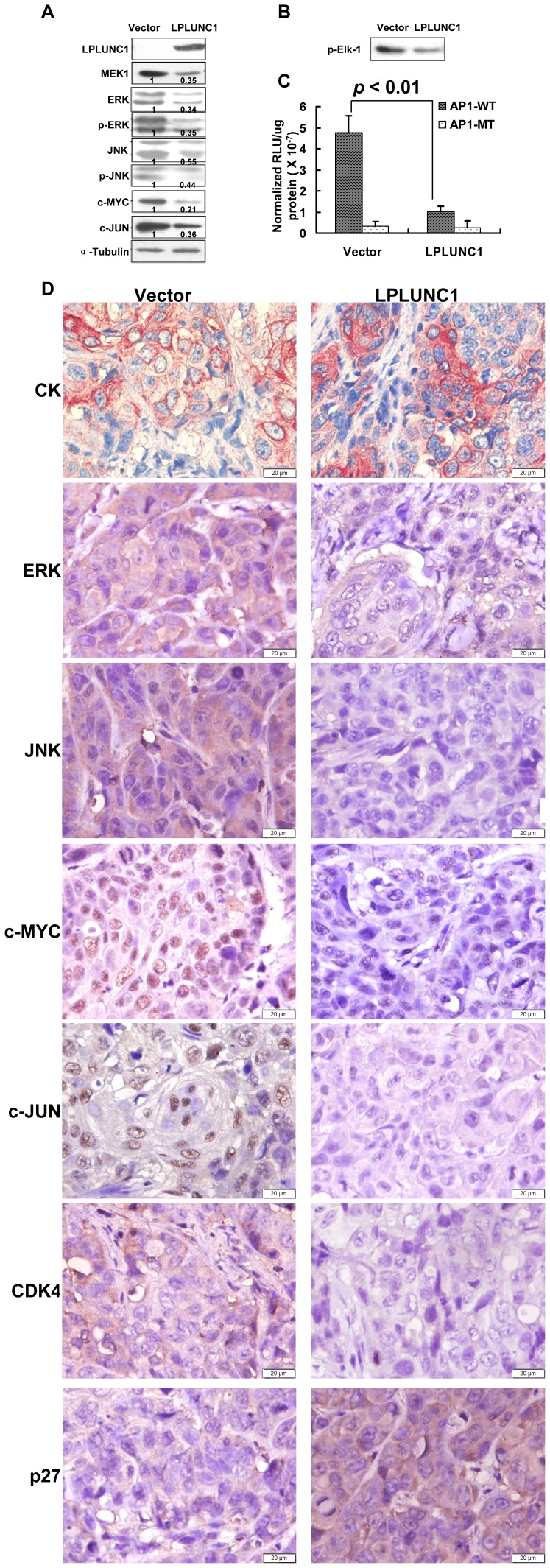
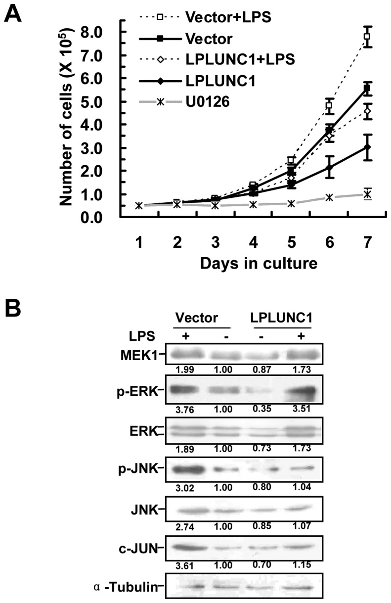
Long-palate, lung and nasal epithelium clone 1 (LPLUNC1) gene expression is relatively tissue specific. It is highly expressed in nontumor nasopharyngeal epithelial tissues, but its expression is reduced in nasopharyngeal carcinoma (NPC), indicating that LPLUNC1 may be associated with the tumorigenesis of NPC. To study the effects of LPLUNC1 on NPC tumorigenesis, a full-length LPLUNC1 expression plasmid was stably transfected into the NPC cell line, 5-8F. Our data indicated that LPLUNC1 inhibited NPC cell proliferation in vitro and tumor formation in vivo. LPLUNC1 also delayed cell cycle progression from G1 to S phase and inhibited the expression of cyclin D1, cyclin-dependent kinase 4 (CDK4) and phosphorylated Rb. To further investigate the molecular mechanisms underlying the suppressive effects of LPLUNC1 on NPC tumorigenesis, cDNA microarray was performed. These studies revealed that LPLUNC1 inhibited the expression of certain mitogen-activated protein (MAP) kinases (MAPK) kinases and cell cycle-related molecules. Western blotting confirmed that the expression of MEK1, phosphorylated ERK1/2, phosphorylated JNK1/2, c-Myc and c-Jun were inhibited by LPLUNC1. Furthermore, the transcriptional activity of AP-1 was down-regulated by LPLUNC1, suggesting that the MAPK signaling pathway is regulated by LPLUNC1. Taken together, the present study indicates that LPLUNC1 delays NPC cell growth by inhibiting the MAPK and cyclin D1/E2F pathways and suggests that LPLUNC1 may represent a promising candidate tumor suppressor gene associated with NPC.


Conflict of interest statement

Competing Interests: The authors have declared that no competing interests exist.

Figures

Figure 1
Figure 1. The expression of LPLUNC mRNA in nontumor nasopharyngeal epithelial tissues and NPC biopsies.
LPLUNC1 was highly expressed in nontumor nasopharyngeal epithelial tissues but was absent or expressed at low levels in NPC biopsies. N, nontumor nasopharyngeal epithelial tissues; T, NPC biopsies. The housekeeping gene, GAPDH, was used as an internal control. A. Expression of LPLUNC mRNA in nasopharyngeal epithelial and NPC samples was detected by Northern blotting. B. Expression of LPLUNC mRNA in nasopharyngeal epithelial and NPC samples was measured by RT-PCR; p<0.001. C. Expression of LPLUNC1 protein in nasopharyngeal epithelial and NPC samples was measured by immunohistochemistry; p<0.001.
Figure 2
Figure 2
LPLUNC1 inhibits 5-8F NPC cell growth and proliferation. A. Western blot analysis of LPLUNC1 expression in 5-8F/vector and 5-8F/LPLUNC1 cells using the anti-GFP primary antibody revealed that 5-8F/LPLUNC1 cells expressed the GFP tagged LPLUNC1 protein, whereas 5-8F/vector cells expressed GFP protein alone. B. MTT assay of 5-8F/LPLUNC1 cells. A total of 1×10⁴ cells were seeded into 96-well, flat-bottom plates and cultured for 72 h, and 10 µl of MTT (5 mg/ml) was added. LPLUNC1 expression significantly inhibited cell proliferation; p<0.01. C. Growth curve of the LPLUNC1 stably transfected 5-8F cells. A total of 1×10⁴ cells were seeded into 24-well plates, and the number of cells were counted with a hemocytometer every 24 h. The expression of LPLUNC1 markedly inhibited cell growth; p<0.01. D. Soft agar colony formation assay of 5-8F/LPLUNC1 cells. Colonies were manually counted and imaged using microscopy and photographed two weeks later (magnification = 100×, Bar = 100 µm). E. The number of colonies per plate in the colony formation assay was derived from three independent experiments, with duplicates in each experiment. The data are presented as the mean ± SD; p<0.05. Both the size and number of the colonies formed in soft agar were decreased in 5-8F/LPLUNC1 cells compared with 5-8F/vector cells.
Figure 3
Figure 3. Over-expression of LPLUNC1 inhibited NPC tumor formation in vivo.
A. Tumor formation in nude mice. The tumors that formed following injection of 2×10⁶ cells were much smaller in the 5-8F/LPLUNC1 group than tumors from the 5-8F/vector group after 40 days. B. Average tumor volume. The average volume of tumors in the 5-8F/LPLUNC1 group was significantly decreased compared with the 5-8F/vector group. The data are presented as the mean ± SD; *p<0.01.
Figure 4
Figure 4. Over-expression of LPLUNC1 decreased 5-8F cell proliferation.
A. Flow cytometry analysis showed that LPLUNC1 transfection increased the proportion of cells in G1 phase from 53.7% to 69.9% and decreased the proportion of cells in S phase from 31.6% to 19.2%; p<0.01. B and C. LPLUNC1 transfection decreased the number of cells positive for BrdU staining; p<0.01. Cells were treated with 30 nM BrdU for 16 h. Fixation and immunocytochemistry were conducted to assess BrdU incorporation.
Figure 5
Figure 5. Over-expression of LPLUNC1 inhibited the expression of MAPKs and their downstream molecules in 5-8F cells.
A. Western blotting analysis of MAPKs. Levels of MEK1, ERK, JNK, phosphorylated ERK, phosphorylated JNK, c-MYC and c-JUN were inhibited by LPLUNC1. α-Tubulin served as a loading control. The data shown are representative of at least three separate experiments. Quantification of protein levels by densitometric analyses of band intensities are marked under each band. B. ERK1/2 kinase activity was measured by p44/42 MAP kinase assay kit. Elf-1 phosphorylation was detected using phosphor-Elf-1 antibody by western blotting. The right lane in LPLUNC1-transfected cells resulted in lower ERK1/2 MAP kinase-induced phosphorylation of Elk-1 compared with the left lane in control vector transfected cells reflecting that LPLUNC1 inhibited ERK kinase activity. C. Luciferase assay measuring AP-1 transcriptional activity. The luciferase reporter plasmid containing wild type AP-1 (AP1-WT) responsive sites was co-transfected with pCMV-myc-LPLUNC1 or empty vector plasmid into 5-8F cells. The luciferase reporter plasmid containing a mutant sequence (AP1-MT) was used as a control. LPLUNC1 over-expression significantly inhibited the promoter activity of AP-1. The data are presented as the mean ± SD; p<0.01. D. Immunohistochemical staining of components of the MAPK pathway in xenograft tumor tissues showed that LPLUNC1 decreased ERK, JNK, CDK4, c-MYC and c-JUN protein expression and increased p27 expression. The epithelia cell marker cytokeratin (CK) was used to validate that xenograft tumors were poorly differentiated NPC (magnification = 400×, Bar = 20 µm).
Figure 6
Figure 6. LPLUNC1 attenuates LPS activated MAP kinase pathway.
A. Growth curve of 5-8F cells treated by LPS and LPLUNC1 transfection. LPS promoted NPC cell proliferation, whereas LPLUNC1 hindered LPS stimulated NPC cell proliferation. MEK1/2 inhibitor U0126 was also used to treat 5-8F cells, and the inhibition of the MAP kinase pathway by U0126 strongly decreased NPC cell growth. B. Western blotting analysis of MAPKs in 5-8F cells treated by LPS and LPLUNC1 transfection. The levels of MEK1, ERK, JNK, phosphorylated ERK and JNK, and c-JUN were increased after LPS stimulation. However LPLUNC1 inhibited the MAPK activation stimulated by LPS. Quantification of protein levels by densitometric analyses of band intensities are marked under each band.
Figure 7
Figure 7. LPLUNC1 inhibited the expression of important molecules in the cell cycle pathway.
A. FACS analysis of the relative expression of the cyclin family. The relative expression levels of cyclin D1 and cyclin E1 were decreased in 5-8F/LPLUNC1 cells compared with the control; p<0.01 and p<0.05, respectively. There was no difference in the expression levels of cyclin A and B between 5-8F/LPLUNC1 and 5-8F/vector cells. B. Western blotting analysis of cell cycle molecules showed that the expression levels of CDK4, cyclin D1 and phosphorylated Rb were decreased in 5-8F/LPLUNC1 compared with 5-8F/vector cells. C. and D. Luciferase assay to assess cyclin D1 promoter (C) and E2F transcriptional activity (D). The luciferase reporter plasmids containing cyclin D1 promoter (C) or E2F responsive sites (D, E2F-WT: wild type, E2F-MT: mutated) were co-transfected with pCMV-myc-LPLUNC1 or empty vector plasmid into 5-8F cells. LPLUNC1 overexpression significantly inhibited the promoter activity of cyclin D1 (C) and E2F transcriptional activity (D). The results are representative of 3 independent experiments. The data are presented as the mean ± SD; p<0.01.

References

    1. Hildesheim A, Levine PH (1993) Etiology of nasopharyngeal carcinoma: a review. Epidemiol Rev 15: 466–485. - PubMed
    1. Zeng Z, Huang H, Zhang W, Xiang B, Zhou M, et al. (2011) Nasopharyngeal carcinoma: advances in genomics and molecular genetics. Sci China Life Sci 54: 966–975. - PubMed
    1. Feng BJ, Huang W, Shugart YY, Lee MK, Zhang F, et al. (2002) Genome-wide scan for familial nasopharyngeal carcinoma reveals evidence of linkage to chromosome 4. Nat Genet 31: 395–399. - PubMed
    1. Xiong W, Zeng ZY, Xia JH, Xia K, Shen SR, et al. (2004) A susceptibility locus at chromosome 3p21 linked to familial nasopharyngeal carcinoma. Cancer Res 64: 1972–1974. - PubMed
    1. Deng L, Jing N, Tan G, Zhou M, Zhan F, et al. (1998) A common region of allelic loss on chromosome region 3p25.3–26.3 in nasopharyngeal carcinoma. Genes Chromosomes Cancer 23: 21–25. - PubMed

Publication types

MeSH terms