

Task-free functional MRI in cervical dystonia reveals multi-network changes that partially normalize with botulinum toxin
- PMID: 23650536
- PMCID: PMC3641096
- DOI: 10.1371/journal.pone.0062877
Task-free functional MRI in cervical dystonia reveals multi-network changes that partially normalize with botulinum toxin
Abstract
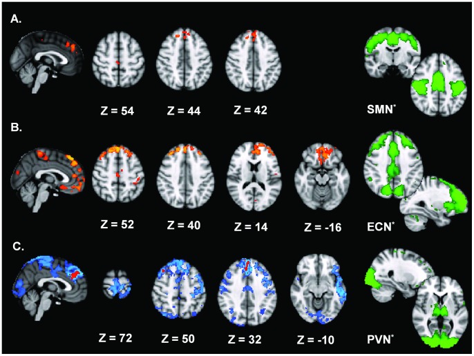
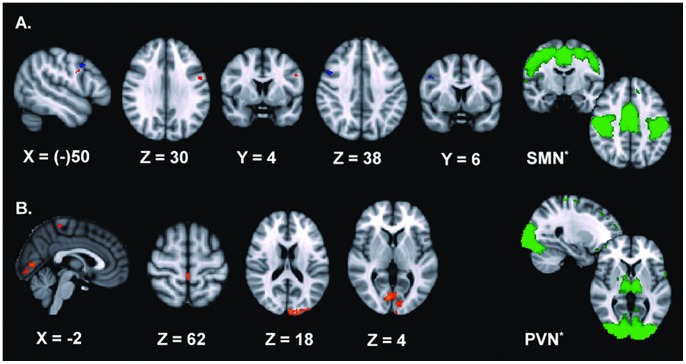
Cervical dystonia is characterized by involuntary, abnormal movements and postures of the head and neck. Current views on its pathophysiology, such as faulty sensorimotor integration and impaired motor planning, are largely based on studies of focal hand dystonia. Using resting state fMRI, we explored whether cervical dystonia patients have altered functional brain connectivity compared to healthy controls, by investigating 10 resting state networks. Scans were repeated immediately before and some weeks after botulinum toxin injections to see whether connectivity abnormalities were restored. We here show that cervical dystonia patients have reduced connectivity in selected regions of the prefrontal cortex, premotor cortex and superior parietal lobule within a distributed network that comprises the premotor cortex, supplementary motor area, primary sensorimotor cortex, and secondary somatosensory cortex (sensorimotor network). With regard to a network originating from the occipital cortex (primary visual network), selected regions in the prefrontal and premotor cortex, superior parietal lobule, and middle temporal gyrus areas have reduced connectivity. In selected regions of the prefrontal, premotor, primary motor and early visual cortex increased connectivity was found within a network that comprises the prefrontal cortex including the anterior cingulate cortex and parietal cortex (executive control network). Botulinum toxin treatment resulted in a partial restoration of connectivity abnormalities in the sensorimotor and primary visual network. These findings demonstrate the involvement of multiple neural networks in cervical dystonia. The reduced connectivity within the sensorimotor and primary visual networks may provide the neural substrate to expect defective motor planning and disturbed spatial cognition. Increased connectivity within the executive control network suggests excessive attentional control and while this may be a primary trait, perhaps contributing to abnormal motor control, this may alternatively serve a compensatory function in order to reduce the consequences of the motor planning defect inflicted by the other network abnormalities.
Conflict of interest statement
Figures
Similar articles
-
Functional activity of the sensorimotor cortex and cerebellum relates to cervical dystonia symptoms.Hum Brain Mapp. 2017 Sep;38(9):4563-4573. doi: 10.1002/hbm.23684. Epub 2017 Jun 8. Hum Brain Mapp. 2017. PMID: 28594097 Free PMC article.
-
Network-specific resting-state connectivity changes in the premotor-parietal axis in writer's cramp.Neuroimage Clin. 2017 Oct 14;17:137-144. doi: 10.1016/j.nicl.2017.10.001. eCollection 2018. Neuroimage Clin. 2017. PMID: 29085775 Free PMC article.
-
Altered functional connectivity in blepharospasm/orofacial dystonia.Brain Behav. 2017 Dec 18;8(1):e00894. doi: 10.1002/brb3.894. eCollection 2018 Jan. Brain Behav. 2017. PMID: 29568690 Free PMC article.
-
Botulinum toxin treatment of axial and cervical dystonia.Disabil Rehabil. 2007 Dec 15;29(23):1769-77. doi: 10.1080/01421590701568262. Disabil Rehabil. 2007. PMID: 18033602 Review.
-
Botulinum toxin for the treatment of cervical dystonia.Expert Opin Pharmacother. 2001 Dec;2(12):1985-94. doi: 10.1517/14656566.2.12.1985. Expert Opin Pharmacother. 2001. PMID: 11825330 Review.
Cited by
-
Current Opinions and Areas of Consensus on the Role of the Cerebellum in Dystonia.Cerebellum. 2017 Apr;16(2):577-594. doi: 10.1007/s12311-016-0825-6. Cerebellum. 2017. PMID: 27734238 Free PMC article.
-
The role of dopamine and dopaminergic pathways in dystonia: insights from neuroimaging.Tremor Other Hyperkinet Mov (N Y). 2015 Jan 29;5:280. doi: 10.7916/D8J101XV. eCollection 2015. Tremor Other Hyperkinet Mov (N Y). 2015. PMID: 25713747 Free PMC article. Review.
-
Hemodynamic responses are abnormal in isolated cervical dystonia.J Neurosci Res. 2020 Apr;98(4):692-703. doi: 10.1002/jnr.24547. Epub 2019 Nov 6. J Neurosci Res. 2020. PMID: 31692015 Free PMC article.
-
Alterations of Interhemispheric Functional Connectivity and Degree Centrality in Cervical Dystonia: A Resting-State fMRI Study.Neural Plast. 2019 Apr 24;2019:7349894. doi: 10.1155/2019/7349894. eCollection 2019. Neural Plast. 2019. PMID: 31178903 Free PMC article.
-
Contemporary clinical neurophysiology applications in dystonia.J Neural Transm (Vienna). 2021 Apr;128(4):509-519. doi: 10.1007/s00702-021-02310-6. Epub 2021 Feb 16. J Neural Transm (Vienna). 2021. PMID: 33591454 Review.
References
-
- Defazio G, Abbruzzese G, Livrea P, Berardelli A (2004) Epidemiology of primary dystonia. Lancet Neurol 3: 673–678. - PubMed
-
- Cakmur R, Donmez B, Uzunel F, Aydin H, Kesken S (2004) Evidence of widespread impairment of motor cortical inhibition in focal dystonia: a transcranial magnetic stimulation study in patients with blepharospasm and cervical dystonia. Adv Neurol 94: 37–44. - PubMed
-
- Hanajima R, Ugawa Y, Terao Y, Sakai K, Furubayashi T, et al. (1998) Cortico-cortical inhibition of the motor cortical area projecting to sternocleidomastoid muscle in normals and patients with spasmodic torticollis or essential tremor. Electroencephalogr Clin Neurophysiol 109: 391–396. - PubMed
-
- Siggelkow S, Kossev A, Moll C, Dauper J, Dengler R, et al. (2002) Impaired sensorimotor integration in cervical dystonia: a study using transcranial magnetic stimulation and muscle vibration. J Clin Neurophysiol 19: 232–239. - PubMed
-
- Naumann M, Magyar-Lehmann S, Reiners K, Erbguth F, Leenders KL (2000) Sensory tricks in cervical dystonia: perceptual dysbalance of parietal cortex modulates frontal motor programming. Ann Neurol 47: 322–328. - PubMed
Publication types
MeSH terms
Substances
LinkOut - more resources
Full Text Sources
Other Literature Sources
Medical
