. 2012 Jul 20:2:187-90.
doi: 10.1016/j.fob.2012.07.003. Print 2012.

CD248 expression on mesenchymal stromal cells is required for post-natal and infection-dependent thymus remodelling and regeneration

Affiliations

CD248 expression on mesenchymal stromal cells is required for post-natal and infection-dependent thymus remodelling and regeneration

Siân Lax et al. FEBS Open Bio. .

Abstract

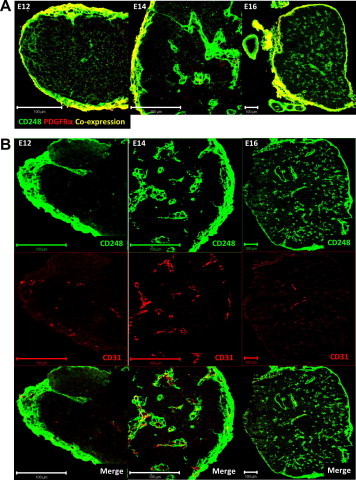
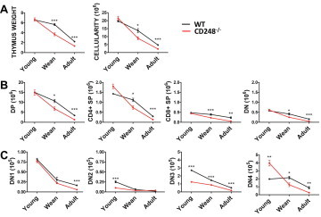
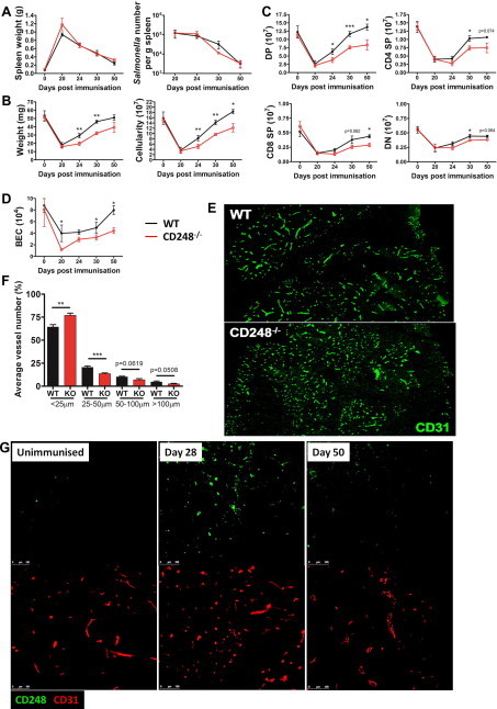
The role of mesenchymal stromal cells (MSCs) in regulating immune responses in the thymus is currently unclear. Here we report the existence and role of a MSC population in the thymus that expresses the pericyte and MSC marker CD248 (endosialin). We show using a CD248-deficient mouse model, that CD248 expression on these cells is required for full post-natal thymus development and regeneration post-Salmonella infection. In CD248(-/-) mice the thymus is hypocellular and regeneration is poorer, with significant loss of all thymocyte populations. This identifies the requirement of CD248 to maintain optimal thymic cellularity post-partum and infection.

Keywords: CD248; Endosialin; MSC; Thymus.


Figures

Fig. 1
Fig. 1
Location of thymic CD248 expressing cells. Frozen sections of E12, 14 and 16 thymuses were co-stained with CD248 (green) and either PDGFRα (A) or CD31 (B) (red). Bar = 100 µm. (For interpretation of the references to colour in this figure legend, the reader is referred to the web version of this article.)
Fig. 2
Fig. 2
CD248 is required for efficient post-natal thymic development. Thymic weight and cellularity are reduced in young (1–2 weeks old), weaning (3–4 weeks old) and adult (9–10 weeks old) CD248−/− mice (A). Flow cytometry revealed a decrease in mature (B) and DN thymocyte populations (C) in CD248−/− mice.
Fig. 3
Fig. 3
CD248 is required for infection-dependent thymic regeneration. Salmonella infection in CD248−/− mice, as assessed by splenomegaly and the number of bacteria per spleen, is similar to that of WT (A). Thymic weight and cellularity are reduced during Salmonella infection-dependent regeneration in CD248−/− mice (B). Significant loss of thymocyte populations was observed in CD248−/− mice (C). Re-vascularisation of regenerating thymuses was dysregulated in CD248−/− mice as assessed by flow cytometry of BEC number (D), and endothelial vessel length in thymuses taken 30 days post-Salmonella infection (E and F). CD248 (green) is upregulated temporally during thymic regeneration (G). (For interpretation of the references to colour in this figure legend, the reader is referred to the web version of this article.)

References

    1. Anderson G., Jenkinson E.J. Lymphostromal interactions in thymic development and function. Nat. Rev. Immunol. 2001;1:31–40. - PubMed
    1. Jenkinson W.E., Rossi S.W., Parnell S.M., Jenkinson E.J., Anderson G. PDGFRα-expressing mesenchyme regulates thymus growth and the availability of intrathymic niches. Blood. 2007;109:954–960. - PubMed
    1. Odaka C. Localization of mesenchymal cells in adult mouse thymus: their abnormal distribution in mice with disorganization of thymic medullary epithelium. J. Histochem. Cytochem. 2009;57:373–382. - PMC - PubMed
    1. Suniara R.K., Jenkinson E.J., Owen J.J. An essential role for the thymic mesenchyme in early T cell development. J. Exp. Med. 2000;191:1051–1056. - PMC - PubMed
    1. Foster K., Sheridan J., Veiga-Fernandas H., Roderick K., Pachnis V., Adams R., Blackburn C., Kioussis D., Coles M. Contribution of neural crest-derived cells in the embryonic and adult thymus. J. Immunol. 2008;180:3183–3189. - PubMed