. 2013 Jun 4;52(22):3939-48.
doi: 10.1021/bi400036z. Epub 2013 May 20.

SV40 late protein VP4 forms toroidal pores to disrupt membranes for viral release

Affiliations

SV40 late protein VP4 forms toroidal pores to disrupt membranes for viral release

Smita Raghava et al. Biochemistry. .

Abstract

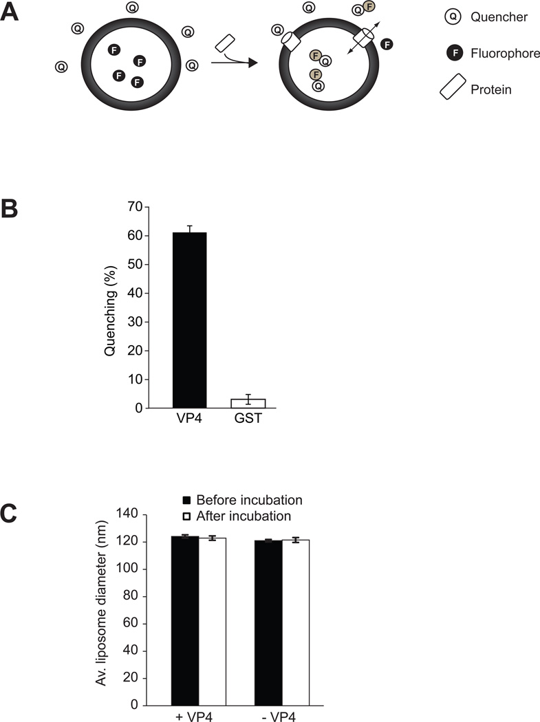
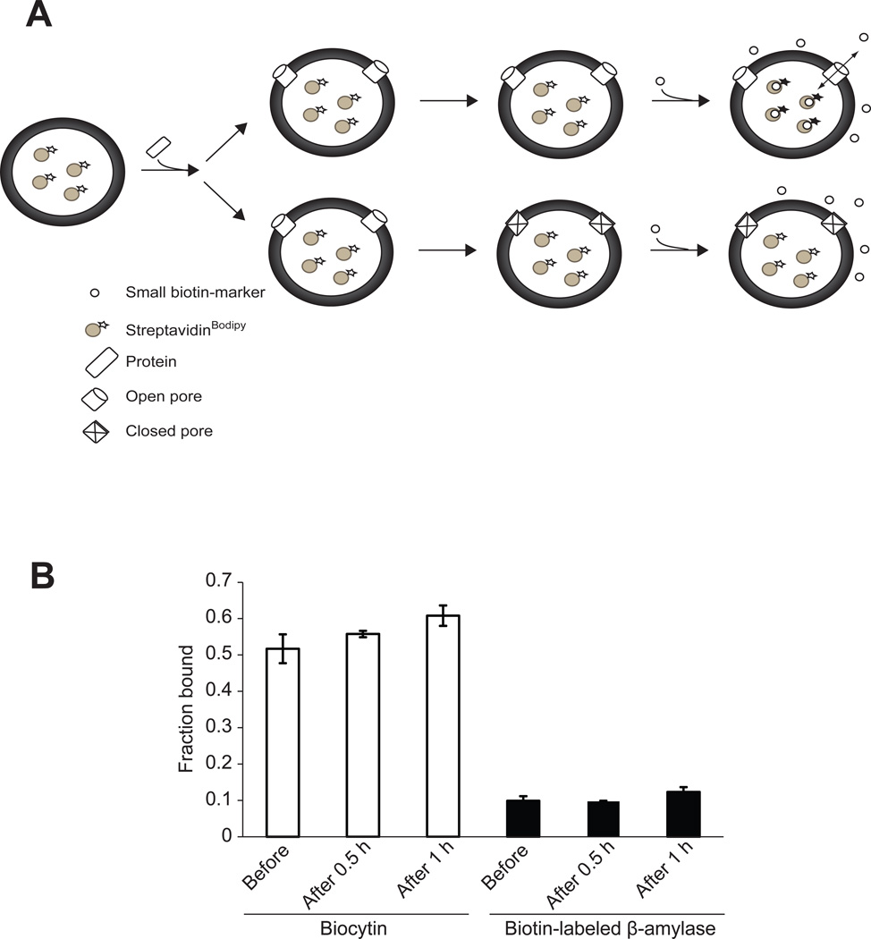
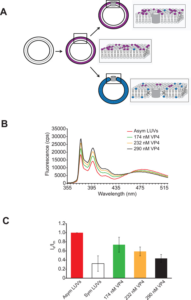
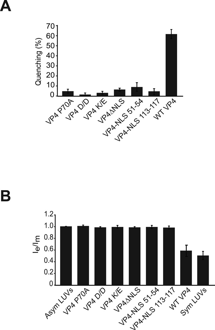
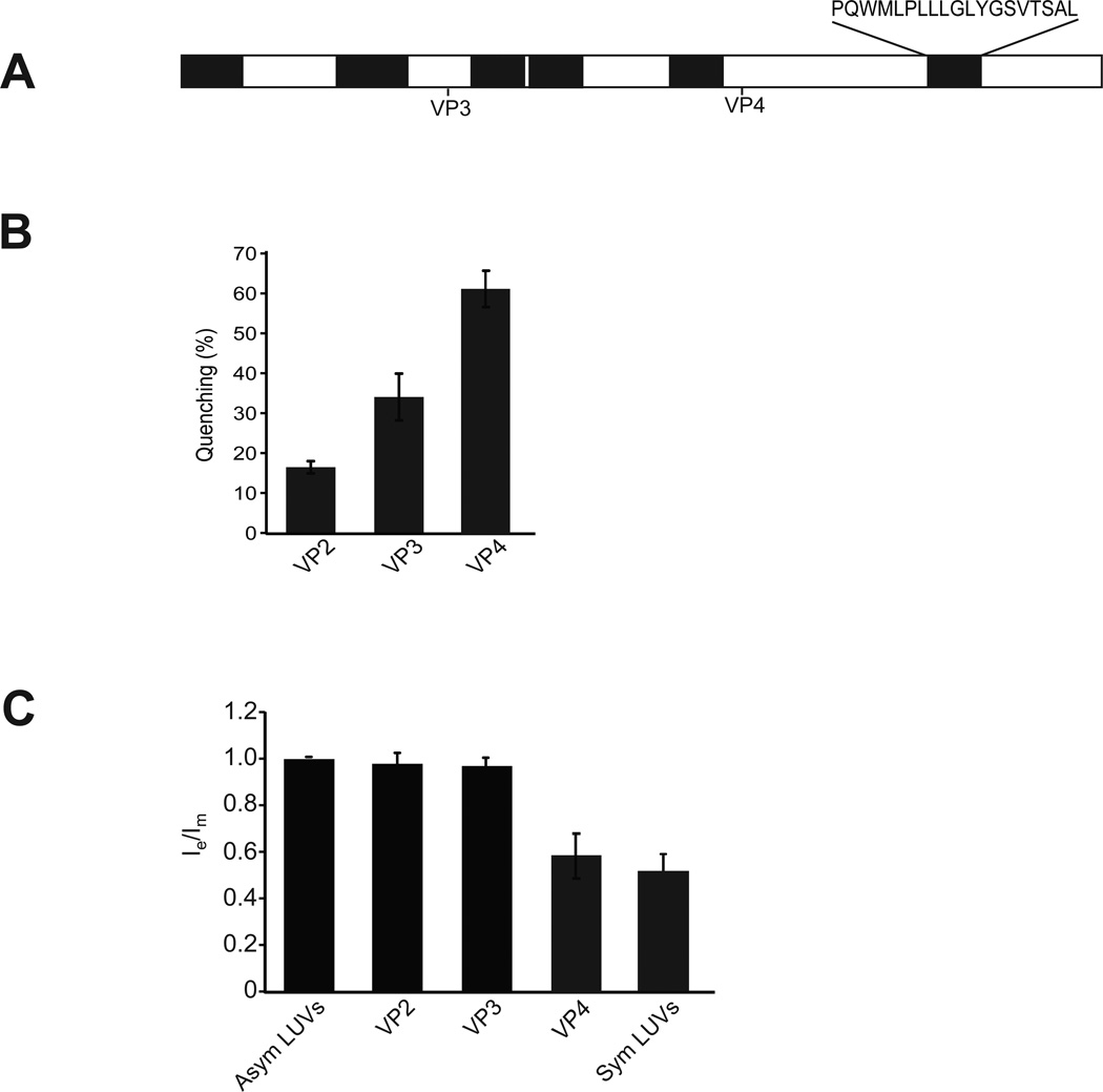
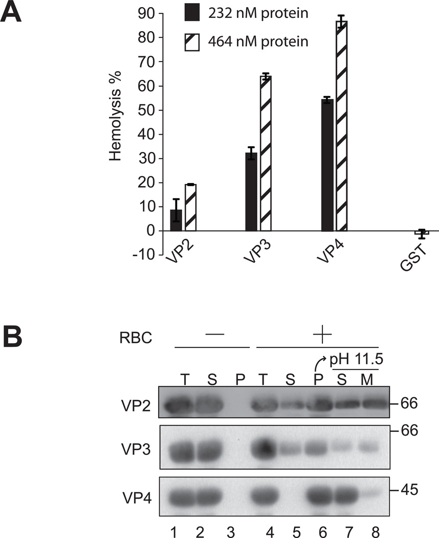
Nonenveloped viruses are generally released from the cell by the timely lysis of host cell membranes. SV40 has been used as a model virus for the study of the lytic nonenveloped virus life cycle. The expression of SV40 VP4 at later times during infection is concomitant with cell lysis. To investigate the role of VP4 in viral release and its mechanism of action, VP4 was expressed and purified from bacteria as a fusion protein for use in membrane disruption assays. Purified VP4 perforated membranes as demonstrated by the release of fluorescent markers encapsulated within large unilamellar vesicles or liposomes. Dynamic light scattering results revealed that VP4 treatment did not cause membrane lysis or change the size of the liposomes. Liposomes encapsulated with 4,4-difluoro-5,7-dimethyl-4-bora-3a,4a-diaza-3-indacene-labeled streptavidin were used to show that VP4 formed stable pores in membranes. These VP4 pores had an inner diameter of 1-5 nm. Asymmetrical liposomes containing pyrene-labeled lipids in the outer monolayer were employed to monitor transbilayer lipid diffusion. Consistent with VP4 forming toroidal pore structures in membranes, VP4 induced transbilayer lipid diffusion or lipid flip-flop. Altogether, these studies support a central role for VP4 acting as a viroporin in the disruption of cellular membranes to trigger SV40 viral release by forming toroidal pores that unite the outer and inner leaflets of membrane bilayers.


Figures

Figure 1
Figure 1. VP4 disrupts liposomal membranes
(A) Scheme showing the liposome disruption assay employed. Membrane disruption was examined by encapsulating [Tb(DPA) 33−] fluorophore into LUVs. When these LUVs were suspended in a solution containing EDTA (quencher), protein mediated membrane disruption was monitored by the quenching of [Tb(DPA) 33−] emission as the encapsulated molecules were released, and terbium ions were chelated by EDTA. (B) VP4 disrupts LUVs. LUVs were prepared to examine the membrane disruption activity of VP4. Liposome disruption was evaluated using LUVs and the percentage of fluorophore quenched is indicated. Each data point shows the average of at least two independent measurements with error bars representing standard deviations. (C) Average diameter of LUVs before and after 30 min incubation with VP4 as determined by DLS. Mock LUVs were incubated in absence of any protein. Each data point shows the average of at least two independent measurements with error bars representing standard deviations.
Figure 2
Figure 2. VP4 forms stable and size selective membrane pores
(A) Scheme showing the pore stability assay employed. Pore stability was analyzed by employing a fluorescence-based spectroscopic assay, which detected the binding of streptavidinBodipy with fluorescence enhancers after the addition of the enhancer after incubation with VP4. The passage of biocytin (~1 nm diameter), biotin-labeled β-amylase (~5 nm diameter), or streptavidinBodipy (~5 nm diameter) through the pores formed by GST-VP4 was measured as detailed in Experimental Procedures. When the liposomes encapsulating streptavidinBodipy are used and the biotin-labeled molecules are externally added following incubation with VP4, fluorescence enhancement will be detected only if VP4 formed stable pores thereby allowing interaction of streptavidinBodipy and the biotin markers (top scenario). On the other hand, if VP4 does not form stable pores and closes after a brief period of time then the fluorescence will not increase when the enhancer is added after incubation with GST-VP4 (bottom scenario). The size of the pore will dictate which molecules can cross the membrane. (B) The stability of the pores was examined by measuring the increase in the fluorescence intensity of encapsulated streptavidinBodipy when the enhancers biocytin or biotin-labeled β-amylase were present in the external buffer solution either before the addition of VP4, added after incubation for 0.5 hr or 1 hr with VP4. Only biocytin was able to diffuse through the formed pores. The pores formed by VP4 remained open after incubation for 1 hr. The total lipid concentration was 100 µM, and the concentration of the protein was 232 nM. Each data point shows the average of at least two independent measurements with error bars representing standard deviations.
Figure 3
Figure 3. VP4 induces lipid flip-flop
(A) Scheme showing the lipid flip-flop assay employed. The method is based on the dilution of the pyrene probe (pyPC) as a result of transbilayer diffusion. Lipid flip-flop was detected by using symmetrically and asymmetrically pyrene-labeled large unilamellar vesicles (LUVs). Addition of VP4 to asymmetric LUVs will result in no significant dilution of the pyrene probe from one leaflet to the other leaflet of LUVs if barrel-stave pores are formed (top). However, formation of toroidal pores by VP4 will result in transbilayer lipid movement resulting in dilution of the pyrene probe from outer leaflet to the inner leaflet of the membrane bilayer (bottom). The fluorescence spectrum of pyrene is characterized by two signals, one arising from monomer molecules (shown in blue) and the other from excited dimer (excimer) molecules (shown in purple). After incorporation of pyrene probe into the outer leaflet of membrane, the redistribution of pyrene to the inner leaflet is accompanied by a change of the analogue concentration in each leaflet and, thus, by a change of the intensity ratio between the excimer and the monomer signals (Ie/Im). (B) Fluorescence spectra of pyrene-labeled LUVs (PC-PG-pyPC, 87:10:3). The lipid concentration of the pyrene-labeled LUVs was 100 µM. (C) The ratio of transbilayer diffusion of pyrene probe (pyPC) is shown. A decrease in the ratio of Ie to Im reflects transbilayer diffusion of pyPC. The ratio Ie/Im has been normalized to a value of 1 with asymmetric LUVs alone. The extent of the transbilayer diffusion was negligible for LUVs in the absence of protein. The error bars correspond to standard deviations of measurements carried out in three different batches of liposomes.
Figure 4
Figure 4. Mutating VP4 abolishes transbilayer lipid diffusion
(A) VP4 mutants are deficient in disruption of LUVs. LUVs were prepared to examine the membrane disruption activity of VP4 and its mutants as in Figure 1. Liposome disruption was evaluated and the percentage of fluorophore quenched is indicated. Each data point shows the average of at least two independent measurements with error bars representing standard deviations. (B) The ratio of transbilayer diffusion of pyrene probe (pyPC). The lipid concentration of the pyrene-labeled LUVs was 100 µM. The ratio Ie/Im has been normalized to a value of 1 with asymmetric LUVs alone. The extent of the transbilayer diffusion was negligible for LUVs in the absence of protein. The error bars correspond to standard deviations of measurements carried out with three different batches of liposomes.
Figure 5
Figure 5. VP2 and VP3 do not possess lipid flip-flop activity
(A) Schematic representation of the SV40 protein VP2 displaying predicted hydrophobic domains (27). These hydrophobic domains, which were identified by using a variety of algorithms, may act as transmembrane domains. Start sites for VP3 and VP4 are indicated. The amino acid sequence of the last HD that is shared by VP2, VP3 and VP4 is indicated. (B) VP2 and VP3 show membrane disruptive activity. The membrane disruption activity of VP2, VP3 and VP4 was examined as in Figure 1. The percentage of fluorophore quenched is indicated. Each data point shows the average of at least two independent measurements with error bars representing standard deviations. (C) The ratio of transbilayer diffusion of pyrene probe (pyPC) was determined as in Figure 3. The lipid concentration of the pyrene-labeled LUVs was 100 µM. The ratio Ie/Im has been normalized to a value of 1 with asymmetric LUVs alone. The extent of the transbilayer diffusion was negligible for LUVs in the absence of protein. The error bars correspond to standard deviations of measurements carried out with three different batches of liposomes.
Figure 6
Figure 6. VP2 and VP3 integrate into the lipid bilayer unlike VP4
(A) VP2, VP3, and VP4 possess hemolytic activity. VP2, VP3, and VP4 were incubated with bovine RBCs for 30 min at 37 °C. Released hemoglobin was measured by the A414 of the supernatant after centrifugation and the removal of unlysed cells. GST was used as a control to rule out its contribution to the hemolytic activity of minor viral proteins. The error bars represent the standard deviation from three independent experiments. (B) Hemolysis reaction mixtures (lane 4, Total) containing bovine RBCs and viral proteins were incubated at 37 °C for 30 min. RBC bound (lane 6, Pellet) and unbound (lane 5, Supernatant) proteins were separated by centrifugation. Membrane fractions (lane 6) were alkaline extracted with 0.1 M Na2CO3, pH 11.5 and ultracentrifuged to separate the soluble (S) and membrane (M) fractions (lane 7 and 8). Samples resolved by reducing SDS-PAGE were immunoblotted with antibody against GST. Separate reactions were performed in the absence of RBCs (lanes 1–3).

References

    1. Bieniasz PD. Late budding domains and host proteins in enveloped virus release. Virology. 2006;344:55–63. - PubMed
    1. Chen BJ, Lamb RA. Mechanisms for enveloped virus budding: can some viruses do without an ESCRT? Virology. 2008;372:221–232. - PMC - PubMed
    1. Aldabe R, Barco A, Carrasco L. Membrane permeabilization by poliovirus proteins 2B and 2BC. J Biol Chem. 1996;271:23134–23137. - PubMed
    1. Han Z, Harty RN. The NS3 protein of bluetongue virus exhibits viroporin-like properties. J Biol Chem. 2004;279:43092–43097. - PubMed
    1. Nieva JL, Madan V, Carrasco L. Viroporins: structure and biological functions. Nat Rev Microbiol. 2012;10:563–574. - PMC - PubMed

Publication types

MeSH terms

LinkOut - more resources