. 2013 Jun;88(6):1176-93.
doi: 10.1111/mmi.12250. Epub 2013 May 20.

PTS phosphorylation of Mga modulates regulon expression and virulence in the group A streptococcus

Affiliations

PTS phosphorylation of Mga modulates regulon expression and virulence in the group A streptococcus

Elise R Hondorp et al. Mol Microbiol. 2013 Jun.

Erratum in

  • Mol Microbiol. 2014 Aug;93(4):834

Abstract

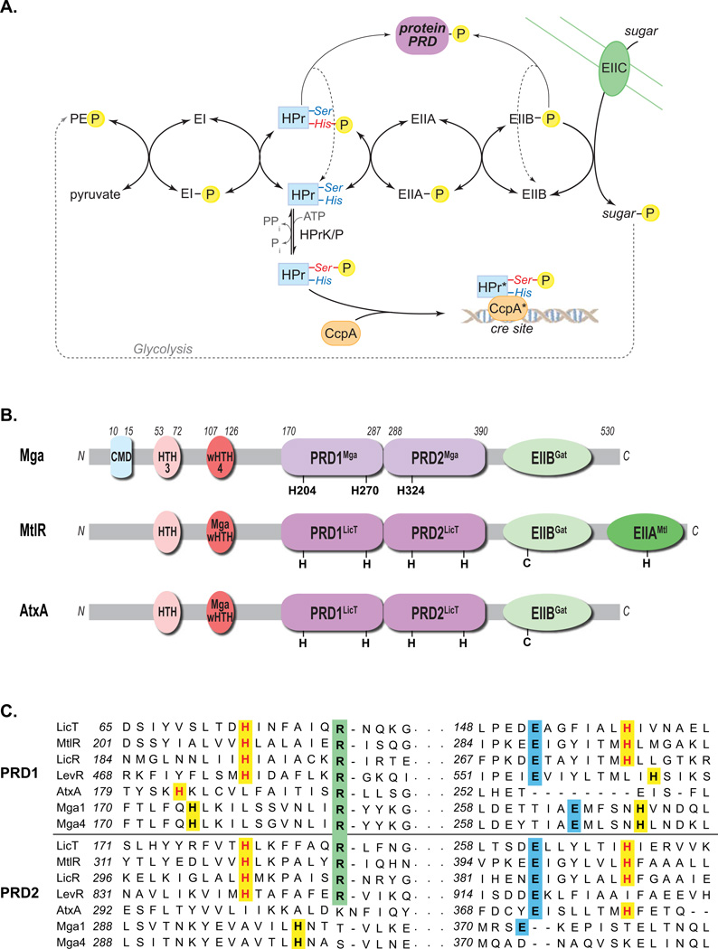
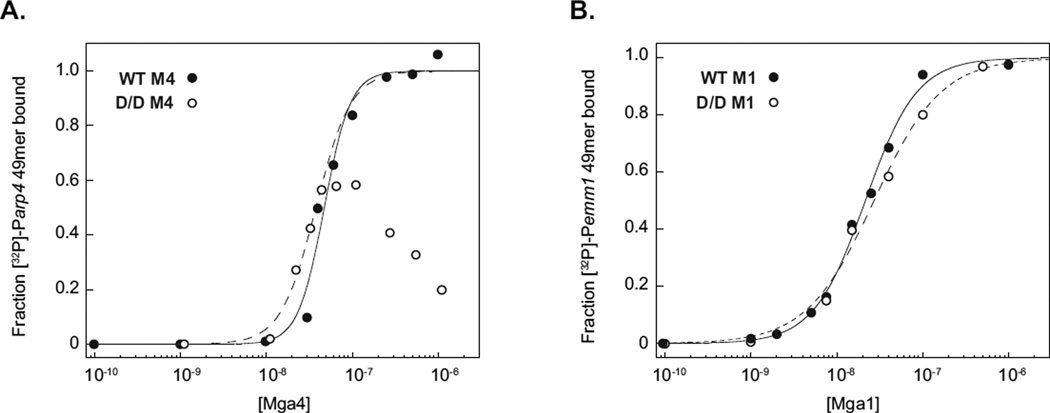
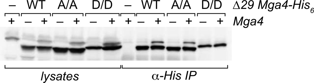
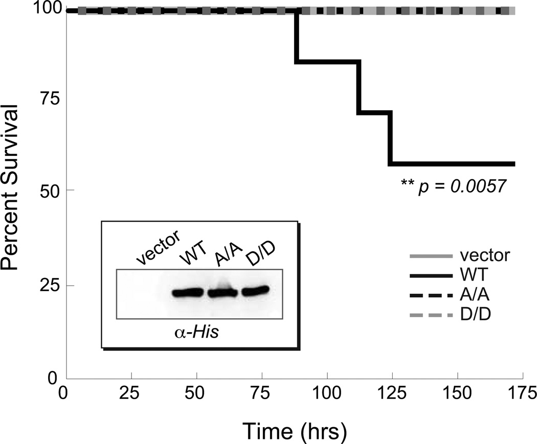
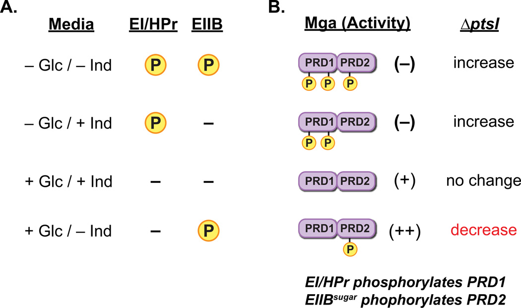
The ability of a bacterial pathogen to monitor available carbon sources in host tissues provides a clear fitness advantage. In the group A streptococcus (GAS), the virulence regulator Mga contains homology to phosphotransferase system (PTS) regulatory domains (PRDs) found in sugar operon regulators. Here we show that Mga was phosphorylated in vitro by the PTS components EI/HPr at conserved PRD histidines. A ΔptsI (EI-deficient) GAS mutant exhibited decreased Mga activity. However, PTS-mediated phosphorylation inhibited Mga-dependent transcription of emm in vitro. Using alanine (unphosphorylated) and aspartate (phosphomimetic) mutations of PRD histidines, we establish that a doubly phosphorylated PRD1 phosphomimetic (D/DMga4) is completely inactive in vivo, shutting down expression of the Mga regulon. Although D/DMga4 is still able to bind DNA in vitro, homo-multimerization of Mga is disrupted and the protein is unable to activate transcription. PTS-mediated regulation of Mga activity appears to be important for pathogenesis, as bacteria expressing either non-phosphorylated (A/A) or phosphomimetic (D/D) PRD1 Mga mutants were attenuated in a model of GAS invasive skin disease. Thus, PTS-mediated phosphorylation of Mga may allow the bacteria to modulate virulence gene expression in response to carbohydrate status. Furthermore, PRD-containing virulence regulators (PCVRs) appear to be widespread in Gram-positive pathogens.


Figures

Figure 1
Figure 1. PTS pathway and alignment of Mga with PRD-containing regulators
(A) The phosphotransferase system (PTS) in Gram-positive bacteria couples the phosphorylation and import of sugars. The general cytoplasmic enzymes (EI and HPr) and sugar-specific EII components form a phosphorelay to transfer phosphate from phosphoenol pyruvate (PEP) produced by glycolysis to the incoming sugar. Carbon catabolite repression (CCR) results from HPrK/P phosphorylation of serine 46 of HPr, which then complexes with CcpA to repress target promoters via cre sites. HPr-His∼P and EIIB∼P can also phosphorylate PTS regulatory domains (PRDs) of sugar regulators, thereby modulating their activity. (B) Domain alignment of GAS Mga with B. subtilis MtlR and B. anthracis AtxA. EII (green), PRD (purple), DNA-binding (red) and conserved (blue) domains are indicated with conserved histidines (H) and cysteines (C). PRDMga and PRDLicT refer to Pfam 08270 and Pfam 00874, respectively. (C) Amino acid sequence alignment of the PRDs from B. subtilis LicT, LicR, LevR, G. stearothermophilus MtlR, B. anthracis AtxA, and GAS Mga (M1 and M4 serotypes), highlighting the regions containing the conserved histidine (highlighted yellow), arginine (highlighted green), and aspartate (highlighted blue) residues. Histidine residues previously shown to be phosphorylated are colored red. (Supplemental Figure S1 contains the full alignment.)
Figure 2
Figure 2. A ∆ptsI mutant of GAS is altered in PTS-dependent growth and Mga regulon expression
(A) Growth curves of wild type GA40634 (closed circles) and GA40634∆ptsI (open circles) in THY or C medium as indicated. Data are representative of three independent experiments. (B) Growth curves of wild type GA40634 (closed circles) and GA40634∆ptsI (open circles) in CDM supplemented with either 0.5% (v/v) glucose or 1% (v/v) fructose as indicated. Data are representative of three independent experiments. (C) Transcript levels of ptsI (black), arp (striped), and sof (grey) were measured by qRT-PCR from late logartithmic phase cultures grown in THY and C medium for GA40634 ∆ptsI compared to GA40634. Two-fold differences in expression (dashed line) were considered significant. Standard error was determined from three biological replicates.
Figure 3
Figure 3. In vitro phosphorylation and inactivation of Mga by the PTS
(A, B) Phosphorylation was assessed by incubation of [32P]-PEP with purified His6-EI, His6-HPr, and Mga4-His6. Reactions were then subjected to SDS-PAGE and phosphorimager analysis. (A) Proteins included in each reaction are indicated; HD refers to heat-denatured Mga4-His6(B) Wild type and various PRD Mga4-His6 mutants were analyzed, alongside heat-denatured Mga4-His6, His6-MBP, MetE, ∆139Mga4-His6, and Mga1-His6 controls as indicated. (C) The ability of Mga1-His6 to activate transcription of the constitutive PrpsL and Mga-regulated Pemm promoters was assessed in vitro immediately following phosphorylation reactions (containing or lacking PEP). Products of the in vitro transcription assays were then separated on a 6% sequencing acrylamide gel and subjected to phosphorimager analysis.
Figure 4
Figure 4. The conserved PRD1 histidines are important for M4 Mga activity in vivo
(A)In vivo Mga activity was assessed by real-time RT-PCR analysis of arp (dark grey), sof (striped) and mga (light grey) mRNA. Transcript levels that were greater than 2-fold different compared to wild type (dotted lines) were considered significant. Single mutations in Mga are indicated, along with H204A/H270A (A/A), H204D/H270D (D/D), H204A/H270A/H324A (A/A/A), H204D/H270D/H324D (D/D/D), and an empty vector (vector) in the mga4-inactivated GAS strain KSM547.4. An isogenic wild type M4 strain GA40634 (WT) with vector was also assayed to show endogenous Mga4 (end WT) activity. (B) Protein levels in cell lysates were determined by immunoblotting with α-His and α-Mga4 antibodies; the α-Hsp60 antibody was employed as a loading control.
Figure 5
Figure 5. The PRD1 D/D Mga phosphomimetic mutant is able to bind target DNA in vitro
Filter-binding assays were employed to analyze protein-DNA associations for M4 (A) and M1 (B) wild type (closed circles) and PRD1 D/D phosphomimetic (open circles) Mga-His6 proteins. Proteins were incubated with [32P]-labeled 49-bp double-stranded oligonucleotides containing the Mga-binding sites of the arp(A) or emm(B) promoters and then filtered through nitrocellulose. The protein-bound radiolabel was quantified by phosphorimager analyses and the calculated fraction of DNA bound was plotted versus Mga protein concentrations.
Figure 6
Figure 6. PRD1 phosphomimetic inhibits Mga oligomerization
Co-immunoprecipitation of Mga4 with PRD1 mutant proteins. Wild type (WT), PRD1 A/A (A/A), and PRD1 D/D phosphomimetic (D/D) versions of the His-tagged 29-residue truncated Mga4 (∆29Mga4-His6) were expressed in isogenic M4 GAS strains containing (+) or lacking (−) endogenous Mga4 (GA40634 and KSM547.4, respectively). His-tagged ∆29 proteins from exponentially growing GAS cell lysates were immunoprecipitated with an α-His antibody, and coimmunoprecipitation of endogenous Mga4 was assessed by SDS-PAGE and immunoblot analysis with an α-Mga4 antibody (right). Western blot analysis of whole cell lysates prior to immunoprecipitation is shown alongside for comparison (left).
Figure 7
Figure 7. PTS phosphorylation of PRD1 is important for GAS virulence
Survival curve for CD-1 mice infected subcutaneously with an M1T1 MGAS5005 Mga mutant (KSM165-L.5005) containing an empty vector (light grey solid, 1.4 × 10⁸ cfu) or Mga1-His6 proteins (WT, black solid, 1.5 × 10⁸ cfu; A/A, black dashed, 2.1 × 10⁸ cfu; D/D, light grey dashed, 2.0 × 10⁸ cfu) expressed from the native Pmga1 promoter. Data shown represent 8–10 mice for each strain. Significance was determined using Kaplan-Meier survival analysis and log rank test. Western blot analysis of GAS cell lysates is shown (inset).
Figure 8
Figure 8. Model for PTS/Mga interactions in vivo
(A) Predicted availability of phosphate for transfer to Mga by either EI/Hpr or a sugar-specific EIIB component when growing in the presence or absence of a preferred sugar source (i.e., glucose) and/or an EIIB-specific inducer sugar. In the absence of glucose, EI/Hpr can phosphorylate PRD domains, whereas it cannot in its presence. Phosphorylation of PRD domains by a cognate EIIB protein would only occur in the absence of inducer. (B) Proposed role of PTS-mediated Mga phosphorylation on activity based upon sugar source in (A). In the absence of glucose, Mga would be inactivated through phosphorylation of PRD1. Inactivation of PTS (∆ptsI) would be expected to increase Mga-regulated expression. With both glucose and inducer present, Mga is not phosphorylated at either PRD and is active. Loss of PTS (∆ptsI) would have no effect. In the presence of glucose only (THY, C media, or CDM + glucose only), phosphorylation of Mga PRD2 by inducer-specific EIIB leads to enhancement of activity. In this case, loss of PTS ((∆ptsI) would result in a decrease in Mga regulon expression.

Similar articles

Cited by

References

    1. Almengor AC, Kinkel TL, Day SJ, McIver KS. The catabolite control protein CcpA binds to Pmga and influences expression of the virulence regulator Mga in the group A streptococcus. J Bacteriol. 2007;189:8405–8416. - PMC - PubMed
    1. Almengor AC, McIver KS. Transcriptional activation of sclA by Mga requires a distal binding site in Streptococcus pyogenes. J Bacteriol. 2004;186:7847–7857. - PMC - PubMed
    1. Almengor AC, Walters MS, McIver KS. Mga is sufficient to activate transcription in vitro of sof/sfbX and other Mga-regulated virulence genes in the group A streptococcus. J Bacteriol. 2006;188:2038–2047. - PMC - PubMed
    1. Andreeva A, Howorth D, Brenner SE, Hubbard TJ, Chothia C, Murzin AG. SCOP database in 2004: refinements integrate structure and sequence family data. Nucleic Acids Res. 2004;32:D226–D229. - PMC - PubMed
    1. Bessen DE, Manoharan A, Luo F, Wertz JE, Robinson DA. Evolution of transcription regulatory genes is linked to niche specialization in the bacterial pathogen Streptococcus pyogenes. J Bacteriol. 2005;187:4163–4172. - PMC - PubMed

Publication types

MeSH terms

Substances