. 2013 Aug;169(7):1612-23.
doi: 10.1111/bph.12243.

Resistance to BH3 mimetic S1 in SCLC cells that up-regulate and phosphorylate Bcl-2 through ERK1/2

Affiliations

Resistance to BH3 mimetic S1 in SCLC cells that up-regulate and phosphorylate Bcl-2 through ERK1/2

Yubo Liu et al. Br J Pharmacol. 2013 Aug.

Abstract

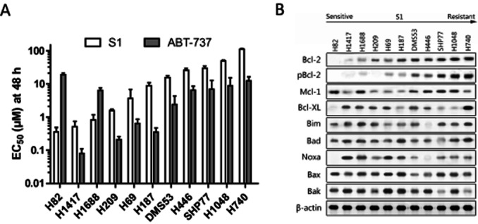
Background and purpose: B cell lymphoma 2 (Bcl-2) is a central regulator of cell survival that is overexpressed in the majority of small-cell lung cancers (SCLC) and contributes to both malignant transformation and therapeutic resistance. The purpose of this work was to study the key factors that determine the sensitivity of SCLC cells to Bcl-2 homology domain-3 (BH3) mimetic S1 and the mechanism underlying the resistance of BH3 mimetics.

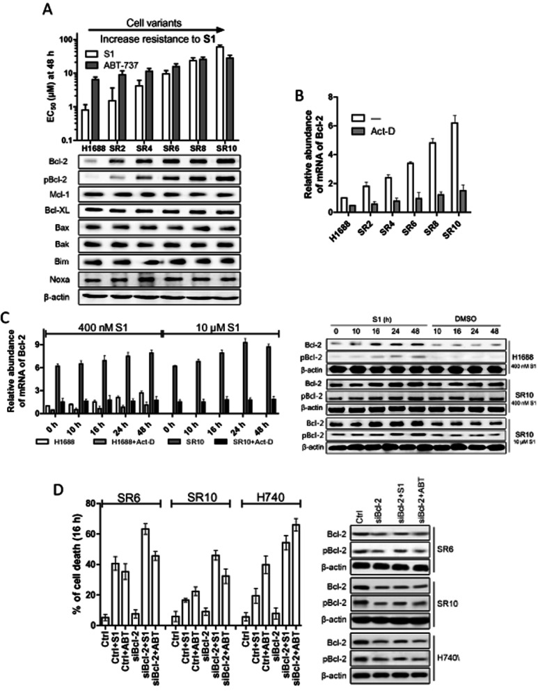
Experimental approaches: Western blot was used to evaluate the contribution of Bcl-2 family members to the cellular response of SCLC cell lines to S1. Acquired resistant cells were derived from initially sensitive H1688 cells. Quantitative PCR and gene silencing were performed to investigate Bcl-2 up-regulation.

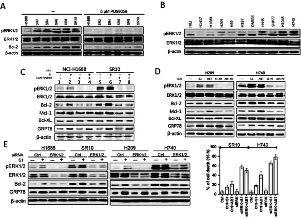
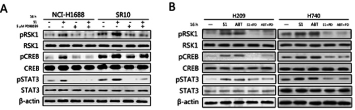
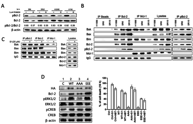
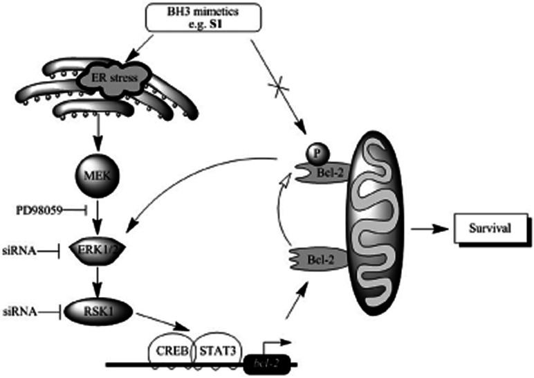
Key results: A progressive increase in the relative levels of Bcl-2 and phosphorylated Bcl-2 (pBcl-2) characterized the increased de novo and acquired resistance of SCLC cell lines. Furthermore, acute treatment of S1 induced Bcl-2 expression and phosphorylation. We showed that BH3 mimetics, including S1 and ABT-737, induced endoplasmic reticulum (ER) stress and then activated MAPK/ERK pathway. The dual function of MAPK/ERK pathway in defining BH3 mimetics was illustrated; ERK1/2 activation leaded to Bcl-2 transcriptional up-regulation and sustained phosphorylation in naïve and acquired resistant SCLC cells. pBcl-2 played a key role in creating resistance of S1 and ABT-737 not only by sequestrating pro-apoptotic proteins, but also sequestrating a positive feedback to promote ERK1/2 activation.

Conclusions and implications: These results provide significant novel insights into the molecular mechanisms for crosstalk between ER stress and endogenously apoptotic pathways in SCLC following BH3 mimetics treatment.

Keywords: BH3 mimetics; Bcl-2 phosphorylation; ERK1/2; SCLC.


Figures

Figure 1
Figure 1
Cytotoxic activity of S1 against a panel of SCLC cell lines associated with Bcl-2 expression and phosphorylation. (A) Cell viability EC50 values of various SCLC cell lines following treatment for 48 h with S1 or ABT-737 were determined by MTS assay. Columns, average (n = 3) of triplicate experiments; bars, SD. (B) Western blot analysis of the Bcl-2 family proteins indicated in the 11 cell lines. Cell lines are arranged according to their resistance to S1.
Figure 2
Figure 2
Bcl-2 is transcriptionally up-regulated in H1688-acquired resistant cells. (A) EC50 values (S1 and ABT-737 48 h) and expressions of Bcl-2 family proteins of parental and S1-acquired resistant H1688 cells. Columns, average (n = 3) of triplicate experiments; bars, SD. (B) Bcl-2 transcript level was analysed by quantitative PCR in H1688-derived cells. H1688-derived resistant cells were cultured in the absence of S1 for 3 weeks and cells were treated or not with 3 μg·mL−1 actinomycin D (Act-D) for 1 h before RNA was isolated. (C) Bcl-2 is up-regulated in both transcriptional, protein and phosphorylation levels in parental H1688 and resistant H1688-SR10 cells after transient treatment with S1. H1688 and H1688-SR10 cells were treated with S1 (400 nM for H1688 and H1688-SR10, 10 μM for H1688-SR10) for indicated time and Bcl-2 transcript level was analysed by quantitative PCR. H1688-SR10 cells were removed from S1-containing media for 3 weeks. H1688 and H1688-SR10 cells were treated with S1 (400 nM for H1688, 10 μM for H1688-SR10) or DMSO for indicated time. H1688 and H1688-SR10 cells were treated actinomycin D (3 μg·mL−1) for 1 h before the addition of S1. Whole-cell lysates were made after treatment and analysed by immunoblot. Columns, average (n = 3) of triplicate experiments; bars, SD. (D) The indicated cell lines were transfected with control (Ctrl) or Bcl-2 siRNA for 48 h and then treated with and without 10 μM S1 or ABT-737 for an additional 16 h. Protein levels were examined by Western blot. Cell viability was assessed by MTS assay.
Figure 3
Figure 3
S1 induces up-regulation of Bcl-2 by an ER stress-induced ERK1/2 activation-dependent mechanism. (A) H1688-derived cells were incubated with 5 μM PD98059 for 16 h. DMSO treatment (left) was used as a control. Whole cell lysates were prepared and Western blots were probed using specific antibodies against phosphorylated ERK1/2, whole ERK1/2 and Bcl-2. Blot shows ERK phosphorylation correlated with up-regulation of Bcl-2 (left). (B) Whole-cell lysates from 11 SCLC cell lines were analysed with Western blot for the levels of phosphorylated ERK1/2 and ERK1/2. Cell lines are arranged according to their resistance to S1. (C) H1688 and H1688-SR10 cells were treated with S1 (400 nM for H1688 and 10 μM for H1688-SR10) alone or together with 5 μM PD98059 for 16 h. Protein levels were examined by Western blot. H1688-SR10 cells were cultured in the absence of S1 for 3 weeks before used. (D) H209 and H740 cells were treated with 400 nM S1 alone or together with 5 μM PD98059 for 16 h. ABT-737 (100 nM) was tested in parallel. DMSO was used as a control. Whole cell lysates were prepared and Western blots were probed using indicated antibodies. (E) The indicated cell lines were transfected with control (Ctrl) or ERK1/2 siRNA for 48 h and then treated with and without 10 μM S1 or ABT-737 for an additional 16 h. Protein levels were examined by Western blot. H1688-SR10 and H740 cells viability were assessed by MTS assay.
Figure 4
Figure 4
CREB and STAT3 were activated upon S1 treatment in SCLC cell lines. (A) H1688 and H1688-SR10 cells were treated with S1 (400 nM for H1688 and 10 μM for H1688-SR10) alone or together with 5 μM PD98059 for 16 h. The activations of RSK1, CREB and STAT3 were examined by Western blot. (B) H209 and H740 cells were treated with 400 nM S1 alone or together with 5 μM PD98059 for 16 h. ABT-737 (100 nM) was tested in parallel. DMSO was used as a control. Whole cell lysates were prepared and Western blots were probed using indicated antibodies.
Figure 5
Figure 5
Bcl-2 is phosphorylated by ERK1/2 in H1688-derived cells and contributes to the resistance to S1. (A) PD98059 was able to block Bcl-2 phosphorylation and reduce the pBcl-2/Bcl-2 ratios. H1688-derived H1688-SR6, H1688-SR10 cell, H1048 and H740 cells were incubated with 5 μM PD98059 for 16 h. DMSO was used as a control. Total protein extracts (50 μg) from these cells were analysed by Western blot for pBcl-2 and Bcl-2 expression. The mean of pBcl-2/Bcl-2 ratio is shown for each cell line. (B) pBcl-2 sequestrated more pro-apoptotic proteins in acquired resistant cells. Immunoprecipitation of H1688-derived cell lysates. Mcl-1, Bcl-2 and pBcl-2 immunoprecipitations were performed, and immunoprecipitated fractions were analysed by Western blotting for the indicated proteins. (C) H1688-SR10 cells were treated with 20 μM S1 for 16 h. Mcl-1, Bcl-2 and pBcl-2 immunoprecipitations were performed, and immunoprecipitated fractions were analysed by Western blotting for the indicated proteins. (D) Phosphorylated Bcl-2 activates ERK1/2. Parental H1688 cells and H1688-expressing WT-Bcl-2, AAA-Bcl-2 or EEE-Bcl-2 were treated with and without 10 μM S1 or ABT-737 for 16 h and then lysed. Protein levels were examined by immunoblotting. Cell viability was assessed by MTS assay.
Figure 6
Figure 6
Schematic working model for ER stress/ERK1/2-mediated signalling of BH3 mimetics (e.g. S1)-induced Bcl-2 expression and phosphorylation. BH3 mimetics, such as S1 and ABT-737, stimulate ER stress then induce MAPK/ERK cascades with respect to Bcl-2 transcriptional up-regulation (solid arrowhead) and phosphorylation (hollow arrowhead). Excess pBcl-2 leads to cell survival by promoting ERK1/2 activation and sequestrating more pro-apoptotic members, which BH3 mimetics cannot release.

Similar articles

Cited by

References

    1. Bassik MC, Scorrano L, Oakes SA, Pozzan T, Korsmeyer SJ. Phosphorylation of BCL-2 regulates ER Ca2+ homeostasis and apoptosis. EMBO J. 2004;23:1207–1216. - PMC - PubMed
    1. Bhattacharya S, Ray RM, Johnson LR. STAT3-mediated transcription of Bcl-2, Mcl-1 and c-IAP2 prevents apoptosis in polyamine-depleted cells. Biochem J. 2005;392(Pt 2):335–344. - PMC - PubMed
    1. Breton C, Story MD, Meyn RE. Bcl-2 expression correlates with apoptosis induction but not loss of clonogenic survival in small cell lung cancer cell lines treated with etoposide. Anticancer Drugs. 1998;9:751–757. - PubMed
    1. Burlacu A. Regulation of apoptosis by Bcl-2 family proteins. J Cell Mol Med. 2007;7:249–257. - PMC - PubMed
    1. Chao DT, Korsmeyer SJ. BCL-2 family: regulators of cell death. Ann Rev Immunol. 1998;16:395–419. - PubMed

Publication types

MeSH terms