. 2013 Jul 15;73(14):4383-94.
doi: 10.1158/0008-5472.CAN-12-3385. Epub 2013 May 7.

Therapeutic destruction of insulin receptor substrates for cancer treatment

Affiliations

Therapeutic destruction of insulin receptor substrates for cancer treatment

Hadas Reuveni et al. Cancer Res. .

Abstract

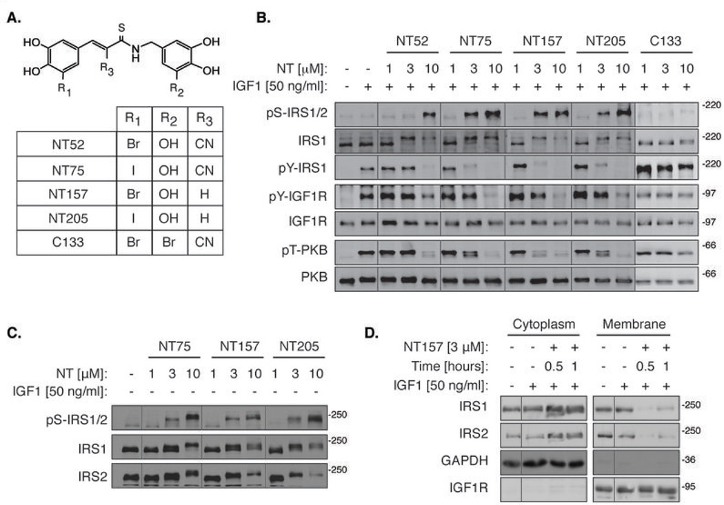
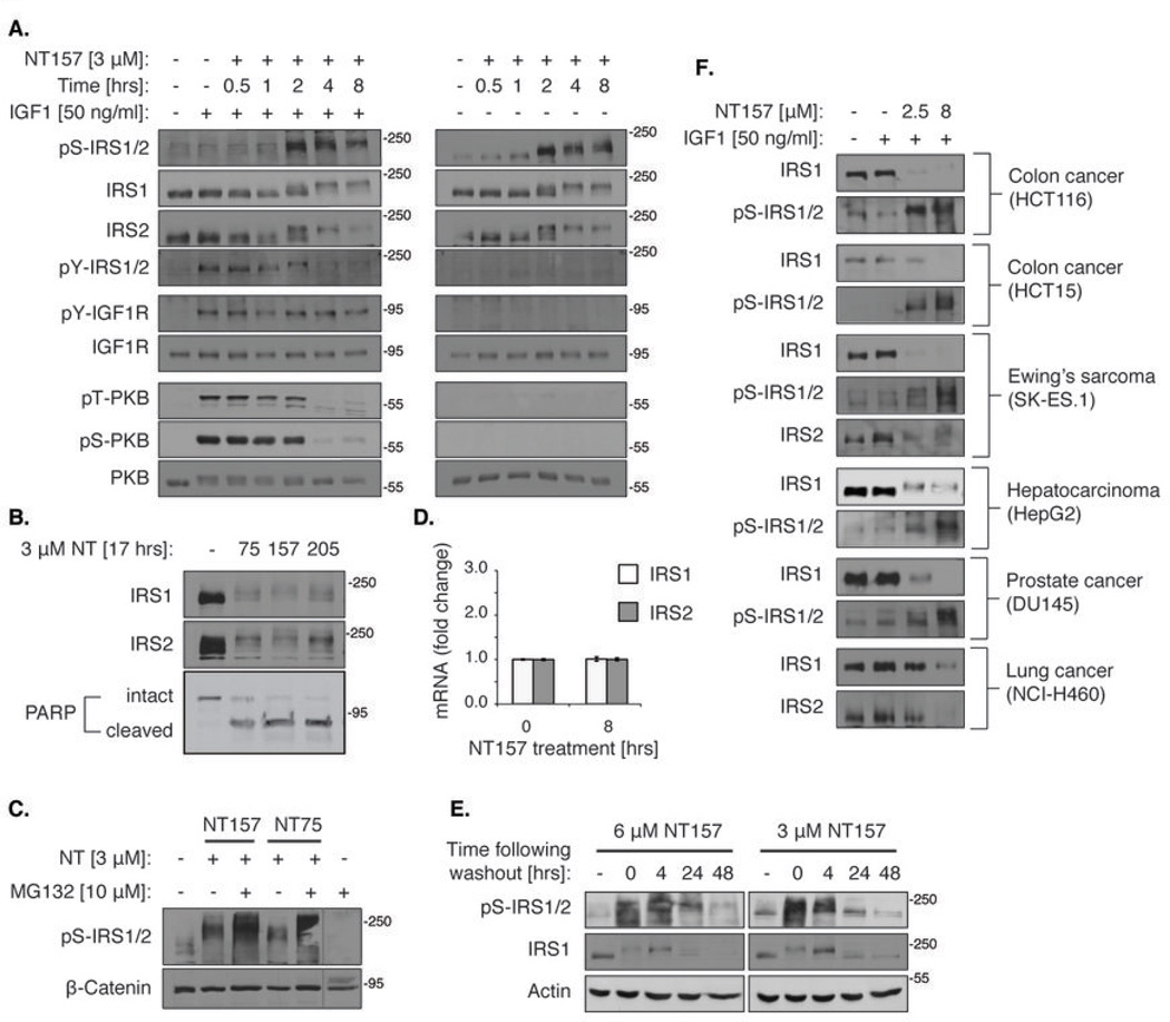
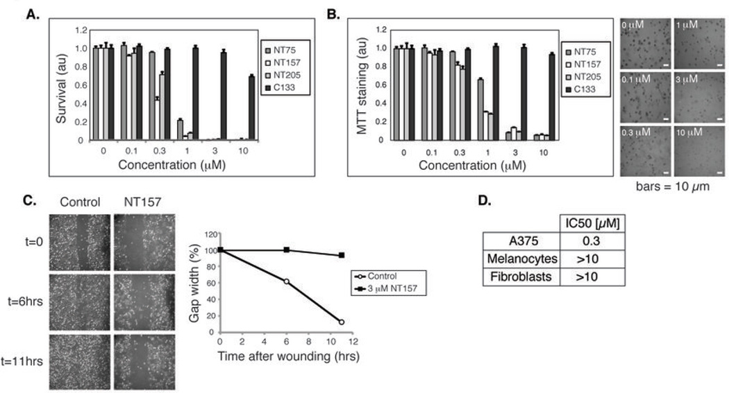
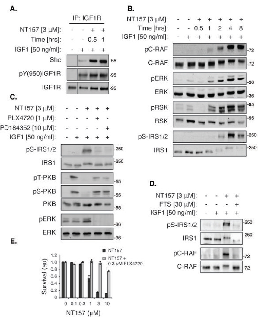
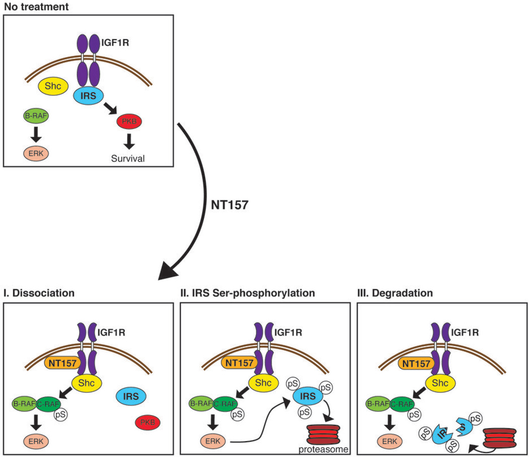
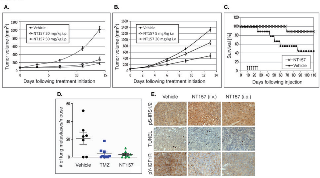
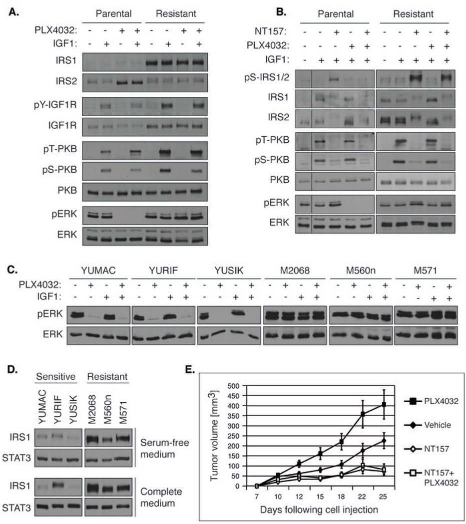
Insulin receptor substrates 1 and 2 (IRS1/2) mediate mitogenic and antiapoptotic signaling from insulin-like growth factor 1 receptor (IGF-IR), insulin receptor (IR), and other oncoproteins. IRS1 plays a central role in cancer cell proliferation, its expression is increased in many human malignancies, and its upregulation mediates resistance to anticancer drugs. IRS2 is associated with cancer cell motility and metastasis. Currently, there are no anticancer agents that target IRS1/2. We present new IGF-IR/IRS-targeted agents (NT compounds) that promote inhibitory Ser-phosphorylation and degradation of IRS1 and IRS2. Elimination of IRS1/2 results in long-term inhibition of IRS1/2-mediated signaling. The therapeutic significance of this inhibition in cancer cells was shown while unraveling a novel mechanism of resistance to B-RAF(V600E/K) inhibitors. We found that IRS1 is upregulated in PLX4032-resistant melanoma cells and in cell lines derived from patients whose tumors developed PLX4032 resistance. In both settings, NT compounds led to the elimination of IRS proteins and evoked cell death. Treatment with NT compounds in vivo significantly inhibited the growth of PLX4032-resistant tumors and displayed potent antitumor effects in ovarian and prostate cancers. Our findings offer preclinical proof-of-concept for IRS1/2 inhibitors as cancer therapeutics including PLX4032-resistant melanoma. By the elimination of IRS proteins, such agents should prevent acquisition of resistance to mutated-B-RAF inhibitors and possibly restore drug sensitivity in resistant tumors.


Conflict of interest statement

The authors declare no conflict of interests.

Figures

Figure 1
Figure 1. NT inhibitors lead to the dissociation of IRS1/2 from IGF1R, and induce Ser-phosphorylation of IRS1/2
(A) Chemical structures of the NT compounds. The active NT compounds all possess an OH at the 5’ position of the aminobenzyl moiety. The control compound C133 possesses a Br substitution at this position. (B) NT compounds, but not C133, induce IRS1/2 Ser-phosphorylation and strongly inhibit IGF1-induced PKB activation. Serum-starved A375 cells were treated with NT inhibitors for 5 hrs, then stimulated with IGF1 for 5 min and lysed. (C) NT-induced IRS1/2 Ser-phosphorylation is IGF1-independent. Serum-starved A375 cells were treated with NT inhibitors for 5 hrs and lysed without IGF1 stimulation. (D) NT157 induces the translocation of IRS1/2 to the cytosol. Serum-starved A375 cells were treated with NT157 as indicated. Cells were stimulated with IGF1 for 5 min, lysed and fractionated by differential centrifugation.
Figure 2
Figure 2. NT inhibitors irreversibly direct Ser-phosphorylated IRS proteins to degradation and induce apoptosis
(A) NT157 induces IRS1/2 Ser-phosphorylation and PKB inhibition in a time-dependent manner. Serum-starved A375 cells were treated with 3 µM NT157 as indicated, and either stimulated with IGF1 for 5 min (left panel) or not (right panel) before lysis. (B) Treatment with NT inhibitors for 17 hrs leads to IRS1 and IRS2 elimination and to A375 cell apoptosis. Serum-starved A375 cells were treated with 3 µM NT75, NT157 or NT205 for 17 hrs, lysed and analyzed by western blot. Apoptosis was detected by PARP cleavage. (C) The NT-induced elimination of Ser-phosphorylated IRS1/2 is mediated by the proteasome. Serum-starved A375 cells were treated with 3 µM NT157 or NT75 with or without 10 µM MG132 for 17 hrs and lysed. (D) IRS1/2 mRNA levels are not affected by NT157 treatment. Serum-starved A375 cells were treated with 3 µM NT157 and then RNA was extracted and subjected to reverse transcription. PCR amplification was performed and IRS1/2 mRNA levels were normalized to the levels of the ribosomal protein HuPO. (E) The effects of NT inhibitors on the Ser-phosphorylation and degradation of IRS1/2 are maintained long after removal of the inhibitors. Serum-starved A375 cells were treated with NT157 (3 or 6 µM) for 4 hrs. Cells were then washed, grown without serum for 4, 24 or 48 hrs, and lysed. (F) NT157 induces the Ser-phosphorylation and degradation of IRS1/2 in multiple cell lines. Cells were treated with 2.5 or 8 µM NT157. The Ser-phosphorylation and levels of IRS1/2 were tested 5 hrs and 24 hrs following treatment, respectively. For HepG2 cells, the levels of IRS1 were tested 4 hrs following treatment.
Figure 3
Figure 3. The NT compounds inhibit the migration and anchorage-dependent and - independent growth of A375 human melanoma cells
(A) NT compounds, but not C133, potently block the proliferation of A375 cells. Cells were treated with inhibitors for 72 hrs and survival was quantified by methylene blue staining. (B) NT compounds inhibit A375 anchorage-independent growth. A375 cells were seeded in 0.3% agar and treated with inhibitors for 7 days. Colony formation was quantified by MTT staining. Representative images from unstained, NT157-treated cells are shown. (C) NT157 inhibits the migration of A375 cells. A375 cells were treated or not treated with NT157 for 18 hrs, and then the cell monolayer was wounded at t=0. Representative images were taken at the specified time-points. (D) NT157 displays a minor effect on the proliferation of normal melanocytes and fibroblasts. Cells were treated with NT157 as in (A). IC50 values are presented. au= arbitrary units
Figure 4
Figure 4. NT157 shifts IGF1R complexation from IRS1/2 to Shc and leads to ERKMAPK-dependent Ser-phosphorylation of IRS1/2 in A375 melanoma cells
(A) NT157 promotes recruitment of Shc to IGF1R. Serum-starved A375 cells were treated with NT157, stimulated with IGF1, and lysed. IGF1R was immunoprecipitated (IP) and Shc levels in the IP samples were detected. (B) Time-dependent activation of the ERKMAPK pathway. Serum-starved A375 cells were treated with NT157 for the indicated times, stimulated with IGF1 for 5 min, and lysed. The kinase activity of ERK was assessed by analysis of the phosphorylation of its direct substrate, RSK. (C) The ERKMAPK pathway mediates the NT157-induced Ser-phosphorylation of IRS1/2. Serum-starved A375 cells were treated with an inhibitor of mutated B-RAF (PLX4720) or of MEK1/2 (PD184352) for 30 min, and then NT157 was added for 4 hrs. Cells were then stimulated with IGF1 for 5 min and lysed. (D) The NT157-induced C-RAF activation is Ras-dependent. Serum-starved A375 cells were treated with an inhibitor of Ras (FTS) for 10 min and then NT157 was added for 4 hrs. Cells were stimulated with IGF1 for 5 min and lysed. (E) The ERKMAPK pathway mediates the anti-proliferative effect of NT157. A375 cells were treated with PLX4720 and NT157 for 72 hrs and then survival was quantified by methylene blue staining.
Figure 5
Figure 5. A three-step model for the inhibitory effects of NT157 on IRS1 in human melanoma A375 cells
In human melanoma A375 cells, where B-RAF is constitutively active, IGF1R signals mainly through the IRS1/2-PKB axis, inducing cellular survival and proliferation. NT157 binds to an allosteric site on IGF1R and induces a conformational change, leading to the dissociation of IRS1/2 from the receptor (I). This allows the receptor to interact more strongly with the adaptor protein Shc, and leads to the activation of C-RAF and to enhanced signaling to ERKMAPK. In the next step, cytoplasmic IRS1/2 undergoes extensive Ser-phosphorylation, which is mediated by the ERKMAPK pathway (II). Ser-phosphorylated IRS1/2 is targeted for degradation by the proteasome (III), and IGF1R signaling becomes severely impaired. Thus, NT157 leads to long-lasting IGF1R inhibition, cancer cell apoptosis, and potent anti-tumor effects.
Figure 6
Figure 6. NT157 inhibits tumor growth and metastasis of melanoma in mice
(A) Growth inhibition of established human melanoma tumors by NT157. A375 cells were injected s.c. into nude mice. Once tumor size reached ~75mm3, mice were treated daily for 12 days by i.p. administration of vehicle or NT157 (20 and 50 mg/kg). (B) Dose-dependent growth inhibition of established A375 tumors by NT157. The experiment was performed as in (A). Vehicle or NT157 (5 and 20 mg/kg) were administered i.v. (C) NT157 increases the survival of mice bearing A375 tumors. Cells were injected i.v. into nude mice. Administration of vehicle or NT157 (100 mg/kg) i.v. three times a week, for 2 weeks (arrows) was initiated 10 days later. (D) NT157 treatment results in a decreased number of pulmonary A375SM metastases in mice. Cells were injected i.v. into nude mice. Treatment was initiated three days later: vehicle or NT157 (70 mg/kg) were administered i.v. three times a week, and Temozolomide (TMZ, 100 mg/kg) was administered i.v. twice a week. Mice were sacrificed after 4 weeks and the number of tumor nodules on the surface of the lungs was counted. (E) Treatment of nude mice harboring A375SM tumors with NT157 results in increased IRS1/2 Ser-phosphorylation, increased apoptosis, and decreased IGF1R phosphorylation. Cells were injected s.c. into nude mice. Administration of NT157 (70 mg/kg) i.v. or i.p. 3 times a week, for 4 weeks, was initiated 10 days later. Samples for immunohistochemistry and TUNEL were taken 48 hrs following the last treatment.
Figure 7
Figure 7
(A) IRS1 levels and downstream signaling are increased in PLX4032-resistant human melanoma 451-Lu-BR cells, as compared to PLX4032-sensitive human melanoma 451-Lu cells. Serum-starved 451-Lu-BR cells (Resistant) and 451-Lu cells (Parental) were treated with 1 µM PLX4032 for 18 hrs, stimulated with IGF1 for 5 min and lysed. (B) NT157 induces Ser-phosphorylation and degradation of IRS1 in 451-Lu and 451-Lu-BR cells in the presence or absence of PLX4032. Serum-starved cells were treated with 5 µM NT157, and 3 hrs later 1 µM PLX4032 was added for an additional hour. The cells were stimulated with IGF1 for 5 min and lysed. (C) Responsiveness of the ERKMAPK pathway to PLX4032 in patient-derived melanoma cells. B-RAFV600E/K-carrying cells derived from six melanoma patients were serum-starved, treated with 1 µM PLX4032 for 24 hrs and lysed. (D) Cells derived from PLX4032-resistant melanoma patients demonstrate increased levels of IRS1 compared to cells derived from melanoma patients not treated with PLX4032. Patient-derived cells were grown with complete growth medium or under starvation conditions and lysed. IRS1 levels were analysed, and STAT3 levels served as loading controls. (E) NT157 inhibits the growth of PLX4032-resistant tumors in the presence or in the absence of PLX4032. 451-Lu-BR cells were injected s.c. into nude mice (10 mice/group), and administration of vehicle, PLX4032 (12.5 mg/kg; 5 times per week; p.o.), NT157 (70 mg/kg; 3 times per week; i.p.), or PLX4032+NT157 was initiated 10 days later.

Similar articles

Cited by

References

    1. Chen W, Wang S, Tian T, Bai J, Hu Z, Xu Y, Dong J, Chen F, Wang X, Shen H. Phenotypes and genotypes of insulin-like growth factor 1, IGF-binding protein-3 and cancer risk: evidence from 96 studies. Eur J Hum Genet. 2009;17:1668–1675. - PMC - PubMed
    1. Dalmizrak O, Wu A, Chen J, Sun H, Utama FE, Zambelli D, Tran TH, Rui H, Baserga R. Insulin receptor substrate-1 regulates the transformed phenotype of BT-20 human mammary cancer cells. Cancer Res. 2007;67:2124–2130. - PubMed
    1. Mitsiades CS, Mitsiades NS, McMullan CJ, Poulaki V, Shringarpure R, Akiyama M, Hideshima T, Chauhan D, Joseph M, Libermann TA, Garcia-Echeverria C, Pearson MA, Hofmann F, Anderson KC, Kung AL. Inhibition of the insulin-like growth factor receptor-1 tyrosine kinase activity as a therapeutic strategy for multiple myeloma, other hematologic malignancies, and solid tumors. Cancer Cell. 2004;5:221–230. - PubMed
    1. Pollak MN, Schernhammer ES, Hankinson SE. Insulin-like growth factors and neoplasia. Nat Rev Cancer. 2004;4:505–518. - PubMed
    1. Ryan PD, Goss PE. The emerging role of the insulin-like growth factor pathway as a therapeutic target in cancer. Oncologist. 2008;13:16–24. - PubMed

Publication types

MeSH terms