. 2013:4:1825.
doi: 10.1038/ncomms2837.

Select interneuron clusters determine female sexual receptivity in Drosophila

Affiliations
Free PMC article

Select interneuron clusters determine female sexual receptivity in Drosophila

Akira Sakurai et al. Nat Commun. 2013.
Free PMC article

Abstract

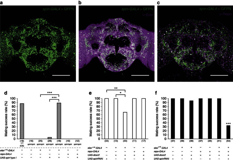
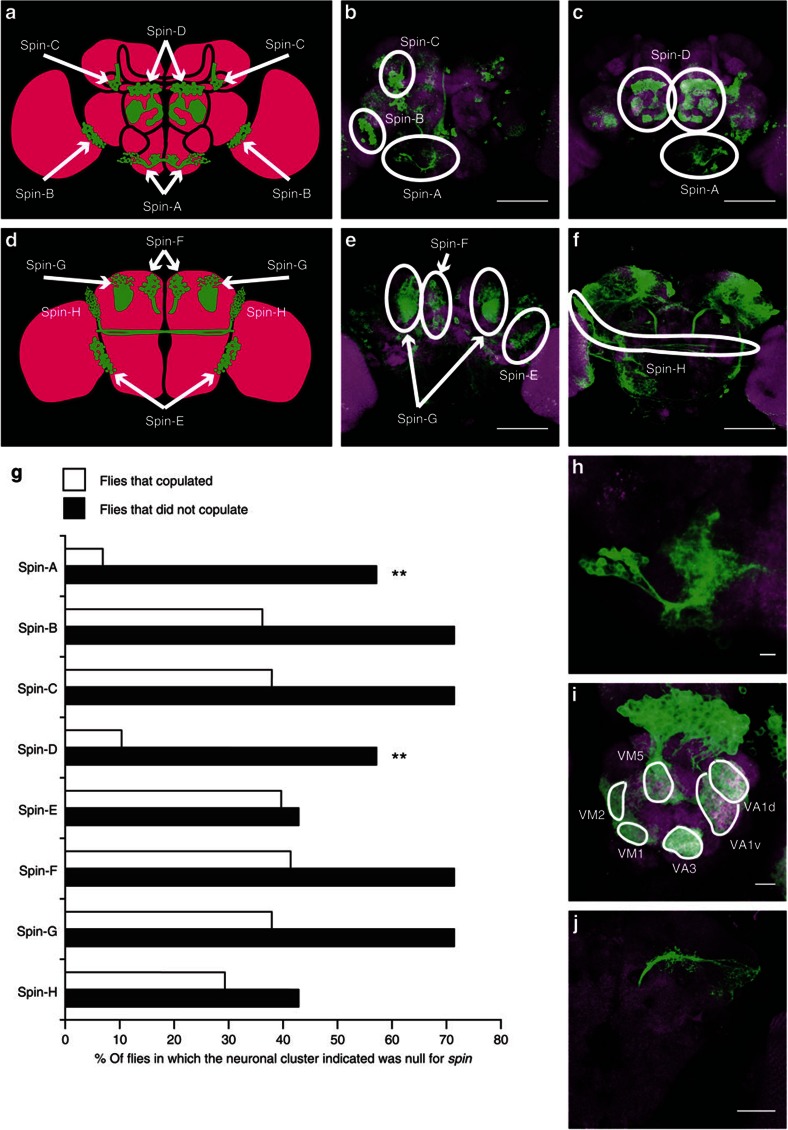
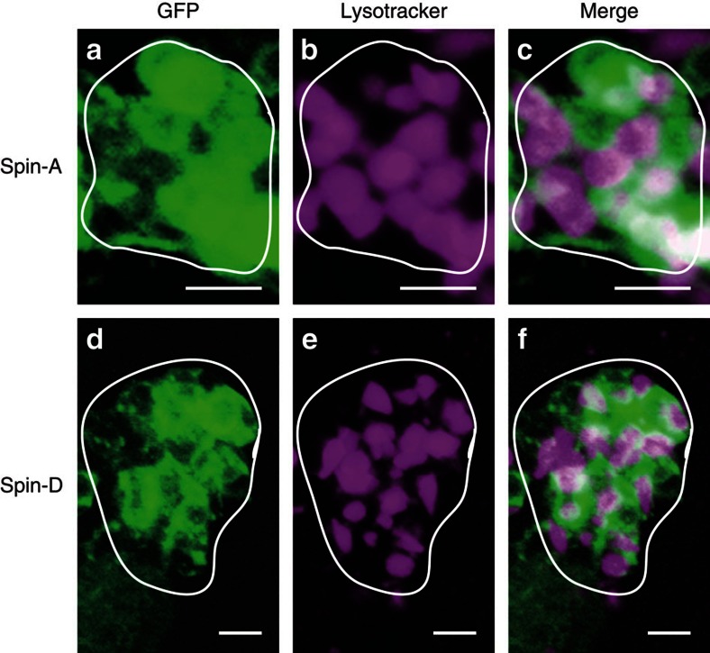
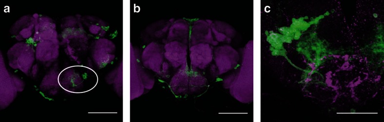
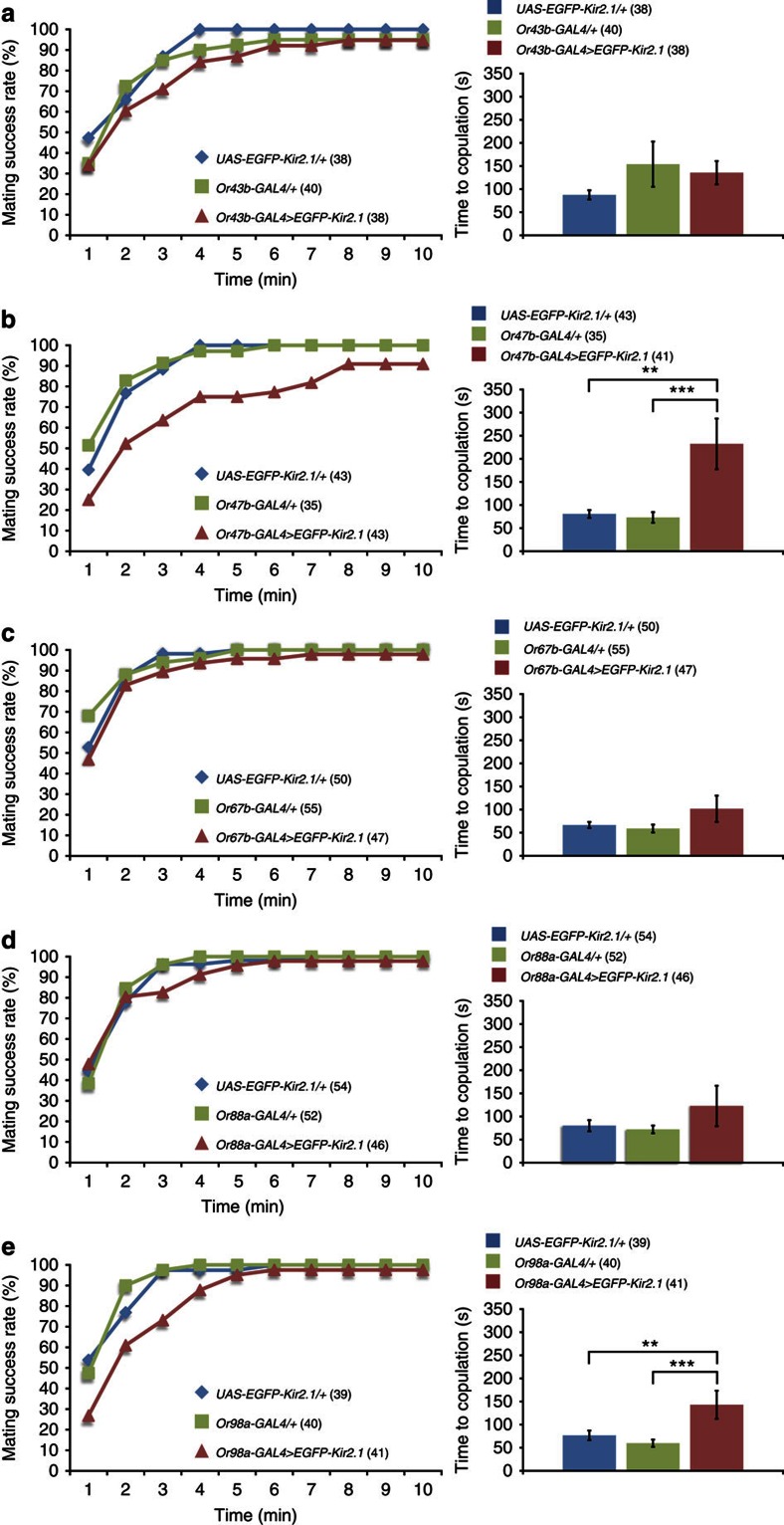
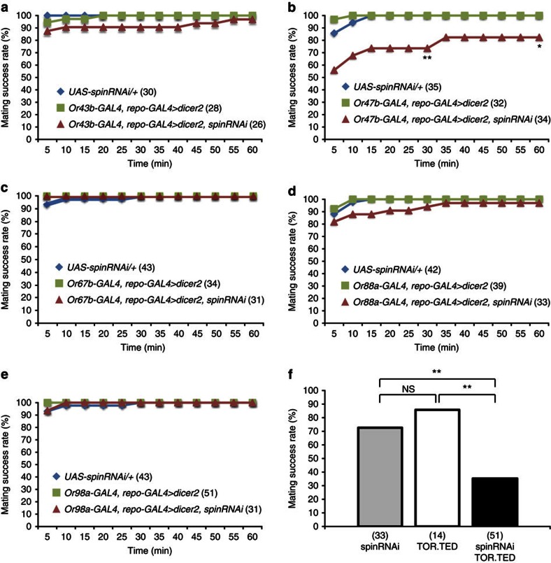
Female Drosophila with the spinster mutation repel courting males and rarely mate. Here we show that the non-copulating phenotype can be recapitulated by the elimination of spinster functions from either spin-A or spin-D neuronal clusters, in the otherwise wild-type (spinster heterozygous) female brain. Spin-D corresponds to the olfactory projection neurons with dendrites in the antennal lobe VA1v glomerulus that is fruitless-positive, sexually dimorphic and responsive to fly odour. Spin-A is a novel local neuron cluster in the suboesophageal ganglion, which is known to process contact chemical pheromone information and copulation-related signals. A slight reduction in spinster expression to a level with a minimal effect is sufficient to shut off female sexual receptivity if the dominant-negative mechanistic target of rapamycin is simultaneously expressed, although the latter manipulation alone has only a marginal effect. We propose that spin-mediated mechanistic target of rapamycin signal transduction in these neurons is essential for females to accept the courting male.


Figures

Figure 1
Figure 1. Contributions of neuronal and glial Spin to female sexual receptivity.
(a) spin-GAL4 expression in the adult female brain was detected with nuclear-targeted GFP (GFPN). (b,c) spin-GAL4-expressing cells are composed of anti-Elav-positive neurons (magenta in b) and anti-Repo-positive glia (magenta in c) in the adult female brain. (d) The effectiveness of UAS-spin+ type I in rescuing the non-copulating phenotype of spinP1 mutant females was estimated by expressing it in neurons with elavc155 or in glia with repo-GAL4. Wild-type (CS, Canton-S) and other control flies were also examined. The spin gene produces five isoforms, Spin I—V, only two of which (Spin I and Spin V) have been demonstrated to rescue the sexual receptivity phenotype. Transgenes the fly carried are shown at the bottom; + and — indicate the presence and absence of the indicated transgene in each fly group. The number of flies examined is shown in parentheses. The female sexual receptivity was measured by the percentage of pairs copulating within a 1-h observation period at 25 °C. (e) The effect of spin knockdown in either neurons or glia on sexual receptivity when the flies were raised at 29 °C to attain higher levels of transgene expression. Dicer2 was also used to enhance the RNAi effect. Neuronal but not glial expression of RNAi was effective in reducing sexual receptivity. (f) When the flies were raised at 25 °C, transgene expression levels would be lower than at 29 °C, spin RNAi expression in either neurons or glia had no discernible effect on receptivity, while its simultaneous expression in both neurons and glia reduced sexual receptivity. The statistical significance of differences was evaluated by the Fisher’s exact probability test with Bonferroni correction (***P<0.001; **P<0.01; *P<0.05). Simultaneous spin knockdown in neurons and glia was not possible at 29 °C because practically no adult flies were recovered for lethality. Scale bar, 100 μm for ac.
Figure 2
Figure 2. Two neuronal clusters reduced receptivity when homozygous for spin in the otherwise spin heterozygous brain.
(af) Eight neural clusters repeatedly labelled as spin mutant cells were present in non-copulating mosaic females. Four clusters (spin-A to -D) in the anterior brain shown as a schematic (a) or reconstructed images (b,c). Another four clusters (spin-E to -H) in the posterior brain shown as a schematic (d) or reconstructed images (e,f). Scale bar, 100 μm. (g) The proportions of flies in which the indicated clusters were spin homozygous in the two fly groups, that is, copulating flies (open bars) and non-copulating flies (filled bars). Female flies that did not copulate within a 1-h observation period were classified as ‘non-copulating.’ By examining 902 mosaic females, we recovered seven non-copulating females that were then used to estimate the spin-homozygous ratio for each neuron cluster. To determine the neuronal spin-homozygous ratio for copulating flies, the brains from 58 flies were scored. These 58 flies were arbitrarily chosen from fly pools that yielded a non-copulating mosaic fly. Pairwise comparisons of the spin mutant ratio between the copulating and non-copulating groups for each cell cluster revealed statistically significant differences only in the spin-A and -D clusters. The statistical significance of differences was evaluated by the Fisher’s exact probability test (**P<0.01). (h) The structure of spin-A cluster neurons labelled as a MARCM clone. (i,j) The structure of spin-D cluster neurons labelled as a MARCM clone, which are shown for somata and dendrites in antennal lobe glomeruli (VA1d, VA1v, VA3, VM1, VM2 and VM5; i) and for axons projecting to the mushroom body and lateral horn (j). Scale bar, 10 μm (h,i) and 50 μm (j).
Figure 3
Figure 3. Spin-A and -D MARCM clones examined for the expression of lysotracker.
GFP fluorescence monitoring spin-GAL4 expression (left-hand side panel), lysotracker signals (middle panel) and their merged image (right-hand side panel) are shown for spin-A (ac) and spin-D (df) clusters that are spin mutant clones generated by MARCM. Scale bar, 5 μm.
Figure 4
Figure 4. Anatomical and neurochemical characteristics of spin-A cluster neurons.
(a) The spin-A cluster was labelled with mCD8::GFP (circled). (b) The spin-A cluster was never labelled when the fly carried Chat-GAL80, which inhibits the GAL4 function in cholinergic neurons. Anterior view. Scale bar, 100 μm. (c) Spin-A cluster neurons have arborizations interdigitating with ppk-positive axon terminals. The ppk-mCherry fusion gene was used to label ppk neurons in the brain that carried a spin-A MARCM clone marked with mCD8::GFP. Scale bar, 50 μm. Green: spin-GAL4-positive cells (ac). Magenta: neuropil staining with nc82 (a,b) or mCherry expression (c).
Figure 5
Figure 5. Differential effects of blocking olfactory receptor neuronal activity on sexual receptivity.
The cumulative number of pairs copulating over time (relative to the total pairs observed: left-hand side graphs) and the time to copulation (mean±s.e.m: right-hand side graph) were compared among the females in which olfactory receptor neurons expressing either Or43b (a), Or47b (b), Or67b (c), Or88a (d) or or98a (e) were prevented from firing. The statistical significance of differences was evaluated by the Kruskal–Wallis analysis of variance followed by Scheffe’s F test for the measure of time to copulation (***P<0.001; **P<0.01). The number of flies examined is shown in parentheses.
Figure 6
Figure 6. The effect of spin RNAi expressed in olfactory receptor neurons and of mTOR inhibition on sexual receptivity.
(ae) The cumulative number of pairs copulating over time (relative to the total pairs observed) was compared among the females in which spin RNAi was expressed in olfactory receptor neurons expressing either Or43b (a), Or47b (b), Or67b (c), Or88a (d) or Or98a (e). Note that repo-GAL4 and UAS-dicer2 were used to enhance the effect of spin RNAi on sexual receptivity. The statistical significance of differences was evaluated by the Fisher’s exact probability test with Bonferroni correction for the cumulative copulation success (**P<0.01; *P< 0.05). (f) Synergy of mTor and spin in the regulation of female receptivity. Although the mating success rate in a 1-h observation period was only moderately reduced by the elav-GAL4-mediated expression of either dominant-negative mTor (TOR.TED, 85.7%; n=14) or spin RNAi (spinRNAi; 72.7%; n=33), simultaneous expression of these two (TOR.TED+spinRNAi) resulted in a marked decrease in the receptivity (35.3%; n=51). The statistical differences were evaluated by the Fisher’s exact probability test with Bonferroni correction, or by the χ2-test for the comparison in which the expectation frequency exceeded 5 in all parameters (**P<0.01; not significant (NS) P>0.05). The number of flies examined is shown in parentheses.

Similar articles

Cited by

References

    1. Sander van Doorn G., Edelaar P. & Weissing F. J. On the origin of species by natural and sexual selection. Science 326, 1704–1707 (2009). - PubMed
    1. Manning A. The control of sexual receptivity in female. Drosophila Anim. Behav. 15, 239–250 (1967). - PubMed
    1. Suzuki K., Juni N. & Yamamoto D. Enhanced mate refusal in female Drosophila induced by a mutation in the spinster locus. Appl. Entomol. Zool. 32, 235–243 (1997).
    1. Nakano Y. et al. Mutations in the novel membrane protein Spinster interfere with programmed cell death and cause neural degeneration in Drosophila melanogaster. Mol. Cell Biol. 21, 3775–3788 (2001). - PMC - PubMed
    1. Lee T. & Luo L. Mosaic analysis with a repressible cell marker for studies of gene function in neuronal morphogenesis. Neuron 22, 451–461 (1999). - PubMed

Publication types

MeSH terms

LinkOut - more resources