

Blue-light reflectance imaging of macular pigment in infants and children
- PMID: 23652486
- PMCID: PMC3680006
- DOI: 10.1167/iovs.13-11891
Blue-light reflectance imaging of macular pigment in infants and children
Abstract
Purpose: While the role of the macular pigment carotenoids in the prevention of age-related macular degeneration has been extensively studied in adults, comparatively little is known about the physiology and function of lutein and zeaxanthin in the developing eye. We therefore developed a protocol using a digital video fundus camera (RetCam) to measure macular pigment optical density (MPOD) and distributions in premature infants and in children.
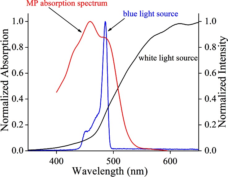
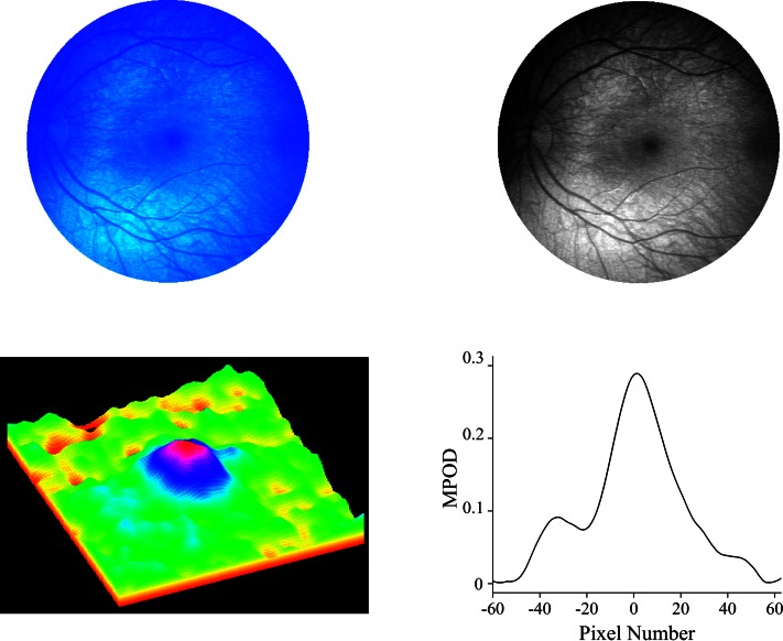
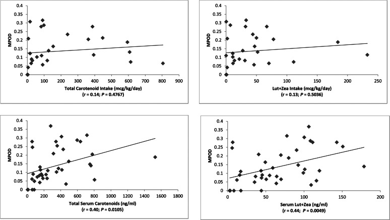
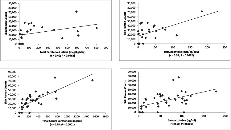
Methods: We used blue light reflectance to image the macular pigment in premature babies at the time of retinopathy of prematurity (ROP) screening and in children aged under 7 years who were undergoing examinations under anesthesia for other reasons. We correlated the MPOD with skin carotenoid levels measured by resonance Raman spectroscopy, serum carotenoids measured by HPLC, and dietary carotenoid intake.
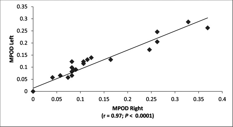
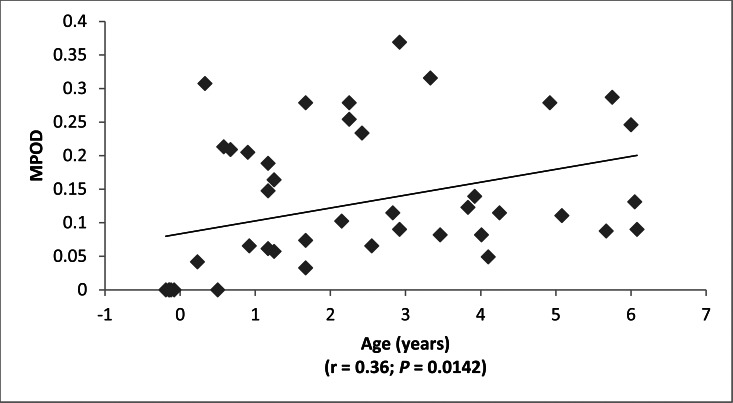
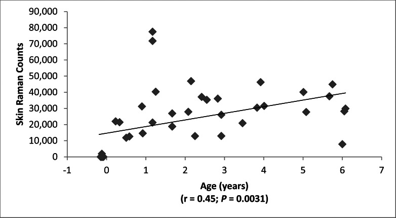
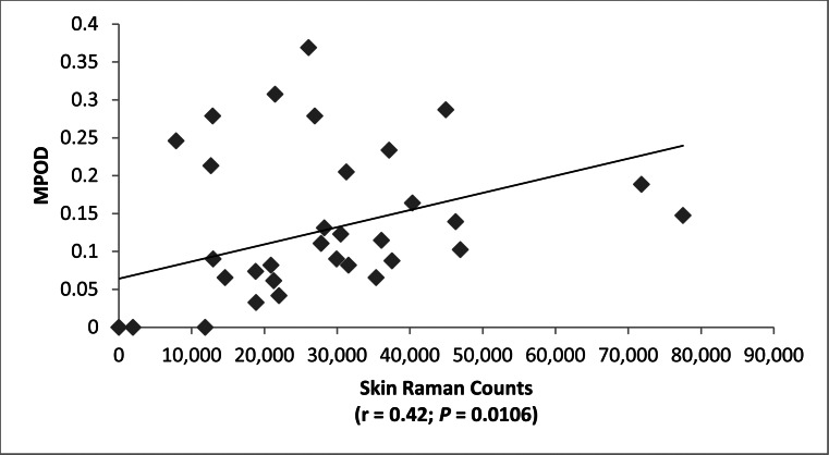
Results: We enrolled 51 infants and children ranging from preterm to age 7 years. MPOD correlated significantly with age (r = 0.36; P = 0.0142), with serum lutein + zeaxanthin (r = 0.44; P = 0.0049) and with skin carotenoid levels (r = 0.42; P = 0.0106), but not with dietary lutein + zeaxanthin intake (r = 0.13; P = 0.50). All premature infants had undetectable macular pigment, and most had unusually low serum and skin carotenoid concentrations.
Conclusions: Our most remarkable finding is the undetectable MPOD in premature infants. This may be due in part to foveal immaturity, but the very low levels of serum and skin carotenoids suggest that these infants are carotenoid insufficient as a consequence of low dietary intake and/or severe oxidative stress. The potential value of carotenoid supplementation in the prevention of ROP and other disorders of prematurity should be a fruitful direction for further investigation.
Keywords: carotenoid; imaging; lutein; macular pigment; zeaxanthin.
Figures
References
-
- Nussbaum JJ, Pruett RC, Delori FC. Historic perspectives. Macular yellow pigment. The first 200 years. Retina. 1981; 1: 296–310 - PubMed
-
- Ma L, Dou HL, Wu YQ, et al. Lutein and zeaxanthin intake and the risk of age-related macular degeneration: a systematic review and meta-analysis. Br J Nutr. 2012; 107: 350–359 - PubMed
-
- Loane E, Nolan JM, O'Donovan O, Bhosale P, Bernstein PS, Beatty S. Transport and retinal capture of lutein and zeaxanthin with reference to age-related macular degeneration. Surv Ophthalmol. 2008; 53: 68–81 - PubMed
-
- Howells O, Eperjesi F, Bartlett H. Measuring macular pigment optical density in vivo: a review of techniques. Graefes Arch Clin Exp Ophthalmol. 2011; 249: 315–347 - PubMed
Publication types
MeSH terms
Substances
Grants and funding
LinkOut - more resources
Full Text Sources
Other Literature Sources
