

Choroid development and feasibility of choroidal imaging in the preterm and term infants utilizing SD-OCT
- PMID: 23652488
- PMCID: PMC3684216
- DOI: 10.1167/iovs.12-11471
Choroid development and feasibility of choroidal imaging in the preterm and term infants utilizing SD-OCT
Abstract
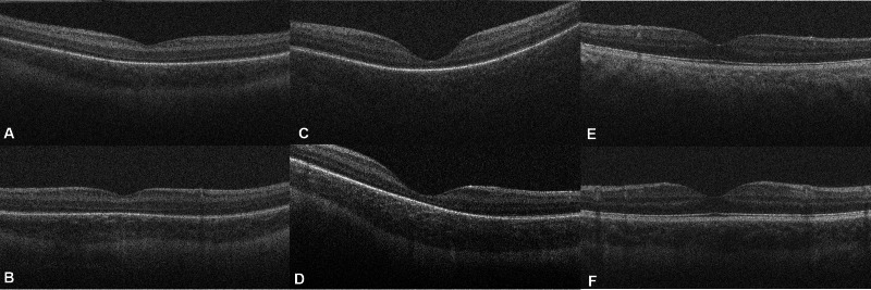
Purpose: To determine whether choroidal imaging is feasible in preterm and term infants using an 840-nm portable spectral domain optical coherence tomography (SD-OCT) system without the use of enhanced-depth imaging techniques and to assess choroidal development by comparing choroidal thickness of preterm infants, term infants, and adults.
Methods: SD-OCT images were obtained from 86 preterm infants, 59 term infants, and nine adults using a portable SD-OCT system plus nine adults using a tabletop system. An unprocessed image across the macula from one randomly selected eye of each participant was selected for determination of whether the choroidal-scleral junction (CSJ) could be visualized and for measurement of choroidal thickness.
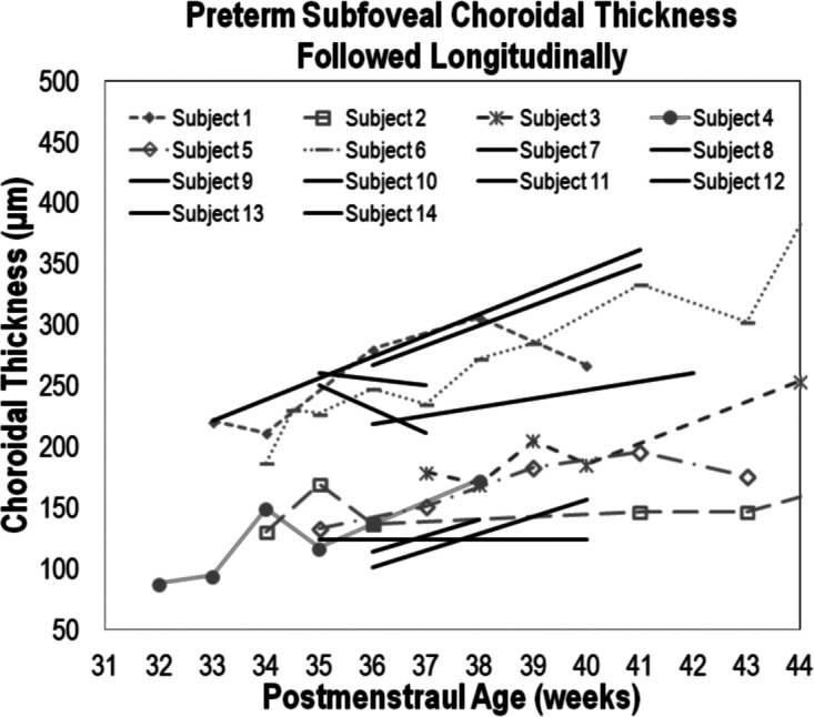
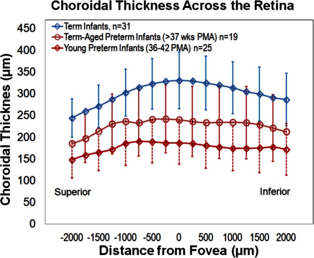
Results: Subfoveal CSJ was visualized in 96% of young-preterm infants (imaged from 30-36 weeks postmenstrual age [PMA]); 78% of term-aged preterm infants (imaged from 37-42 weeks PMA); 49% of term infants; and 39% of adult subjects. Racial pigmentation did not affect CSJ visibility in young-preterm infants (P = 0.57). Subfoveal choroidal thickness (SFCT) in young-preterm infants, term-aged preterm infants, term infants, and adults was 176 ± 53 μm, 289 ± 92 μm, 329 ± 66 μm, and 258 ± 66 μm, respectively, and these were all statistically significantly different from one another except term-aged preterms to adults.
Conclusions: Infant choroid can be imaged with a portable SD-OCT system without enhanced depth imaging. Melanin in the RPE and choroid does not hinder outer choroidal imaging in young-preterm infants without advanced retinopathy of prematurity (ROP). In preterm infants, choroidal thickness increased with age but was thinner when compared to term infants suggesting delayed development due to ROP.
Keywords: choroid; ocular development; optical coherence tomography; retinopathy of prematurity.
Figures

Comment in
-
Choroidal imaging in preterm and term infants.Surv Ophthalmol. 2014 Nov-Dec;59(6):671-2. doi: 10.1016/j.survophthal.2014.01.007. Epub 2014 Mar 11. Surv Ophthalmol. 2014. PMID: 25444366 No abstract available.
References
-
- Yin ZQ, Vaegan , Millar TJ, Beaumont P, Sarks S. Widespread choroidal insufficiency in primary open-angle glaucoma. J Glaucoma. 1997; 6: 23–32 - PubMed
-
- Imamura Y, Fujiwara T, Margolis R, Spaide RF. Enhanced depth imaging optical coherence tomography of the choroid in central serous chorioretinopathy. Retina. 2009; 29: 1469–1473 - PubMed
-
- Switzer DW Jr, Mendonca LS, Saito M, Zweifel SA, Spaide RF. Segregation of ophthalmoscopic characteristics according to choroidal thickness in patients with early age-related macular degeneration. Retina. 2012; 32: 1265–1271 - PubMed
-
- Saint-Geniez M, D'Amore PA. Development and pathology of the hyaloid, choroidal and retinal vasculature. Int J Dev Biol. 2004; 48: 1045–1058 - PubMed
Publication types
MeSH terms
Grants and funding
LinkOut - more resources
Full Text Sources
Other Literature Sources
