Review
. 2013 Sep 15;591(18):4405-13.
doi: 10.1113/jphysiol.2013.251629. Epub 2013 May 7.

Muscle glycogen stores and fatigue

Affiliations
Review

Muscle glycogen stores and fatigue

Niels Ørtenblad et al. J Physiol. .

Abstract

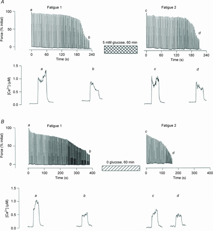
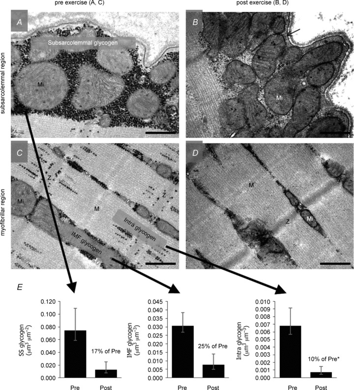
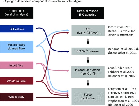
Studies performed at the beginning of the last century revealed the importance of carbohydrate as a fuel during exercise, and the importance of muscle glycogen on performance has subsequently been confirmed in numerous studies. However, the link between glycogen depletion and impaired muscle function during fatigue is not well understood and a direct cause-and-effect relationship between glycogen and muscle function remains to be established. The use of electron microscopy has revealed that glycogen is not homogeneously distributed in skeletal muscle fibres, but rather localized in distinct pools. Furthermore, each glycogen granule has its own metabolic machinery with glycolytic enzymes and regulating proteins. One pool of such glycogenolytic complexes is localized within the myofibrils in close contact with key proteins involved in the excitation-contraction coupling and Ca2+ release from the sarcoplasmic reticulum (SR). We and others have provided experimental evidence in favour of a direct role of decreased glycogen, localized within the myofibrils, for the reduction in SR Ca2+ release during fatigue. This is consistent with compartmentalized energy turnover and distinctly localized glycogen pools being of key importance for SR Ca2+ release and thereby affecting muscle contractility and fatigability.


Figures

Figure 1
Figure 1. Fatigue occurs more rapidly after recovery without glucose
Original data traces of force (upper panels) and [Ca2+]i (lower panels) obtained in single fast-twitch mouse muscle fibres during fatigue induced by repeated tetanic contractions (100 Hz, 350 ms). This type of fatiguing stimulation resulted in marked decrease in glycogen to ∼30% of the control value (Chin & Allen, 1997). Force and [Ca2+]i were well maintained during a second fatigue run after 60 min of recovery in 5.5 mm glucose (A), which restored glycogen to pre-fatigue levels. Conversely, force and [Ca2+]i were not fully restored and fatigue occurred more rapidly after 60 min of recovery without glucose (B), where glycogen stores remained depleted. Adapted from Figs 5 and 6 of Chin & Allen (1997).
Figure 2
Figure 2. Glycogen content in three subcellular localizations in muscle before and after approximately 1 h of exhaustive exercise
A–D, overview showing the typical localization pattern of glycogen particles in the subsarcolemmal region (A, B) and the myofibrillar region (C, D) of a muscle fibre, pre (A, C) and post (B, D) approximately 1 h of exhaustive exercise. Representative images originate from an arm (m. triceps brachii) type I fibre from trained subjects. Glycogen particles are visualized as black dots, with the intermyofibrillar (IMF) glycogen located between the myofibrils and the intramyofibrillar (Intra) glycogen within the myofibrils, mainly located in the I-band. Subsarcolemmal (SS) glycogen is located between the sarcolemma and the outermost myofibril. E, geometric mean glycogen content in the three localizations, with a significantly higher relative utilization of the Intra glycogen (5–15% of total glycogen) during the exercise compared to SS and IMF glycogen (5–15 and 75% of total glycogen, respectively). Mi, mitochondria; Z, Z-line; M, M-band. Scale bars = 0.5 μm. Original ×40,000 magnification. Adapted from Fig. 4 of Nielsen et al. (2011).
Figure 3
Figure 3. Tentative glycogen-dependent components in muscle fatigue
Measurements at the level from organelles (SR vesicles) to whole body experiments indicate a glycogen-dependent role in the E-C coupling failure leading to muscle fatigue. Arrows indicate the level in the series of events in the E-C coupling, where glycogen has been demonstrated to play a significant role.

References

    1. Allen DG, Lamb GD, Westerblad H. Skeletal muscle fatigue: cellular mechanisms. Physiol Rev. 2008;88:287–332. - PubMed
    1. Bangsbo J, Graham TE, Kiens B, Saltin B. Elevated muscle glycogen and anaerobic energy-production during exhaustive exercise in man. J Physiol. 1992;451:205–227. - PMC - PubMed
    1. Barnes M, Gibson LM, Stephenson DG. Increased muscle glycogen content is associated with increased capacity to respond to T-system depolarisation in mechanically skinned skeletal muscle fibres from the rat. Pflugers Arch. 2001;442:101–106. - PubMed
    1. Bergström J, Hermansen L, Hultman E, Saltin B. Diet muscle glycogen and physical performance. Acta Physiol Scand. 1967;71:140–150. - PubMed
    1. Norman B, Sollevi A, Jansson E. Increased IMP content in glycogen-depleted muscle fibres during submaximal exercise in man. Acta Physiol Scand. 1988;133:97–100. - PubMed