

The role of α-adrenergic receptors in mediating beat-by-beat sympathetic vascular transduction in the forearm of resting man
- PMID: 23652594
- PMCID: PMC3731619
- DOI: 10.1113/jphysiol.2013.250894
The role of α-adrenergic receptors in mediating beat-by-beat sympathetic vascular transduction in the forearm of resting man
Abstract
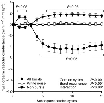
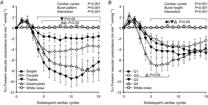
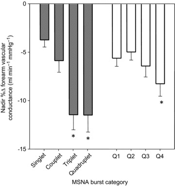
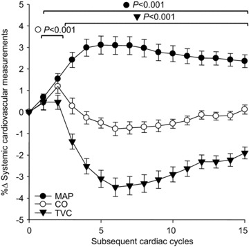
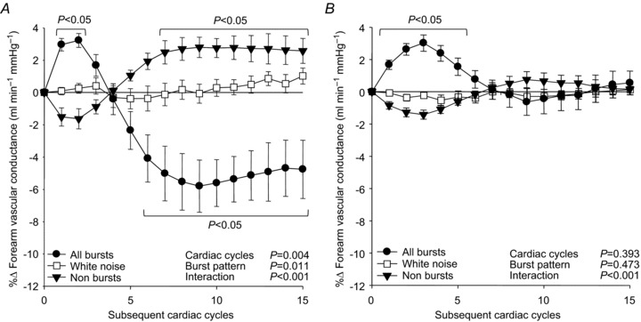
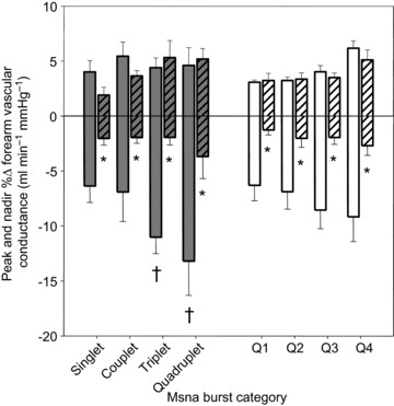
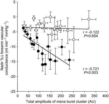
Sympathetic vascular transduction is commonly understood to act as a basic relay mechanism, but under basal conditions, competing dilatory signals may interact with and alter the ability of sympathetic activity to decrease vascular conductance. Thus, we determined the extent to which spontaneous bursts of muscle sympathetic nerve activity (MSNA) mediate decreases in forearm vascular conductance (FVC) and the contribution of local α-adrenergic receptor-mediated pathways to the observed FVC responses. In 19 young men, MSNA (microneurography), arterial blood pressure and brachial artery blood flow (duplex Doppler ultrasound) were continuously measured during supine rest. These measures were also recorded in seven men during intra-arterial infusions of normal saline, phentolamine (PHEN) and PHEN with angiotensin II (PHEN+ANG). The latter was used to control for increases in resting blood flow with α-adrenergic blockade. Spike-triggered averaging was used to characterize beat-by-beat changes in FVC for 15 cardiac cycles following each MSNA burst and a peak response was calculated. Following MSNA bursts, FVC initially increased by +3.3 ± 0.3% (P = 0.016) and then robustly decreased to a nadir of -5.8 ± 1.6% (P < 0.001). The magnitude of vasoconstriction appeared graded with the number of consecutive MSNA bursts; while individual burst size only had a mild influence. Neither PHEN nor PHEN+ANG infusions affected the initial rise in FVC, but both infusions significantly attenuated the subsequent decrease in FVC (-2.1 ± 0.7% and -0.7 ± 0.8%, respectively; P < 0.001 vs. normal saline). These findings indicate that spontaneous MSNA bursts evoke robust beat-by-beat decreases in FVC that are exclusively mediated via α-adrenergic receptors.
Figures
Comment in
-
Alpha males: muscle sympathetic discharge on beat-to-beat forearm vascular conductance.J Physiol. 2013 Sep 15;591(18):4375-6. doi: 10.1113/jphysiol.2013.261743. J Physiol. 2013. PMID: 24037135 Free PMC article. No abstract available.
-
Output vs. outcome: neurovascular transduction at rest.J Physiol. 2014 Jan 15;592(2):257-8. doi: 10.1113/jphysiol.2013.266114. J Physiol. 2014. PMID: 24453351 Free PMC article. No abstract available.
References
-
- Barrett-O’Keefe Z, Witman MA, McDaniel J, Fjeldstad AS, Trinity JD, Ives SJ, Conklin JD, Reese V, Runnels S, Morgan DE, Sander M, Richardson RS, Wray DW. Angiotensin II potentiates α-adrenergic vasoconstriction in the elderly. Clin Sci (Lond) 2013;124:413–422. - PubMed
-
- Cardillo C, Kilcoyne CM, Waclawiw M, Cannon RO, 3rd, Panza JA. Role of endothelin in the increased vascular tone of patients with essential hypertension. Hypertension. 1999;33:753–758. - PubMed
-
- Davis MJ. Perspective: physiological role(s) of the vascular myogenic response. Microcirculation. 2012;19:99–114. - PubMed
-
- Davy KP, Seals DR, Tanaka H. Augmented cardiopulmonary and integrative sympathetic baroreflexes but attenuated peripheral vasoconstriction with age. Hypertension. 1998;32:298–304. - PubMed
Publication types
MeSH terms
Substances
Grants and funding
LinkOut - more resources
Full Text Sources
Other Literature Sources
Medical
Miscellaneous