

doi: 10.1038/jid.2013.218.
Epub 2013 May 7.
Myosin-Va contributes to manifestation of malignant-related properties in melanoma cells
Affiliations
- PMID: 23652798
- PMCID: PMC3806899
- DOI: 10.1038/jid.2013.218
Item in Clipboard
Myosin-Va contributes to manifestation of malignant-related properties in melanoma cells
J Invest Dermatol.
2013 Dec.
No abstract available
Figures
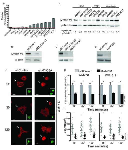
(a) Relative MYO5A mRNA expression detected by qPCR in melanocytes (pMel1 to 4) versus melanoma cells, using β-actin for normalization and mean of all melanocytes as reference value. (b) Western-blot of myosin-Va in a panel of melanoma cell lines of radial growth phase (RGP), vertical growth phase (VGP) and metastasis (M), including two genetic pairs (WM793 and 1205Lu; WM278 and WM1617). (c-e) Western-blots for myosin-Va in (c, d) WM1617 and (e) UACC-257. WM1617 cells were lysed 3 days after transduction with lentiviral vectors carrying shRNAs targeted to bacterial Lac-Z (shControl) or MYO5A (shMYO5A#1), or after stable selection with antibiotics for about 2-3 weeks in the case of shMYO5A#2-3 and respective shControl (Figure S3). UACC-257 cells were lysed 3 days after transfection with siRNA against myosin-Va or control. (f) Confocal images of F-actin stained cells adhered to fibronectin-coated coverslips for the indicated times. Arrows indicate transduced cells visualized by GFP expression (inserts). Scale bar = 20μm. (g) Cells allowed to adhere on fibronectin-coated surface for the indicated times were counted and data were plotted as mean ± SD from 3 independent experiments. (h) Cell spreading. Imaged as in (f) and the areas for 60 cells/time point were measured using ImageJ.

(a-c) Lentiviral transduced WM1617 cells, using three independent shMYO5A or shControls, were used to assess: (a) Colony formation in soft-agar after 30 days of growth. Scale bar = 500μm; (b) Proliferation rates by absorbance measurement of crystal violet staining; (c) Migration in transwell and invasion in transwell-matrigel assay. Cells were kept in starvation conditions 24 hour prior the assay and were then allowed to migrate/invade for 24 hours. Scale bar = 50μm. (d) Migration in 3D collagen. After 24 or 48 hours of incubation - distance from spheroid edge to invasive front was measured and the data from three independent experiments were plotted as a percentage of control. Scale bar = 100μm. (e-f) Transwell invasion and proliferation rates of UACC257 cells transiently transfected with duplex siRNA targeted to MYO5A and irrelevant siRNA. Invasion assay was done as described in c, and proliferation rates were estimated based on ATP measurements. Data were plotted as mean ± SD from 3 independent experiments. Scale bar = 50μm.
References
-
- Alexander S, Friedl P. Cancer invasion and resistance: interconnected processes of disease progression and therapy failure. Trends Mol Med. 2012;18:13–26. - PubMed
-
- Bobrie A, Krumeich S, Reyal F, et al. Rab27a supports exosome-dependent and - independent mechanisms that modify the tumor microenvironment and can promote tumor progression. Cancer Res. 2012;72:4920–30. - PubMed
-
- Dynoodt P, Mestdagh P, Peer GV, et al. Identification of miR-145 as a key regulator of the pigmentary process. J Invest Dermatol. 2013;133(1):201–209. - PubMed
Publication types
MeSH terms
Substances
Grants and funding
LinkOut - more resources
Full Text Sources
Other Literature Sources
Medical
