

Evaluation of protocol before transplantation and after reperfusion biopsies from human orthotopic liver allografts: considerations of preservation and early immunological injury
- PMID: 2365291
- PMCID: PMC3022473
- DOI: 10.1002/hep.1840110605
Evaluation of protocol before transplantation and after reperfusion biopsies from human orthotopic liver allografts: considerations of preservation and early immunological injury
Abstract
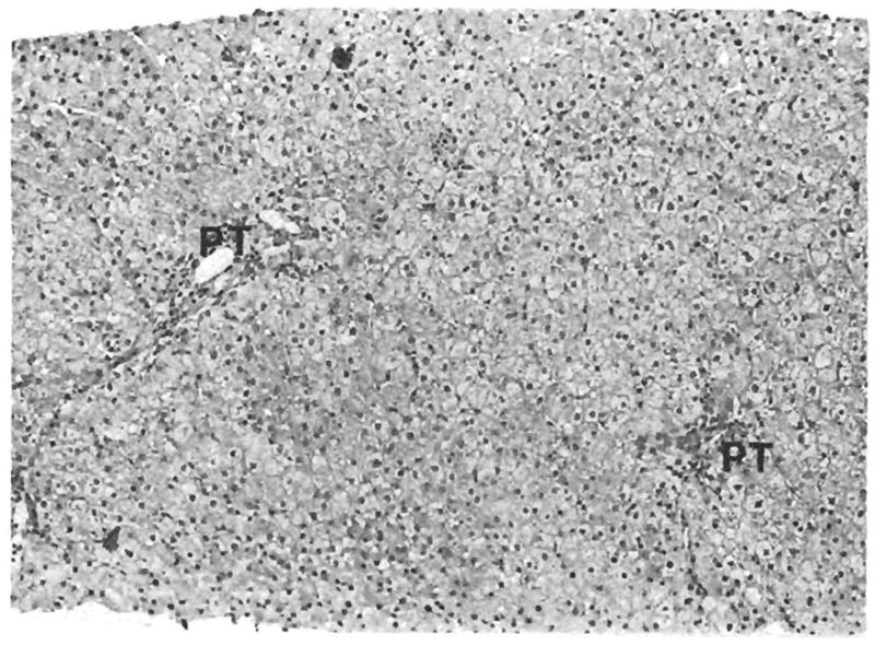
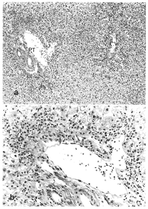
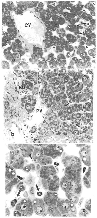
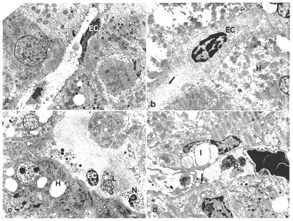
Light microscopic, immunohistochemical and ultrastructural analysis of protocol before transplantation and after reperfusion biopsy specimens from 87 randomly selected patients was performed to assess the contribution of preservation and immunological injury to early graft failure. Most biopsy specimens were essentially normal by light microscopy before transplantation, and no particular feature could be relied on to predict function after transplantation. Ultrastructural examination of biopsy specimens before transplantation demonstrated preferential degeneration of sinusoidal lining cells, but no strict correlation was seen between ultrastructural sinusoidal integrity before transplantation and function after transplantation. The presence of zonal or severe focal necrosis and a severe neutrophilic exudate in biopsy specimens after reperfusion presaged a poor early postoperative course in most, but not all, patients. The presence of preformed lymphocytotoxic antibodies had no effect on the early clinical course, but was associated with Kupffer cell hypertrophy in needle biopsy specimens taken after transplantation. No definite evidence was seen of hyperacute rejection as a result of preformed lymphocytotoxic antibodies as detected in conventional assays. These findings suggest that preservation injury accounts for only a subset of grafts that fail to function after transplantation. Other perioperative or "recipient" factors may be of equal or greater importance in early graft dysfunction or failure.
Figures
Similar articles
-
Role of postreperfusion subcapsular wedge biopsies in predicting initially poor graft function after liver transplantation.Transplant Proc. 2009 Sep;41(7):2747-8. doi: 10.1016/j.transproceed.2009.07.003. Transplant Proc. 2009. PMID: 19765424
-
Liver allograft rejection in sensitized recipients. Observations in a clinically relevant small animal model.Am J Pathol. 1993 May;142(5):1383-91. Am J Pathol. 1993. PMID: 8494042 Free PMC article.
-
Influence of preformed donor-specific antibodies and C4d on early liver allograft function.Scand J Gastroenterol. 2013 Dec;48(12):1444-51. doi: 10.3109/00365521.2013.845795. Epub 2013 Oct 16. Scand J Gastroenterol. 2013. PMID: 24131305
-
The Human Immune Response to Cadaveric and Living Donor Liver Allografts.Front Immunol. 2020 Jun 22;11:1227. doi: 10.3389/fimmu.2020.01227. eCollection 2020. Front Immunol. 2020. PMID: 32655558 Free PMC article. Review.
-
Transplantation pathology.Semin Liver Dis. 2009 Feb;29(1):74-90. doi: 10.1055/s-0029-1192057. Epub 2009 Feb 23. Semin Liver Dis. 2009. PMID: 19235661 Review.
Cited by
-
Timing of arterialization in liver transplantation.Ann Surg. 1994 Nov;220(5):691-8. doi: 10.1097/00000658-199411000-00014. Ann Surg. 1994. PMID: 7979619 Free PMC article.
-
Correlation of Histopathologic Findings of Non-Graft Threatening Preservation/Reperfusion Injury in Time-Zero Liver Needle Biopsies With Short-Term Post-transplantation Laboratory Alterations.Hepat Mon. 2015 Jun 23;15(6):e30008. doi: 10.5812/hepatmon.30008v2. eCollection 2015 Jun. Hepat Mon. 2015. PMID: 26288638 Free PMC article.
-
Does the presence of a measurable blood alcohol level in a potential organ donor affect the outcome of liver transplantation?Alcohol Clin Exp Res. 1991 Mar;15(2):300-3. doi: 10.1111/j.1530-0277.1991.tb01873.x. Alcohol Clin Exp Res. 1991. PMID: 2058808 Free PMC article.
-
The biological basis of and strategies for clinical xenotransplantation.Immunol Rev. 1994 Oct;141:213-44. doi: 10.1111/j.1600-065x.1994.tb00879.x. Immunol Rev. 1994. PMID: 7868154 Free PMC article. Review.
-
Postreperfusion Liver Biopsy as Predictor of Early Graft Dysfunction and Survival After Orthotopic Liver Transplantation.J Clin Exp Hepatol. 2022 Jul-Aug;12(4):1133-1141. doi: 10.1016/j.jceh.2021.12.015. Epub 2022 Jan 4. J Clin Exp Hepatol. 2022. PMID: 35814514 Free PMC article.
References
-
- Shaw BW, Wood RP. Improved results with retransplantation of the liver. Transplant Proc. 1989;21:2407–2488. - PubMed
-
- Paulsen AW, Brajtbord D, Klintmalm GB, Ramsay MA, Swygert TH, Valek TR. Intraoperative measurements related to subsequent hepatic graft failure. Transplant Proc. 1989;21:2337–2338. - PubMed
MeSH terms
Grants and funding
LinkOut - more resources
Full Text Sources
Other Literature Sources
Medical