. 2013 May 7;2(5):e90.
doi: 10.1038/mtna.2013.17.

Vectofusin-1, a new viral entry enhancer, strongly promotes lentiviral transduction of human hematopoietic stem cells

Affiliations

Vectofusin-1, a new viral entry enhancer, strongly promotes lentiviral transduction of human hematopoietic stem cells

David Fenard et al. Mol Ther Nucleic Acids. .

Abstract

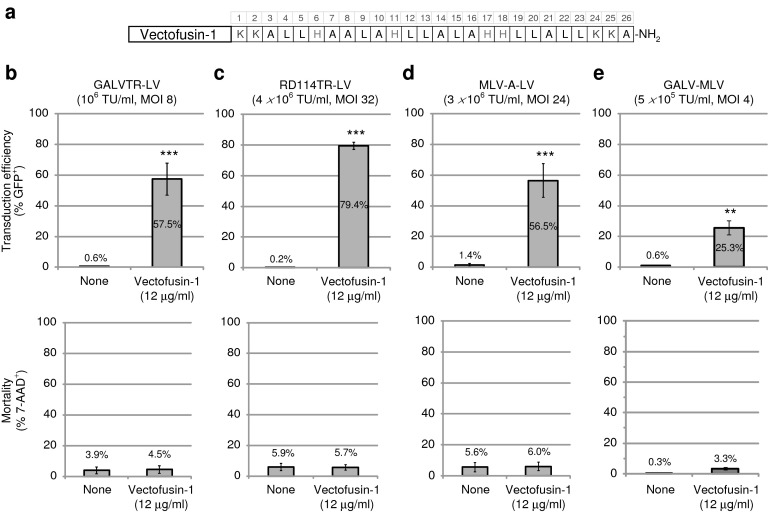
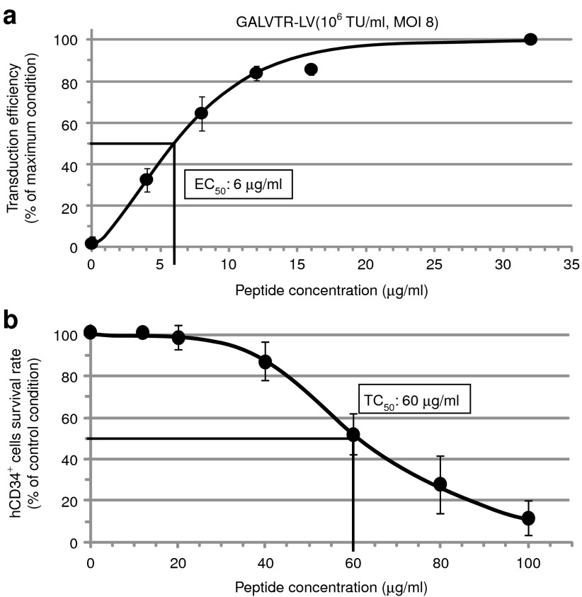
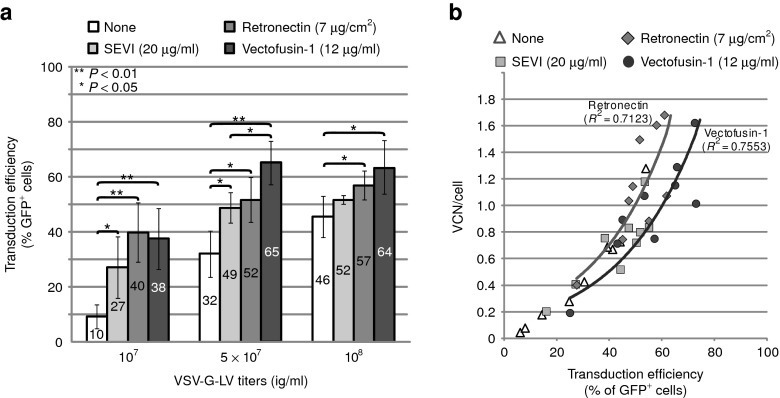
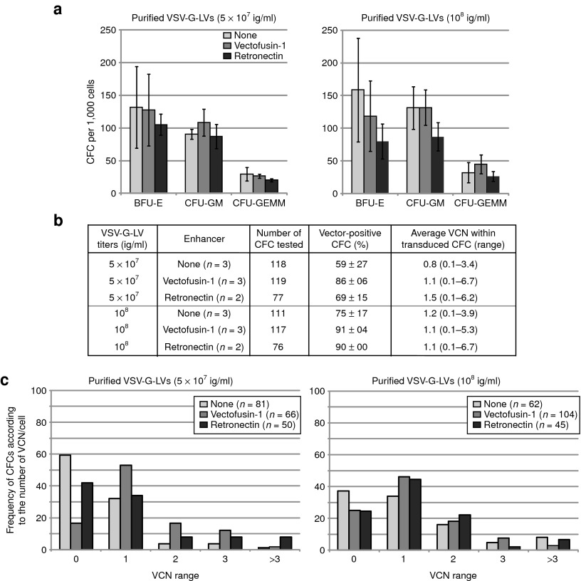
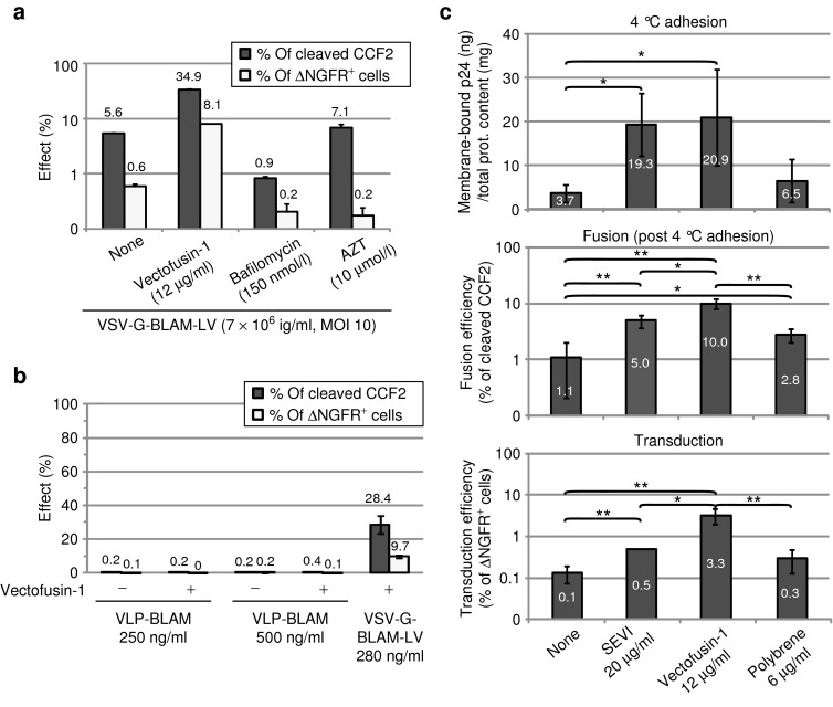
Gene transfer into hCD34(+) hematopoietic stem/progenitor cells (HSCs) using human immunodeficiency virus type 1 (HIV-1)-based lentiviral vectors (LVs) has several promising therapeutic applications. Yet, efficiency, safety, and cost of LV gene therapy could be ameliorated by enhancing target cell transduction levels and reducing the amount of LV used on the cells. Several transduction enhancers already exist such as fibronectin fragments and cationic compounds, but all present limitations. In this study, we describe a new transduction enhancer called Vectofusin-1, which is a short cationic peptide, active on several LV pseudotypes. Vectofusin-1 is used as a soluble additive to safely increase the frequency of transduced HSCs and to augment the level of transduction to one or two copies of vector per cell in a vector dose-dependent manner. Vectofusin-1 acts at the entry step by promoting the adhesion and the fusion between viral and cellular membranes. Vectofusin-1 is therefore a promising additive that could significantly ameliorate hCD34(+) cell-based gene therapy.Molecular Therapy-Nucleic Acids (2013) 2, e90; doi:10.1038/mtna.2013.17; published online 7 May 2013.


Figures

Figure 1
Figure 1
Vectofusin-1 enhances CD34+ hematopoietic stem/progenitor cells transduction with various lentiviral or retroviral pseudotypes. (a) Schematic representation of the primary sequence of Vectofusin-1 peptide composed of four different types of residue: lysine (K), histidine (H), leucine (L), and alanine (A); carboxy-terminal amidation (-NH2). (be) A variety of green fluorescent protein (GFP)-encoding vectors (GALVTR-LVs (n = 5), RD114TR-LVs (n = 3), MLV-A-LVs (n = 3), GALV-MLV (n = 3)) were used to transduce hCD34+ cells during 6 hours in the absence (none) or presence of Vectofusin-1 (12 µg/ml). Data are shown as the average percentage of GFP+ or 7-AAD+ cells ± SD from number of umbilical cord blood samples treated in duplicate (**P < 0.01; ***P < 0.001, Student's t-test). 7-AAD, 7-aminoactinomycin D; GALV, gibbon ape leukemia virus; LV, lentiviral vector; MLV-A, amphotropic murine leukemia virus; MOI, multiplicity of infection; TU, transducing unit.
Figure 2
Figure 2
Determination of the half maximal efficient (EC50) and toxic (TC50) concentrations of Vectofusin-1. (a) hCD34+ cells were infected with GALVTR-LVs in the absence or presence of various concentrations of Vectofusin-1. Transduction efficiencies (percentage of GFP+ cells) were obtained 5 days post-transduction (n = 4). Data are normalized to the maximum effect observed ± SD (average maximal value of transduction was 67%). (b) Evaluation of the TC50 of Vectofusin-1. The hCD34+ cells were incubated overnight with the indicated amounts of Vectofusin-1 (n = 6). The survival rate was estimated by counting the number of living cells using the Trypan blue exclusion method under light microscopy. Data are normalized to the control condition ± SD (average value of survival rate in the absence of peptides was 98.9%). GALV, gibbon ape leukemia virus; LV, lentiviral vector; MOI, multiplicity of infection; TU, transducing unit.
Figure 3
Figure 3
Comparison of Vectofusin-1 with other culture additives to promote transduction of hCD34+ cells with purified vesicular stomatitis virus-G-lentiviral vectors (VSV-G-LVs). hCD34+ cells (three umbilical cord blood donors tested in three independent experiments) were infected with increasing concentrations of purified VSV-G-LVs (107, 5 × 10⁷, and 108 ig/ml corresponding to multiplicity of infection (MOI) 80, 400, and 800, respectively) in the absence (none) or presence of indicated transduction enhancers. (a) Transduction was measured in the bulk of cultured cells after 5 days by following the percentage of GFP+ cells ± SD using flow cytometry (**P < 0.01; *P < 0.05, Student's t-test) and (b) average vector copy number (VCN) of the cell population by quantitative PCR. GFP, green fluorescent protein; ig, infectious genome; SEVI, semen enhancer of viral infection.
Figure 4
Figure 4
Lack of in vitro hematopoietic toxicity of Vectofusin-1. (a) Differentiation of transduced hCD34+ cells in colony-forming cell (CFC) assays. Results represent the average number of different types of colonies obtained for 1,000 cells plated after transduction with 5 × 10⁷ and 108 ig/ml (conditions of Figure 3). (b) Transduction was measured in individual CFCs obtained after 2 weeks of culture in methylcellulose. Transduction of CFCs was measured by determining the percentage of GFP+ CFCs using epifluorescence microscopy (“vector-positive CFC (%)”), and the vector copy number (VCN) in each CFC by quantitative PCR. The average VCN per vector-positive CFC and range are listed (right column). (c) Bars represent the percentage of CFCs in each category (VCN range) over the total number of CFCs analyzed. The number of CFCs analyzed is indicated between brackets for each group. BFU-E, burst-forming unit, erythroid; CFU-GEMM, colony-forming unit, granulocyte, erythrocyte, macrophage, megakaryocyte; CFU-GM, colony-forming unit, granulocyte-monocyte; GFP, green fluorescent protein; ig, infectious genome; VSV-G-LV, vesicular stomatitis virus-G-lentiviral vector.
Figure 5
Figure 5
Evaluation of the safety of Vectofusin-1 in the immunodeficient BALB-Rag/γC mouse model. hCD34+ cells were transduced for 6 hours with GALVTR-LVs either in the presence of 12 µg/ml of Vectofusin-1 (squares) or 20 µg/cm2 of Retronectin (circle) and injected into the liver of newborn mice. Controls (NT) included non-infected cells not exposed to any transduction enhancer (triangle). (a,b) Eleven to thirteen weeks post-injection, the engraftment of human transduced cells into HIS (BALB-Rag/γC) mice was measured by flow cytometry in the peripheral blood, the thymus, the spleen, and the bone marrow (BM) using anti-hCD45 antibodies and green fluorescent protein (GFP) expression levels. (c,d) Human T lymphopoiesis was measured in the thymus by monitoring human TCRα/β in the hCD45+ gate, and CD4 and CD8 marker expression in CD3+ low or high (hi) gates. Human B lymphoid development, monocytes, natural killer cells, granulocytes, and hematopoietic progenitors were determined in the (e,f) spleen or the (g,h) BM by monitoring, respectively, the human CD19, CD14, CD56, CD11b, and CD34 surface markers in the hCD45+ gate. GALV, gibbon ape leukemia virus; HIS, human immune system; LV, lentiviral vector; NT, not transduced; TCRα/β, T-cell receptor α/β.
Figure 6
Figure 6
Influence of Vectofusin-1 and other culture additives on the adhesion and fusion step of vesicular stomatitis virus-G-lentiviral vectors (VSV-G-LVs). (a) Viral fusion (percentage of cleaved CCF2) and level of transduction after 9 days (percentage of ΔNGFR+ cells) are represented as the average of duplicates ± SD. Data are representative of two different experiments. (b) Fusion and transduction assay on hCD34+ cells in the presence of BLAM-VLPs (250 or 500 ng/ml) or the positive control VSV-G-BLAM-LVs (280 ng/ml) in the absence or presence of Vectofusin-1 (12 µg/ml). Data are expressed as the average of two independent experiments performed in duplicate ± SD. (c) Adhesion, fusion, and transduction assays of hCD34+ cells in the presence of VSV-G-BLAM-LVs. Pre-cooled hCD34+ cells were incubated for 2.5–3 hours at 4 °C with the pre-cooled vector/peptide mix. Cells were then washed and half of the cells were lysed to evaluate the total membrane-bound p24 (top panel). The remaining cells were further cultured for 2–2.5 hours at 37 °C to induce viral fusion. Next, an aliquot of cells was used for the BLAM fusion assay (middle panel), whereas the remaining cells were further cultured. After 9 days, the transduction efficiency was evaluated as in a by monitoring ΔNGFR expression (lower panel). Data are expressed as the average of three independent experiments ± SD (**P < 0.01; *P < 0.05, Student's t-test). AZT, azidothymidine; BLAM, β-lactamase; MOI, multiplicity of infection; SEVI, semen enhancer of viral infection; VLP, virus-like particle.

References

    1. D'Costa, J, Mansfield, SG and Humeau, LM (2009). Lentiviral vectors in clinical trials: Current status. Curr Opin Mol Ther 11: 554–564. - PubMed
    1. Naldini, L (2011). Ex vivo gene transfer and correction for cell-based therapies. Nat Rev Genet 12: 301–315. - PubMed
    1. Cronin, J, Zhang, XY and Reiser, J (2005). Altering the tropism of lentiviral vectors through pseudotyping. Curr Gene Ther 5: 387–398. - PMC - PubMed
    1. Merten, OW, Charrier, S, Laroudie, N, Fauchille, S, Dugué, C, Jenny, C et al. (2011). Large-scale manufacture and characterization of a lentiviral vector produced for clinical ex vivo gene therapy application. Hum Gene Ther 22: 343–356. - PubMed
    1. Sandrin, V, Boson, B, Salmon, P, Gay, W, Nègre, D, Le Grand, R et al. (2002). Lentiviral vectors pseudotyped with a modified RD114 envelope glycoprotein show increased stability in sera and augmented transduction of primary lymphocytes and CD34+ cells derived from human and nonhuman primates. Blood 100: 823–832. - PubMed