. 2013 Nov 11;9(21):3704-13.
doi: 10.1002/smll.201202075. Epub 2013 May 8.

Transdermal delivery devices: fabrication, mechanics and drug release from silk

Affiliations

Transdermal delivery devices: fabrication, mechanics and drug release from silk

Waseem K Raja et al. Small. .

Abstract

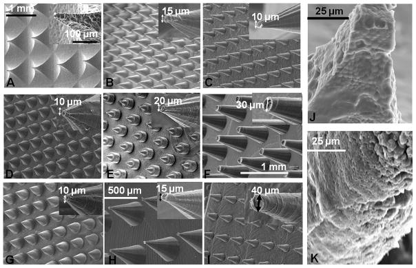
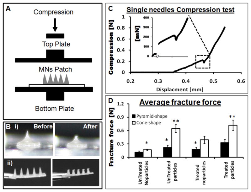
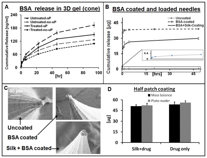
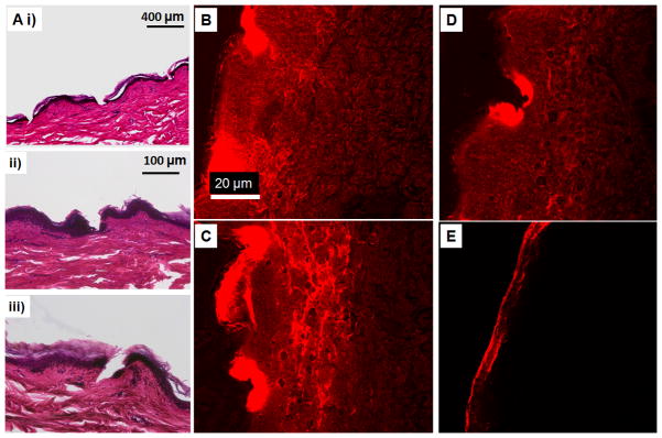
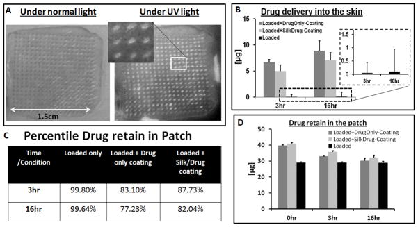
Microneedles are a relatively simple, minimally invasive and painless approach to deliver drugs across the skin. However, there remain limitations with this approach because of the materials most commonly utilized for such systems. Silk protein, with tunable and biocompatibility properties, is a useful biomaterial to overcome the current limitations with microneedles. Silk devices preserve drug activity, offer superior mechanical properties and biocompatibility, can be tuned for biodegradability, and can be processed under aqueous, benign conditions. In the present work, the fabrication of dense microneedle arrays from silk with different drug release kinetics is reported. The mechanical properties of the microneedle patches are tuned by post-fabrication treatments or by loading the needles with silk microparticles, to increase capacity and mechanical strength. Drug release is further enhanced by the encapsulation of the drugs in the silk matrix and coating with a thin dissolvable drug layer. The microneedles are used on human cadaver skin and drugs are delivered successfully. The various attributes demonstrated suggest that silk-based microneedle devices can provide significant benefit as a platform material for transdermal drug delivery.

Keywords: micromachining; microneedles; microparticles; silk; transdermal drug delivery.


Figures

Figure 1
Figure 1
A) (i – iv) Process flow of mold fabrication, showing a machinable wax mold. Left Bottom left is showing SEM images of drilling bits with different tip sizes and bottom right is the fabricated negative MN mold in machinable wax material. B) (i) Schematic of epoxy casting onto the wax mold and optical image of epoxy MN, (ii) Polydimethylsiloxane (PDMS) negative MN mold fabricated from an epoxy-based MN generated from machinable wax, (iii) digital image of pure silk MN prepared in a PDMS mold.
Figure 2
Figure 2
A) SEM digital image of aqueous silk MN (pyramid shape) fabricated by our previous method [43]. B) – F) SEM images of cone shaped MN prepared from an aqueous silk solution with different shapes and geometries. G) – I) SEM images of cone shaped MN made from solvent-based (hexafluoroisopropanol) silk solution with various shapes and sizes. J) and K) Microparticle-loaded aqueous silk MN from pyramid and cone shaped needles, respectively.
Figure 3
Figure 3
A) Schematic of an axial mechanical test setup to assess MN device performance with two horizontal parallel plates. B) (i) and (ii) Digital images of pyramid and a cone shaped needles, respectively, before and after mechanical testing. C) A single needle compression test that breaks around 225 mN during compression. D) Fracture force of all sub-groups of the pyramid and the cone shaped MN fabricated by micromolding. (N = 10, error bars represent standard deviations) Significant effects are indicated by one (p < 0.05) or two stars (p < 0.01), the one start belong to same group and two stars belong to second group on the bases of 95% confidence interval using ANOVA.
Figure 4
Figure 4
BSA release from the silk MN in 3D collagen hydrogels, determined by digestion of collagen in collagenase solution followed by optical spectroscopy of the released BSA. A) Plots showing the release profile from cone shaped microneedles loaded with drug only and also microparticle containing same drug. B) Release kinetics of Drug loaded and coated MN patches. C) SEM digital images of the MN before and after coating with different drug solutions. D) Quantification of coated drug onto MN patch and verified by two separate techniques. (N = 3, error bars represent standard deviations).
Figure 5
Figure 5
A) H&E micrographs of human cadaver skin treated with cone shaped silk MN, (i) MN loaded with microparticles and no post treatment, (ii) MN with no microparticles and post treatment with water vapor and (iii) MN loaded with microparticles and post treated with water vapor. B) Sulforhodamine released into the skin from the cone shaped silk MN loaded with the microparticles and no further post fabrication treatment. C) and D) Sulforhodamine released into human cadaver skin from MN patch with no post fabrication treatment and water vapor annealing treatment, respectively. The needles contained no microparticles. E) Human cadaver skin treated with the control patch (silk film without microneedles).
Figure 6
Figure 6
A) Optical image of human cadaver skin treated with MN patch under normal and UV light, inserted image showing the magnified view of the position of needles fluorescent under UV light. B) Amount of drug delivered into the cadaver skin, inserted plot is the drug delivery from drug loaded patches without coating. C) Table of the drug retains in the MN patches after cadaver skin application. D) Total amount of drug per patch at different time point.

References

    1. Donnelly RF, Majithiya R, Singh TR, Morrow DI, Garland MJ, Demir YK, Migalska K, Ryan E, Gillen D, Scott CJ, Woolfso1n AD. Pharm Res. 2011;28:41–57. - PMC - PubMed
    1. Kochhar JS, Goh WJ, Chan SY, Kang L. Drug Dev Ind Pharm. 2012;39:299–309. - PubMed
    1. McAllister DV, Wang PM, Davis SP, Park JH, Canatella PJ, Allen MG, Prausnitz MR. Proc Natl Acad Sci U S A. 2003;100:13755–60. - PMC - PubMed
    1. Park JH, Allen MG, Prausnitz MR. Pharm Res. 2006;23:1008–19. - PubMed
    1. Coulman SA, Anstey A, Gateley C, Morrissey A, McLoughlin P, Allender C, Birchall JC. Int J Pharm. 2009;366:190–200. - PubMed

Publication types

LinkOut - more resources