. 2013 Jun 14;288(24):17441-50.
doi: 10.1074/jbc.M113.469841. Epub 2013 May 6.

Structural analysis of collagen type I interactions with human fibronectin reveals a cooperative binding mode

Affiliations

Structural analysis of collagen type I interactions with human fibronectin reveals a cooperative binding mode

Michèle C Erat et al. J Biol Chem. .

Abstract

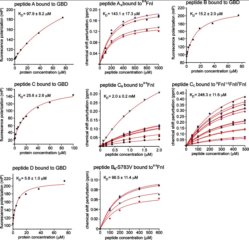
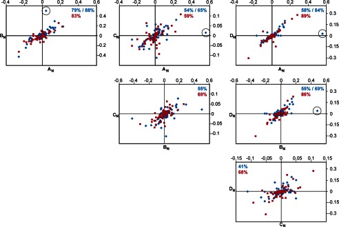
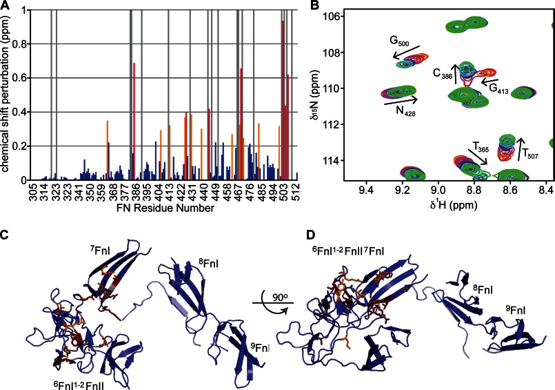
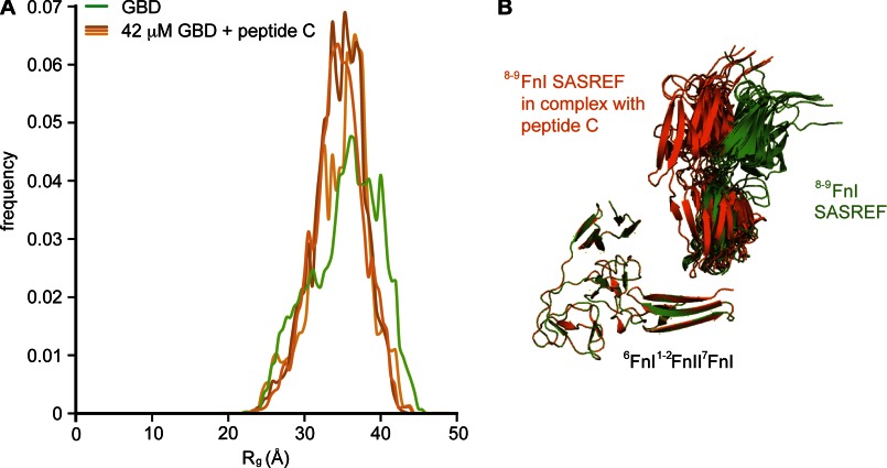
Despite its biological importance, the interaction between fibronectin (FN) and collagen, two abundant and crucial tissue components, has not been well characterized on a structural level. Here, we analyzed the four interactions formed between epitopes of collagen type I and the collagen-binding fragment (gelatin-binding domain (GBD)) of human FN using solution NMR, fluorescence, and small angle x-ray scattering methods. Collagen association with FN modules (8-9)FnI occurs through a conserved structural mechanism but exhibits a 400-fold disparity in affinity between collagen sites. This disparity is reduced in the full-length GBD, as (6)FnI(1-2)FnII(7)FnI binds a specific collagen epitope next to the weakest (8-9)FnI-binding site. The cooperative engagement of all GBD modules with collagen results in four broadly equipotent FN-collagen interaction sites. Collagen association stabilizes a distinct monomeric GBD conformation in solution, giving further evidence to the view that FN fragments form well defined functional and structural units.

Keywords: Collagen; Extracellular Matrix Proteins; Fibronectin; GBD; NMR; SAXS; X-ray Crystallography.


Figures

FIGURE 1.
FIGURE 1.
SAXS data and GBD structure. A, the previously suggested model of a monomeric GBD (blue) based on the crystal structures of 8–9FnI and 6FnI1–2FnII7FnI (7) compared with 10 SASREF-derived GBD models (green). SAXS analysis suggested an ∼90° kink between 7FnI and 8FnI. B, Guinier analysis of the SAXS curve for the GBD yielded an Rg of 3.45 nm and an I0 of 48.77. C, scattering curve of 3 mg/ml GBD overlaid with back-calculated CRYSOL curves from the previously proposed composite GBD model (blue) (7), monomeric and dimeric versions of the GBD crystal structure (dark and light purple) (30), or the GBD SASREF model (green). D, As expected, the SASREF model fits the measured data best, especially in the crucial low angle region.
FIGURE 2.
FIGURE 2.
Ensemble optimization analysis of the GBD. A, three independent ensemble optimization method runs of the GBD SAXS data yielded essentially the same distribution, with an average Rg centered at ∼36 Å. At 2 S.D., the width of the Rg distribution of the GBD alone is 17 Å. Sample GBD models corresponding to the center and tail ends of the distribution for all three runs are shown. B and C, representative back-calculated scattering curves for the best ensembles of the GBD alone (B) and in a 1:1 molar complex with peptide C (C) compared with the respective experimental data. χ values are 0.802 (B) and 0.956 (C). χ values below 1 indicate an acceptable fit to the data.
FIGURE 3.
FIGURE 3.
GBD-binding sites on collagen type I. A, schematic representation of the collagen type I α1 and α2 chains and the two FN-binding sites at 1/10 and 3/4 sequence distance from the collagen N terminus. 8–9FnI-binding sites (peptides AN, BN, CN, and DN) are shown in blue, with the sequences immediately C-terminal thereof (peptides AC, BC, CC, and DC) shown in red. Dissociation constants are indicated. Highlighted in green is the only site where the two GBD subfragments, 6FnI1–2FnII7FnI and 8–9FnI, bind collagen type I cooperatively. B, amino acid sequences of peptides A–D. Conserved positions 2 (Leu) and 9 (Arg) of the 8–9FnI collagen-binding epitope are in shown in boldface; color coding is as in A. The hydrophobic residue-containing triplet that enhances 8–9FnI affinity in the 3/4 sites is indicated in italics, with the crucial hydrophobic residue shown in boldface italics.
FIGURE 4.
FIGURE 4.
Crystal structure of 8–9FnI in complex with a peptide from the collagen α1 1/10 site. A, schematic representation of the crystal structure of 8–9FnI (blue) in complex with the low affinity peptide AN (cyan). B, overlay of the crystal structure of 8–9FnI (red) in complex with the high affinity peptide BN (orange) (26). The antiparallel β-strand mode of binding alongside strand E of 8FnI is conserved, and the primary hydrophobic contacts are indicated. Val783, which plays an important role in increasing the affinity of peptide BN for 8–9FnI but is not part of the consensus 9-mer sequence, is shown. A peptide hairpin just C-terminal of the consensus binding site in both collagen peptides leads to a 90° kink as indicated.
FIGURE 5.
FIGURE 5.
Biophysical studies of collagen interactions with FN modules. Shown here are the protein titration data for interactions summarized in Table 1. For NMR measurements, chemical shift perturbations are plotted against collagen peptide concentration. For fluorescence measurements, we report the polarization of peptide-bound fluorescein against the GBD concentration. All data were fit assuming a single binding event. mP, millipolarization units.
FIGURE 6.
FIGURE 6.
8–9FnI binds collagen through a conserved interaction mode. Shown here are pairwise comparisons of proton (blue) and nitrogen (red) chemical shift changes in 8–9FnI resonances upon addition of collagen peptides AN, BN, CN, and DN. Nitrogen values were divided by a factor of 5 to allow comparison with proton values on the same graph. All values are in ppm. The correlation coefficients (R) of independent linear fits to each data series are indicated in blue (for proton) or red (for nitrogen). In some cases, the coefficients after removal of a single proton data point (circled) are also shown. Note that shift changes for peptides CN and DN are small due to weak binding; thus, experimental errors adversely affect the correlations.
FIGURE 7.
FIGURE 7.
NMR chemical shift analysis of the interaction between 6FnI1–2FnII7FnI and peptide CC. A, combined amide chemical shift differences. Red bars indicate perturbations >2 S.D. from the mean, and orange bars indicate perturbations >1 S.D. Blue bars denote measured chemical shift perturbations < 1 S.D. Gray bars indicate peak disappearance upon titration with the peptide. B, region of a 1H-15N heteronuclear single quantum correlation NMR spectrum showing an overlay of 6FnI1–2FnII7FnI resonances that shift upon addition of peptide CC. C and D, two perpendicular representations of the GBD SASREF model with residues in stick representations and colored according to the chemical shift perturbations found in A. The light blue collagen peptide indicates how the most perturbed residues in 2FnII and 7FnI can form a continuous collagen-binding interface with 8–9FnI.
FIGURE 8.
FIGURE 8.
SAXS analysis of the GBD in complex with collagen. A, ensemble optimization analysis of the GBD alone or in a 1:1 complex with peptide C. Upon complex formation, the Rg distribution narrows through disappearance of minor conformational states. B, schematic representation of 10 SASREF models of the GBD alone (green) or in complex with peptide C (orange). All structures are aligned at the 6FnI1–2FnII7FnI subfragment. Peptide binding does not lead to a major structural rearrangement but stabilizes the pre-existing major conformation.

References

    1. George E. L., Georges-Labouesse E. N., Patel-King R. S., Rayburn H., Hynes R. O. (1993) Defects in mesoderm, neural tube and vascular development in mouse embryos lacking fibronectin. Development 119, 1079–1091 - PubMed
    1. Leitinger B., Hohenester E. (2007) Mammalian collagen receptors. Matrix Biol. 26, 146–155 - PubMed
    1. Campbell I. D., Downing A. K. (1998) NMR of modular proteins. Nat. Struct. Biol. 5, 496–499 - PubMed
    1. Erickson H. P., Carrell N., McDonagh J. (1981) Fibronectin molecule visualized in electron microscopy: a long, thin, flexible strand. J. Cell Biol. 91, 673–678 - PMC - PubMed
    1. Leahy D. J., Aukhil I., Erickson H. P. (1996) 2.0 Å crystal structure of a four-domain segment of human fibronectin encompassing the RGD loop and synergy region. Cell 84, 155–164 - PubMed

Publication types

Associated data

LinkOut - more resources