. 2013 Jul;154(7):2468-80.
doi: 10.1210/en.2012-2109. Epub 2013 May 7.

Progesterone antagonism of neurite outgrowth depends on microglial activation via Pgrmc1/S2R

Affiliations

Progesterone antagonism of neurite outgrowth depends on microglial activation via Pgrmc1/S2R

N Bali et al. Endocrinology. 2013 Jul.

Abstract

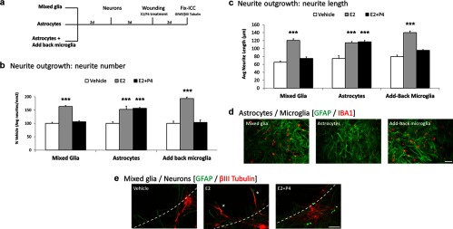
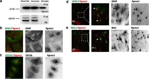
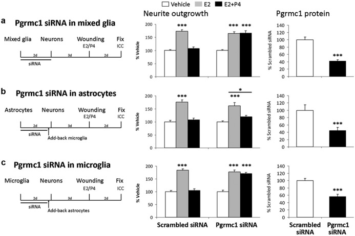
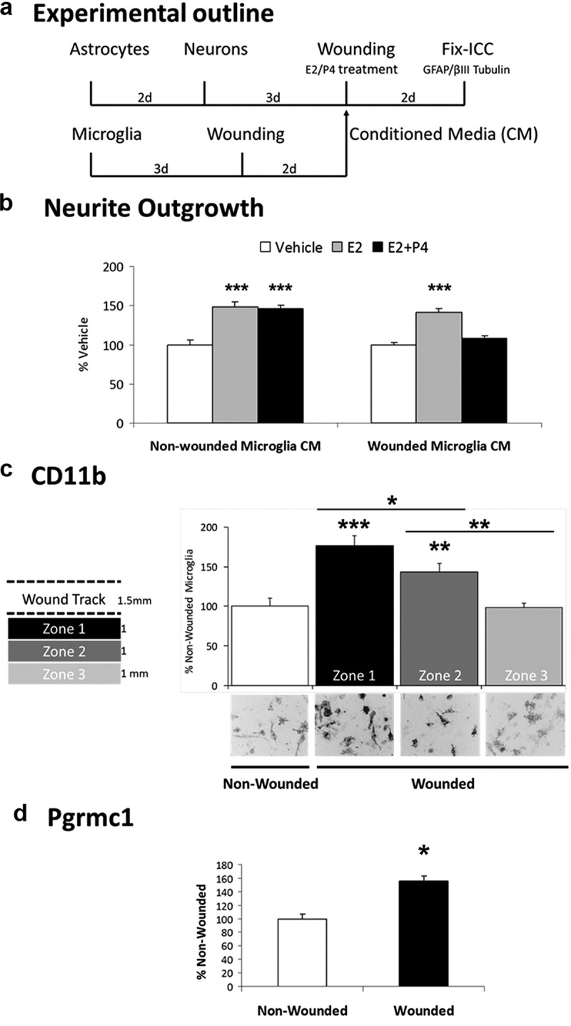
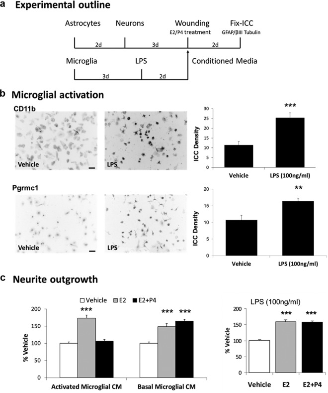
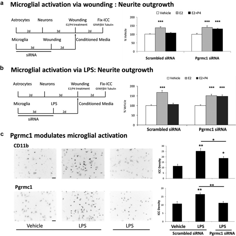
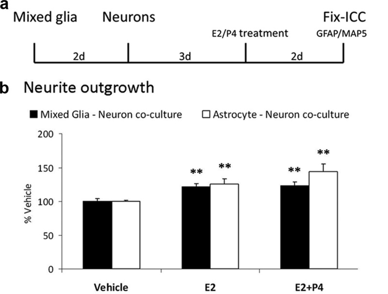
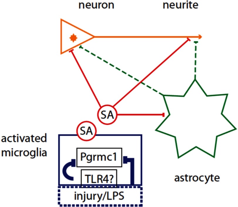
Neuronal plasticity is regulated by the ovarian steroids estradiol (E2) and progesterone (P4) in many normal brain functions, as well as in acute response to injury and chronic neurodegenerative disease. In a female rat model of axotomy, the E2-dependent compensatory neuronal sprouting is antagonized by P4. To resolve complex glial-neuronal cell interactions, we used the "wounding-in-a-dish" model of neurons cocultured with astrocytes or mixed glia (microglia to astrocytes, 1:3). Although both astrocytes and mixed glia supported E2-enhanced neurite outgrowth, P4 antagonized E2-induced neurite outgrowth only with mixed glia, but not astrocytes alone. We now show that P4-E2 antagonism of neurite outgrowth is mediated by microglial expression of progesterone receptor (Pgr) membrane component 1 (Pgrmc1)/S2R, a putative nonclassical Pgr mediator with multiple functions. The P4-E2 antagonism of neurite outgrowth was restored by add-back of microglia to astrocyte-neuron cocultures. Because microglia do not express the classical Pgr, we examined the role of Pgrmc1, which is expressed in microglia in vitro and in vivo. Knockdown by siRNA-Pgrmc1 in microglia before add-back to astrocyte-neuron cocultures suppressed the P4-E2 antagonism of neurite outgrowth. Conditioned media from microglia restored the P4-E2 activity, but only if microglia were activated by lipopolysaccharide or by wounding. Moreover, the microglial activation was blocked by Pgmrc1-siRNA knockdown. These findings explain why nonwounded cultures without microglial activation lack P4 antagonism of E2-induced neurite outgrowth. We suggest that microglial activation may influence brain responses to exogenous P4, which is a prospective therapy in traumatic brain injury.


Figures

Figure 1.
Figure 1.
Microglial Requirement for P4 Antagonism of Neurite Outgrowth. a, Mixed glia or astrocytes alone, or astrocytes with microglia added-back (reconstituted mixed glia) were plated in 4-chamber slides, followed by neurons 2 days later. After 3 days, cocultures were scratch wounded on a defined grid (Materials and Methods). Immediately after wounding, cocultures were treated with either vehicle (Veh), E2 (0.1 nM), or E2+P4 (E2 0.1 nM; P4 100 nM) for 2 days. Slides were immunostained for βIII-tubulin (neuron) and GFAP (astrocyte). Neurite outgrowth was quantified by the number and length of βIII-tubulin immunostained neurites extending into the wound zone. b, In mixed glia-neuron cocultures, E2 increased neurite outgrowth (average neurite number) 63% (P < .0001) and P4 antagonized the E2-induced neurite outgrowth (average neurite number: Veh, 5.2 ± 0.5; E2 9.5 ± 0.8; E2+P4 5.0 ± 0.6; P < .001). In astrocyte-neuron cocultures, E2 increased neurite outgrowth (P < .001), but absent microglia, P4 did not antagonize E2-induced neurite outgrowth (P < .001) (neurite number: Veh, 6.4 ± 0.4; E2 9.8 ± 0.8; E2+P4 10.1 ± 0.2; P < .001). In reconstituted mixed glia (astrocytes with add-back microglia) P4 antagonized E2-induced neurite outgrowth (neurite number: Veh, < 4.3 ± 0.4; E2, 8.3 ± 0.2; E2+P4, 4.5 ± 0.4; P < .0001). c, Neurite length analyses showed similar P4–E2 effects as on neurite numbers (b). In mixed glia, E2 increased the average length of neurites extending into the wound zone 83% (P < .0001) whereas P4 antagonized the E2-induced neurite outgrowth (average neurite length in microns: Veh, 131 ± 5.3; E2, 241 ± 8.2; E2+P4, 150 ± 7.5; P < .0001). P4 did not antagonize neurite length in enriched astrocyte cultures without microglia. E2 induction of average neurite length was similar to neurite number (P < .0001) neurite length in microns: Veh, 149 ± 14.2; E2, 229 ± 6.3; E2+P4, 234 ± 6.3; P < .0001). In add-back microglia cultures, E2 increased neurite length 75% (P < .0001) and P4 antagonized the E2-induced neurite outgrowth (neurite length in microns: Veh, 159 ± 7.4; E2, 280 ± 8.7; E2+P4, 191 ± 3.5; P < .0001). d, Cell composition in cultured mixed glia, enriched astrocytes, and reconstituted mixed glia after add-back microglial add-back; immunostaining of astrocytes for GFAP (green), microglia for IBA1 (red). Microglia-astrocyte: ratios were similar in mixed glia and add-back microglia cultures (1:3); enriched astrocyte cultures had <5% microglia. Scale bars, 50 μm. e, Representative images of wounded mixed glia-neuron cocultures (GFAP green, astrocytes; βIII-tubulin red, neurons) treated with vehicle, E2, or E2+P4. The dotted line shows the wound zone. The E2-induced neurite outgrowth into the wound zone (asterisks) was antagonized by P4. Scale bars, 50 μm. ***, P < .001.
Figure 2.
Figure 2.
Glial Expression of Pgrmc1 and Pgr. a, Western blots with 10 μg total protein from mixed glia, astrocytes, or microglia alone showed Pgrmc1 in both astrocytes and in microglia. No Pgr was detected in microglia with 25 μg total protein. b and c, Pgrmc1 protein in cultured primary astrocytes and microglia by ICC. Double ICC for GFAP and Pgrmc1 detected Pgrmc1 in astrocytes (b). Double ICC using CD11b and Pgrmc1 detected Pgrmc1 in microglia (c). Scale bars, 20 μm. d and e, Pgrmc1 protein in the rat hippocampus dentate gyrus (DG) molecular layer. Double IHC using GFAP and Pgrmc1 detected Pgrmc1 in astrocytes (d). Double IHC using IBA1 and Pgrmc1 detected Pgrmc1 in microglia in vivo (e). Scale bars, 20 μm.
Figure 3.
Figure 3.
Requirement of Pgrmc1 in P4 Antagonism of Neurite Outgrowth. a, Pgrmc1 siRNA in mixed glia. Left panel, experimental design: Mixed glia were treated with scrambled siRNA (control) or Pgrmc1 siRNA. Center panel, neurite outgrowth: in scrambled siRNA controls, E2 increased neurite outgrowth 73% (P < .0001) and P4 antagonized E2-induced neurite outgrowth (average neurite number: Veh, 2.2 ± 0.3; E2, 3.7 ± 0.4; E2+P4, 2.4 ± 0.3; P < .01). After Pgrmc1 knockdown, E2 still induced neurite outgrowth 65% (P < .0001), but P4 did not antagonize E2-induced neurite outgrowth (P < .0001) (neurite number: Veh, 2.2 ± 0.2; E2, 3.3 ± 0.3; E2+P4, 3.3 ± 0.3; P < .03). Right panel, Pgrmc1 knockdown analyzed by Western blot showed 58% reduction in Pgrmc1 protein after 48 hours (day 3) (P < .0001). b, Pgrmc1 siRNA in astrocytes only. Astrocytes were treated with siRNA, and untreated microglia were added back. E2 increased neurite outgrowth 75% in scrambled siRNA-treated cocultures (P < .0001). P4 antagonized E2-induced neurite outgrowth (neurite number: Veh, 4.8 ± 0.3; E2, 8.3 ± 0.2; E2+P4, 5.3 ± 0.4; P < .0001). E2 increased neurite outgrowth 61% even with Pgrmc1 knocked down in astrocytes (P < .0001). However, P4 antagonized E2-induced neurite outgrowth (neurite number: Veh, 4.3 ± 0.5; E2, 6.8 ± 0.6; E2+P4, 4.8 ± 0.5; P < .02). Pgrmc1 knockdown in enriched astrocytes caused 55% reduction in Pgrmc1 protein by ICC on day 6 (P < .0001). c, Pgrmc1 knockdown in microglia only with add-back astrocytes. Microglia were treated with siRNA to which untreated astrocytes were added back. In scrambled siRNA controls, E2 increased neurite outgrowth 84% (P < .0001) and P4 antagonized E2-induced neurite outgrowth (neurite number: Veh, 4.1 ± 0.3; E2, 7.6 ± 0.3; E2+P4, 4.4 ± 0.3; P < .0001). Pgrmc1 knockdown in microglia increased E2-dependent neurite outgrowth by 78% (P < .0001) and abolished P4 antagonism of E2-induced neurite (P < .0001) (neurite number: Veh, 3.9 ± 0.3; E2, 7.1 ± 0.4; E2+P4, 6.8 ± 0.3; P < .0001). Pgrmc1 knockdown caused 43% reduction in Pgrmc1 protein at day 6 (P < .0001). ***, P < .001.
Figure 4.
Figure 4.
Requirement of Microglial Wounding for P4 Antagonism of Neurite Outgrowth. a, Experimental design: microglia were grown in 4-chamber slides, followed by scratch wounding. Cell-free conditioned media (CM) from nonwounded or wounded microglia were added to wounded astrocyte-neuron cocultures with vehicle, E2 (0.1 nM), or E2+P4 (E2, 0.1 nM; P4,100 nM). b, With CM from nonwounded microglia, E2 increased neurite outgrowth by 48% (panel b, left) (P < .0001). P4 did not antagonize E2-induced neurite outgrowth measured by length, or by neurite number: Veh, 7.1 ± 0.4; E2, 10.3 ± 0.5; E2+P4, 10.2 ± 0.4; P < .0001). With wounded CM, E2 increased neurite outgrowth by 42% (P < .0001), which was antagonized by P4 measured by length (panel b, right), or by neurite number: Veh, 8.7 ± 0.5; E2, 12.3 ± 0.7; E2+P4, 9.8 ± 0.6; P < .001). c, Microglial CD11b protein levels by ICC in 3 zones of 1 mm width distal to the wound scratch showed progressively decreasing induction: zone 1, 77% increase relative to nonwounded culture (P < .0001); zone 2, 44% increase (P < .03); zone 3, no increase. Micrographs represent cell fields in each zone (scale bar, 50 μm). d, Pgrmc1 induction by scratch wounding, average of zones 1-3, 55% increase vs nonwounded microglia (P < .0001). *, P < .05; **, P < .01; ***, P < .001.
Figure 5.
Figure 5.
Requirement of Activated Microglial Secreted Factors for P4 Antagonism of Neurite Outgrowth. a, Microglia were plated in 4-chamber slides and grown for 3 days, followed by treatment with either vehicle or LPS (100 ng/ml) for 2 days. Conditioned media (CM) from unactivated and activated microglia was introduced to wounded astrocyte-neuron cocultures with vehicle, E2 (0.1 nM), or E2+P4 (E2, 0.1 nM; P4, 100 nM). b, CD11b protein in vehicle and LPS-treated microglia by ICC was induced by 122% by LPS (P < .0001). Pgrmc1 protein was also increased by LPS (P < .0001). Mean of 3 individual experiments ± SEM. c (left), In the presence of activated microglial CM, P4 antagonized E2-induced neurite outgrowth (neurite number: Veh, 4.9 ± 0.2; E2, 8.5 ± 0.4; E2+P4, 5.2 ± 0.3; P < .0001). Controls included basal microglial CM that did not support P4 antagonism of E2-induced neurite outgrowth (P < .03) (neurite number: Veh, 5.7 ± 0.5; E2, 8.3 ± 0.3; E2+P4, 9.3 ± 0.6; P < .0001). With LPS-activated microglial CM, E2 increased neurite outgrowth 79% (P < .001). c (right), After treatment of astrocyte-neuron cocultures with LPS alone, E2 increased neurite outgrowth 53% (P < .001). LPS alone had no effect on P4 antagonism: P4 did not antagonize E2-induced neurite outgrowth (P < .001) (neurite number: Veh, 6.1 ± 0.2; E2, 9.9 ± 0.2; E2+P4, 9.6 ± 0.2; P < .0001). **, P < .01; ***, P < .001.
Figure 6.
Figure 6.
Requirement of Pgrmc1 in Activated Microglial conditioned media (CM)-Mediated P4 Antagonism. a, Microglia were plated and treated with siRNA followed by scratch wounding. CM were collected from scrambled siRNA and Pgrmc1 siRNA-treated wounded microglia and used for steroid treatment of wounded astrocyte-neuron cocultures. Add-back of CM from wounded/scrambled siRNA-treated microglia to astrocyte-neuron cocultures resulted in P4–E2 antagonism of neurite outgrowth (neurite number: Veh, 11.2 ± 0.4; E2, 15.4 ± 0.9; E2+P4, 12.0 ± 0.4; P < .0001). P4 did not antagonize E2-induced neurite outgrowth (P < .001), whereas E2 still increased neurite outgrowth 40% (P < .0001) in CM from wounded/Pgrmc1 siRNA-treated microglia (neurite number: Veh, 10.3 ± 0.5; E2, 14.4 ± 0.9; E2+P4, 13.5 ± 0.5; P < .001). b, Microglia were treated with Pgrmc1 siRNA followed by LPS (100 ng/mL) to induce microglial activation. CM from activated microglia was introduced before steroid treatment of wounded astrocyte-neuron cocultures. When CM from scrambled siRNA/LPS-treated microglia were added to astrocyte-neuron cocultures, E2 increased neurite outgrowth 69% (P < .0001). P4 antagonized E2-induced neurite outgrowth in this CM (neurite number: Veh, 5.1 ± 0.2; E2, 8.6 ± 0.5; E2+P4, 5.4 ± 0.4; P < .0001). When CM from Pgrmc1 siRNA/LPS-treated microglia were added to astrocyte-neuron cocultures, E2 increased neurite outgrowth 52% (P < .0001). However, P4 did not antagonize E2-induced neurite outgrowth (P < .0001) (neurite number: Veh, 6.6 ± 0.3; E2, 10.0 ± 0.5; E2+P4, 9.8 ± 0.5; P < .0001). c, CD11b protein was induced 120% by LPS treatment (P < .0001). Treatment of microglia with Pgrmc1 siRNA before LPS treatment decreased CD11b protein 56% vs scrambled siRNA/LPS-treated microglia (<0.04). Pgrmc1 protein in vehicle, LPS, and LPS and Pgrmc1 siRNA-treated microglia showed 53% induction after LPS treatment (P < .01). Treatment of LPS-treated microglia with Pgrmc1 siRNA caused 50% decrease in Pgrmc1 protein vs LPS/scrambled siRNA-treated microglia (P < .01). *, P < .05; **, P < .01; ***, P < .001.
Figure 7.
Figure 7.
Nonwounded Mixed Glia-Neuron Cocultures: Absence of P4–E2 Antagonism of Neurite Outgrowth. a, Experimental design: Mixed glia or astrocytes alone were plated in 4-chamber slides, followed by neurons 2 days later. After 3 days, cocultures were treated with either Veh, E2 (0.1 nM), or E2+P4 (E2 0.1 nM; P4 100 nM) for 2 days. Slides were immunostained for microtubule-associated protein (MAP)5 and GFAP. b, E2 increased neurite outgrowth in coculture with astrocytes and mixed glia by 20% (total neurite length; P < .01). P4 did not antagonize E2-induced neurite outgrowth in mixed glia, in contrast to Figure 1. **, P < .01.
Figure 8.
Figure 8.
Targets of soluble activity (SA) from Activated Microglia and Microglial Pgrmc1 That Mediate the Antagonism of P4 on E2-Dependent Neurite Outgrowth. The schema shows 2 possibilities for SA action on neurites: direct effects (solid red lines) of the microglial SA on neurite outgrowth at the neurite growth cone (arrowhead) and/or involving the neuronal nucleus; indirect effects (dashed green lines) via astrocyte secretions on neurite or neuronal nucleus. Microglial activation by LPS is assumed to be mediated by toll-like receptor-4 (TLR4) receptors (36, 37); activation by scratch wounding has unknown pathways. Glial activation by injury or LPS and the SA was blocked by Pgrmc1 knockdown.

Similar articles

Cited by

References

    1. Woolley CS, McEwen BS. Roles of estradiol and progesterone in regulation of hippocampal dendritic spine density during the estrous cycle in the rat. J Comp Neurol. 1993;336:293–306 - PubMed
    1. Wong AM, Rozovsky I, Arimoto JM, et al. Progesterone influence on neurite outgrowth involves microglia. Endocrinology. 2009;150:324–332 - PMC - PubMed
    1. Lefrançois T, Fages C, Peschanski M, Tardy M. Neuritic outgrowth associated with astroglial phenotypic changes induced by antisense glial fibrillary acidic protein (GFAP) mRNA in injured neuron-astrocyte cocultures. J Neurosci. 1997;17:4121–4128 - PMC - PubMed
    1. Sierra A, Gottfried-Blackmore A, Milner TA, McEwen BS, Bulloch K. Steroid hormone receptor expression and function in microglia. Glia. 2008;56:659–674 - PubMed
    1. Giulian D, Baker TJ. Characterization of ameboid microglia isolated from developing mammalian brain. J Neurosci. 1986;6:2163–2178 - PMC - PubMed

Publication types