

Clinical, genetic and environmental factors associated with congenital vertebral malformations
- PMID: 23653580
- PMCID: PMC3638777
- DOI: 10.1159/000345329
Clinical, genetic and environmental factors associated with congenital vertebral malformations
Abstract
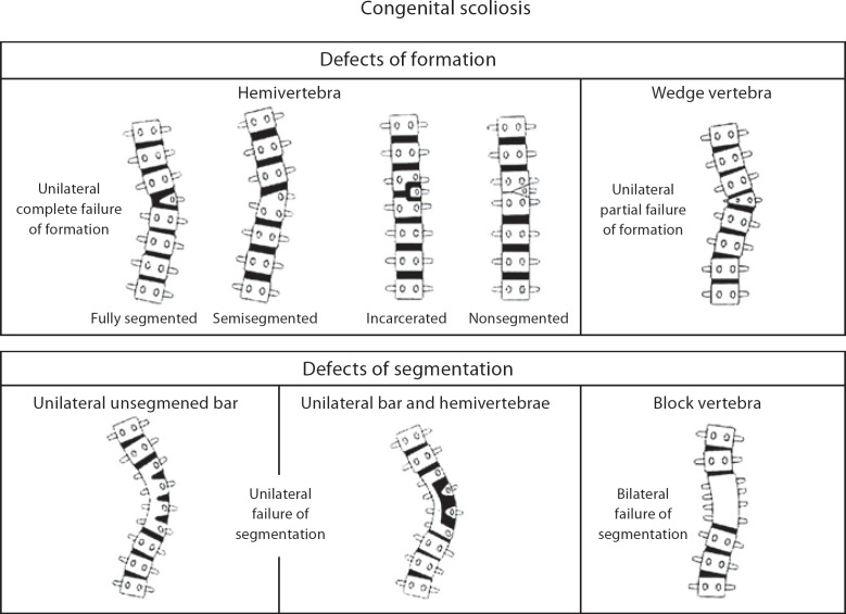
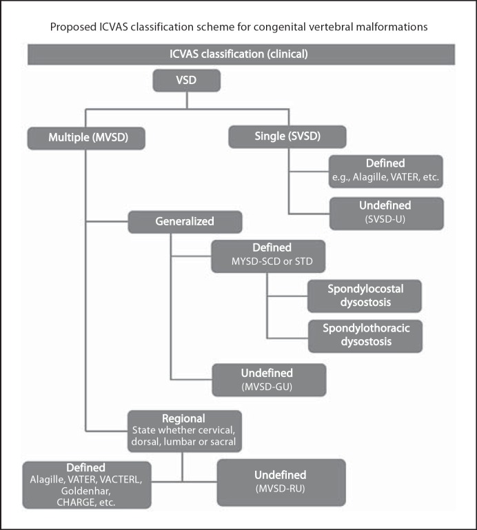
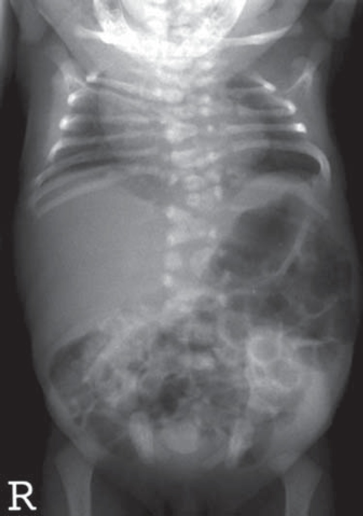
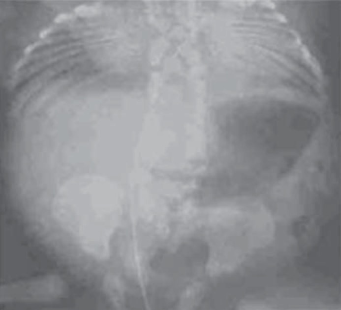
Congenital vertebral malformations (CVM) pose a significant health problem because they can be associated with spinal deformities, such as congenital scoliosis and kyphosis, in addition to various syndromes and other congenital malformations. Additional information remains to be learned regarding the natural history of congenital scoliosis and related health problems. Although significant progress has been made in understanding the process of somite formation, which gives rise to vertebral bodies, there is a wide gap in our understanding of how genetic factors contribute to CVM development. Maternal diabetes during pregnancy most commonly contributes to the occurrence of CVM, followed by other factors such as hypoxia and anticonvulsant medications. This review highlights several emerging clinical issues related to CVM, including pulmonary and orthopedic outcome in congenital scoliosis. Recent breakthroughs in genetics related to gene and environment interactions associated with CVM development are discussed. The Klippel-Feil syndrome which is associated with cervical segmentation abnormalities is illustrated as an example in which animal models, such as the zebrafish, can be utilized to provide functional evidence of pathogenicity of identified mutations.
Keywords: Congenital vertebral malformation; Hemifacial microsomia; Klippel-Feil syndrome; Maternal diabetes; Spondylocostal dysostosis; Spondylothoracic dysostosis; Thoracic insufficiency syndrome; VACTERL syndrome.
Figures

References
-
- Aberg A, Westbom L, Källén B. Congenital malformations among infants whose mothers had gestational diabetes or preexisting diabetes. Early Hum Dev. 2001;61:85–95. - PubMed
-
- Aburakawa K, Harada M, Otake S. Clinical evaluations of the treatment of scoliosis. Orthop Surg Trauma. 1996;39:55–62.
-
- Akbarnia BA, Emans JB. Complications of growth-sparing surgery in early onset scoliosis. Spine (Phila Pa 1976) 2010;35:2193–2204. - PubMed
-
- Alexander PG, Tuan RS. Role of environmental factors in axial skeletal dysmorphogenesis. Birth Defects Res C Embryo Today. 2010;90:118–132. - PubMed
-
- Alexander PG, Chau L, Tuan RS. Role of nitric oxide in chick embryonic organogenesis and dysmorphogenesis. Birth Defects Res A Clin Mol Teratol. 2007;79:581–594. - PubMed
LinkOut - more resources
Full Text Sources
Miscellaneous
