. 2013 May 6:4:46.
doi: 10.3389/fneur.2013.00046. eCollection 2013.

Ataxin-3 and its e3 partners: implications for machado-joseph disease

Affiliations

Ataxin-3 and its e3 partners: implications for machado-joseph disease

Thomas M Durcan et al. Front Neurol. .

Abstract

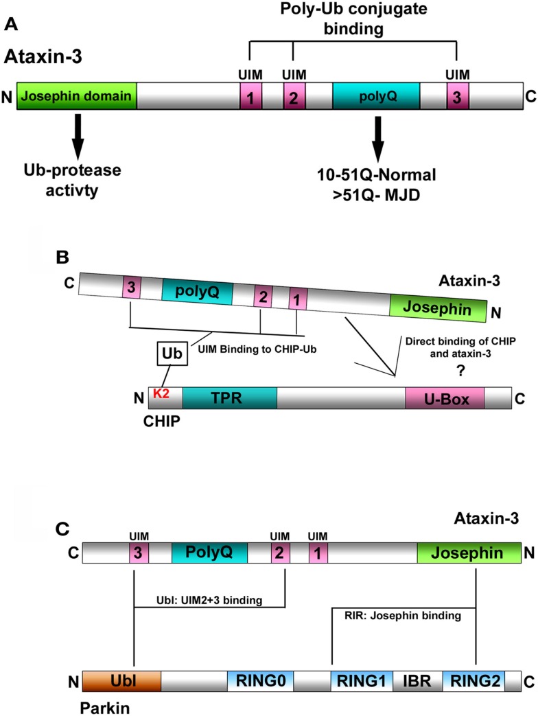
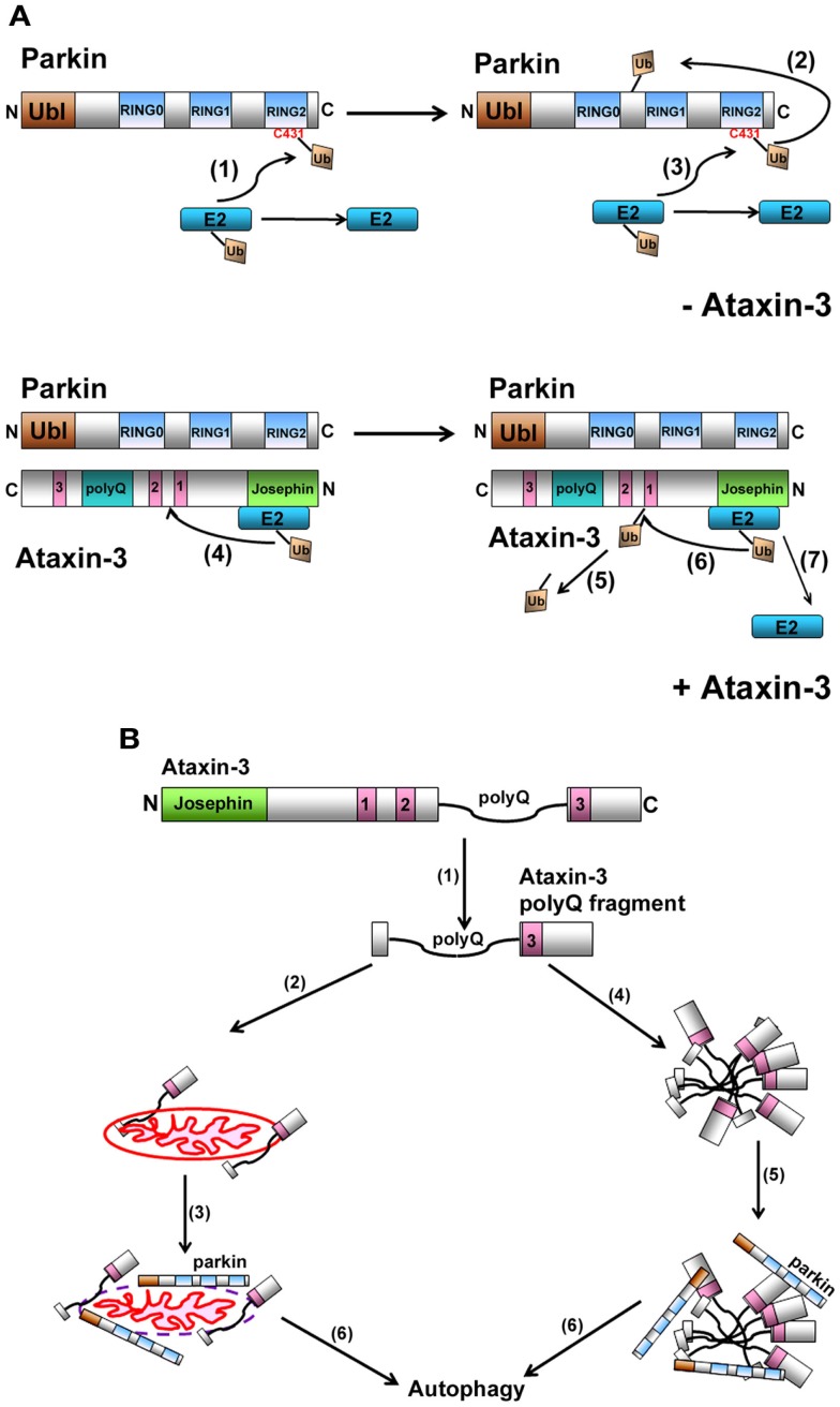
Machado-Joseph disease (MJD) is the most common dominant inherited ataxia worldwide, caused by an unstable CAG trinucleotide expansion mutation within the SCA3 gene resulting in an expanded polyglutamine tract within the ataxin-3 protein. Ataxin-3 functions as a deubiquitinating enzyme (DUB), within the Ub system and whilst many DUBs are known to partner with and deubiquitinate specific E3-Ub ligases, ataxin-3 had no identified E3 partner until recent studies implicated parkin and CHIP, two neuroprotective E3 ligases. MJD often presents with symptoms of Parkinson disease (PD), which led to identification of parkin as a novel E3-Ub ligase whose activity was regulated by ataxin-3-mediated deubiquitination. Findings from these studies also revealed an unexpected convergence upon the E2-Ub-conjugating enzyme in the regulation of an E3/DUBenzyme pair. Moreover, mutant but not wild-type ataxin-3 promotes the clearance of parkin via the autophagy pathway, raising the intriguing possibility that increased turnover of parkin may contribute to the pathogenesis of MJD and help explain some of the Parkinsonian features in MJD. In addition to parkin, the U-box E3 ligase CHIP, a neuroprotective E3 implicated in protein quality control, was identified as a second E3 partner of ataxin-3, with ataxin-3 regulating the ability of CHIP to ubiquitinate itself. Indeed, ataxin-3 not only deubiquitinated CHIP, but also trimmed Ub conjugates on CHIP substrates, thereby regulating the length of Ub chains. Interestingly, when expanded ataxin-3 was present, CHIP levels were also reduced in the brains of MJD transgenic mice, raising the possibility that loss of one or both E3 partners may be a contributing factor in the pathogenesis of SCA3. In this review we discuss the implications from these studies and describe the importance of these findings in helping us understand the molecular processes involved in SCA3 and other neurodegenerative disorders.

Keywords: CHIP; Machado–Joseph disease; Parkinson’s disease; ataxin-3; parkin; polyglutamine expansion.


Figures

Figure 1
Figure 1
(A) Schematic representation of ataxin-3. Ataxin-3 contains an N-terminal Josephin domain, and three Ub-interacting motifs (UIMs) that flank a polyQ tract of variable length. In normal individuals, this tract can be anywhere from 10 to 51Q. In individuals with MJD, this tract has expanded to now contain >51Q. (B) Schematic representation of the interaction between ataxin-3 and CHIP. (1) CHIP contains an N-terminal tetratricopeptide repeat (TPR) domain and a C-terminal U-Box domain that confers CHIP with E3-Ub ligase activity. CHIP monoubiquitinates itself at lysine 2 (K2), which in turn facilitates the interaction between CHIP and ataxin-3. CHIP and ataxin-3 also interact directly although the nature of this interaction has yet to be characterized. (C) Schematic representation of the interaction between ataxin-3 and parkin. Parkin interacts directly with ataxin-3 via its N-terminal Ub-like domain (Ubl) that binds to the UIMs of ataxin-3, and through its inbetweenring-RING2 (IBR-RING2) domain binding to the Josephin domain of ataxin-3.
Figure 2
Figure 2
(A) Model representation of how ataxin-3-mediated deubiquitination regulates parkin self-ubiquitination. When parkin ubiquitinates itself, the charged E2-Ub thioester transfers the Ub onto C431 in parkin to form a transient parkin-Ub thioester complex (1). Parkin now transfers this Ub onto one of its own lysines generating an isopeptide linkage (2). Following the transfer of Ub onto itself, parkin is now free to receive a new Ub and this continues the cycle of parkin self-ubiquitination (3). When ataxin-3 is present, we propose that ataxin-3, through its interaction with the IBR-RING2 domain of parkin, blocks the C431 residue, impeding parkin from forming a parkin-Ub thioester. Instead, ataxin-3 now interacts with the E2, directing the transfer of Ub onto a lysine within ataxin-3 (4). This isopeptide linkage is transient as ataxin-3 can catalyze its removal (5) (unpublished data), allowing ataxin-3 to repeat this cycle of self-ubiquitination/deubiquitination, thereby reducing the ability of parkin to ubiquitinate itself (6 and 7). (B) Proposed model for how the expanded ataxin-3 can promote the autophagic clearance of parkin. In the presence of calcium, calpains cleave full-length ataxin-3, generating fragments containing the expanded polyQ tract (1). These fragments can associate with mitochondria (2), causing mitochondrial damage, parkin recruitment (3), and ultimately parkin-mediated clearance of itself and the mitochondria via the autophagy pathway (6). Alternatively, these expanded polyQ fragments also have a propensity to aggregate (4), recruiting parkin and CHIP (5), which now act to direct these aggregates and themselves for autophagic clearance (6).

Similar articles

Cited by

References

    1. Bettencourt C., Santos C., Coutinho P., Rizzu P., Vasconcelos J., Kay T., et al. (2011). Parkinsonian phenotype in Machado-Joseph disease (MJD/SCA3): a two-case report. BMC Neurol. 11:131.10.1186/1471-2377-11-131 - DOI - PMC - PubMed
    1. Burnett B., Li F., Pittman R. N. (2003). The polyglutamine neurodegenerative protein ataxin-3 binds polyubiquitylated proteins and has ubiquitin protease activity. Hum. Mol. Genet. 12, 3195–320510.1093/hmg/ddg344 - DOI - PubMed
    1. Burnett B. G., Pittman R. N. (2005). The polyglutamine neurodegenerative protein ataxin 3 regulates aggresome formation. Proc. Natl. Acad. Sci. U.S.A. 102, 4330–433510.1073/pnas.0407252102 - DOI - PMC - PubMed
    1. Chai Y., Wu L., Griffin J. D., Paulson H. L. (2001). The role of protein composition in specifying nuclear inclusion formation in polyglutamine disease. J. Biol. Chem. 276, 44889–4489710.1074/jbc.M106575200 - DOI - PubMed
    1. Daviet L., Colland F. (2008). Targeting ubiquitin specific proteases for drug discovery. Biochimie 90, 270–28310.1016/j.biochi.2007.09.013 - DOI - PubMed

LinkOut - more resources